

The DOI matches its Crossref record; 1 of the 1 references it cites were found in a registry or an index.
Characterization of Dye-decolorizing Peroxidase (DyP) from Thermomonospora curvata Reveals Unique Catalytic Properties of A-type DyPs
Overview
The claims and statistics of this paper haven’t been checked yet.
Run the check — free, no account needed. It reads the open-access text and checks the sources it cites.
Integrity signals
Registry
Cites
Statistics
Figures
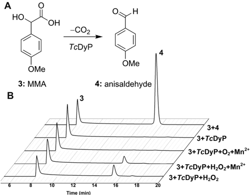
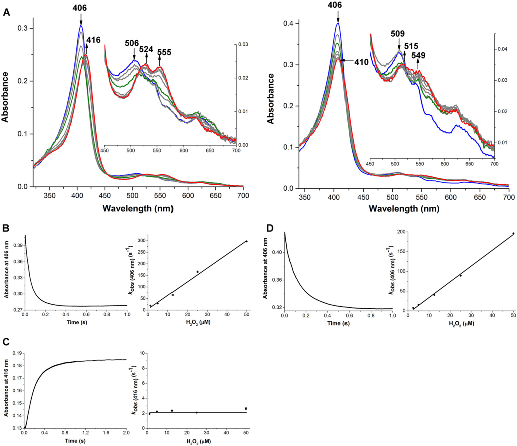
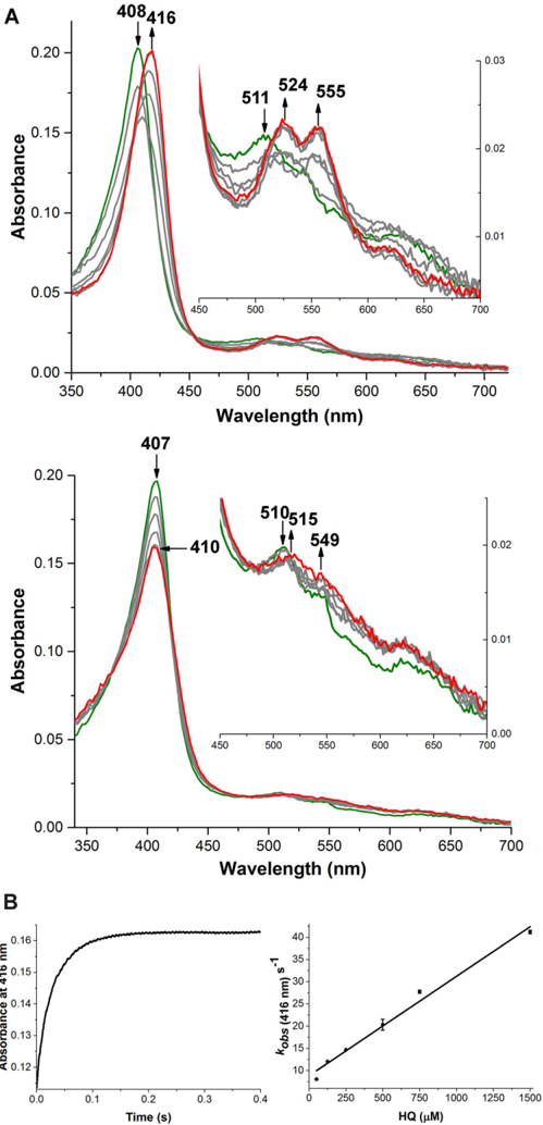
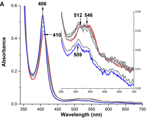
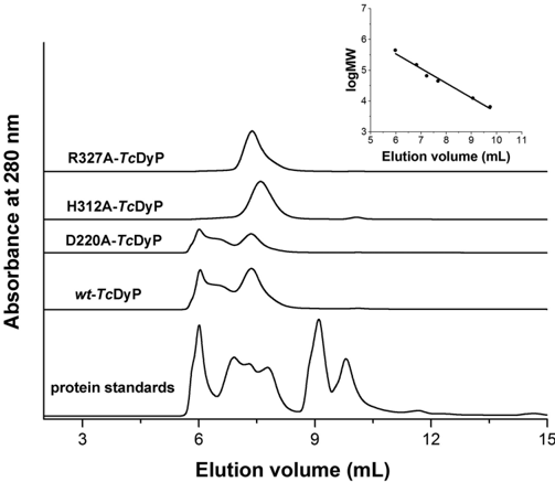
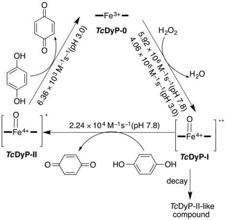
Verification
Claims the paper makes
The claims and statistics of this paper haven’t been checked yet.
Run the check — free, no account needed. It reads the open-access text and checks the sources it cites.
Claims other papers make about this one
Each is checked against this paper’s own text: does it say what the citing paper says it does?
The source backs part of the claim, not all of it — or backs it for a different population, condition or comparison.
In the citing paper
“Although their action mechanism resembles that of other peroxidases, anthraquinone dye degradation is not yet fully mapped 129-134 .”
From Degradation of anthraquinone dyes from effluents: A review focusing on enzymatic dye degradation with industrial potential (2020) · cites this paper as [130]
Experimental — accuracy not yet measured
Self-check
Percentages, sums and reported differences re-computed from the paper’s own numbers.
The self-check needs the paper’s full text.
Citations
Cites
Commercializing lignocellulosic bioethanol: technology bottlenecks and possible remedies
Banerjee, S., Mudliar, S., Sen, R., Giri, B., Satpute, D., Chakrabarti, T., & Pandey, R. A. (2010) · Biofuels, Bioproducts and Biorefining · doi:10.1002/bbb.188 · Cited by 377
Research works printed but not linked: 65
- Paliwal, 2012, Bioligninolysis: recent updates for biotechnological solution, Appl. Biochem. Biotechnol, 167, 1865, 10.1007/s12010-012-9735-3 · DOI
- FitzPatrick, 2010, A biorefinery processing perspective: treatment of lignocellulosic materials for the production of value-added products, Bioresour. Technol, 101, 8915, 10.1016/j.biortech.2010.06.125 · DOI · Paper page
- ten Have, 2001, Oxidative mechanisms involved in lignin degradation by white-rot fungi, Chem. Rev, 101, 3397, 10.1021/cr000115l · DOI
- Brown, 2014, Exploring bacterial lignin degradation, Curr. Opin. Chem. Biol, 19, 1, 10.1016/j.cbpa.2013.11.015 · DOI
- Fennington, 1984, Cellulase biosynthesis in a catabolite repression-resistant mutant of Thermomonospora curvata, Appl. Environ. Microbiol, 47, 201, 10.1128/aem.47.1.201-204.1984 · DOI
- Stutzenberger, 1979, Degradation of cellulosic substances by Thermomonospora curvata, Biotechnol. Bioeng, 21, 909, 10.1002/bit.260210514 · DOI
- Chertkov, 2011, Complete genome sequence of Thermomonospora curvata type strain (B9T), Stand. Genomic Sci, 4, 13, 10.4056/sigs.1453580 · DOI
- Ahmad, 2011, Identification of DypB from Rhodococcus jostii RHA1 as a lignin peroxidase, Biochemistry, 50, 5096, 10.1021/bi101892z · DOI
- Brown, 2012, Identification and characterization of a multifunctional dye peroxidase from a lignin-reactive Bacterium, ACS Chem. Biol, 7, 2074, 10.1021/cb300383y · DOI
- Min, 2015, A dye-decolorizing peroxidase from Bacillus subtilis exhibiting substrate-dependent optimum temperature for dyes and β-ether lignin dimer, Sci. Rep, 5, 10.1038/srep08245 · DOI
- Colpa, 2014, DyP-type peroxidases: a promising and versatile class of enzymes, J. Ind. Microbiol. Biotechnol, 41, 1, 10.1007/s10295-013-1371-6 · DOI · Paper page
- Koua, 2009, PeroxiBase: a database with new tools for peroxidase family classification, Nucleic Acids Res, 37, D261, 10.1093/nar/gkn680 · DOI
- Liu, 2011, Crystal structure and biochemical features of EfeB/YcdB from Escherichia coli O157: ASP(235) plays divergent roles in different enzyme-catalyzed processes, J. Biol. Chem, 286, 14922, 10.1074/jbc.M110.197780 · DOI
- Sugano, 2009, Degradation pathway of an anthraquinone dye catalyzed by a unique peroxidase DyP from Thanatephorus cucumeris Dec 1, Biodegradation, 20, 433, 10.1007/s10532-008-9234-y · DOI · Paper page
- Sugano, 2007, DyP, a unique dye-decolorizing peroxidase, represents a novel heme peroxidase family, J. Biol. Chem, 282, 36652, 10.1074/jbc.M706996200 · DOI
- Roberts, 2011, Characterization of dye-decolorizing peroxidases from Rhodococcus jostii RHA1, Biochemistry, 50, 5108, 10.1021/bi200427h · DOI
- Yang, 2013, Protein-ligand binding site recognition using complementary binding-specific substructure comparison and sequence profile alignment, Bioinformatics, 29, 2588, 10.1093/bioinformatics/btt447 · DOI
- Yang, 2013, BioLiP: a semi-manually curated database for biologically relevant ligand-protein interactions, Nucleic Acids Res, 41, D1096, 10.1093/nar/gks966 · DOI
- Gajhede, 1997, Crystal structure of horseradish peroxidase C at 2.15 angstrom resolution, Nat. Struct. Biol, 4, 1032, 10.1038/nsb1297-1032 · DOI
- Sugano, 2009, DyP-type peroxidases comprise a novel heme peroxidase family, Cell Mol. Life Sci, 66, 1387, 10.1007/s00018-008-8651-8 · DOI
- Singh, 2012, Distal heme pocket residues of B-type dye-decolorizing peroxidase: arginine but not aspartate is essential for peroxidase activity, J. Biol. Chem, 287, 10623, 10.1074/jbc.M111.332171 · DOI
- Qin, 2008, Construction of a series of vectors for high throughput cloning and expression screening of membrane proteins from Mycobacterium tuberculosis, BMC Biotechnol, 8, 51, 10.1186/1472-6750-8-51 · DOI
- Smith, 1985, Measurement of protein using bicinchoninic acid, Anal. Biochem, 150, 76, 10.1016/0003-2697(85)90442-7 · DOI · Paper page
- Berry, 1987, Simultaneous determination of hemes-A, hemes-B, and hemes-C from pyridine hemochrome spectra, Anal. Biochem, 161, 1, 10.1016/0003-2697(87)90643-9 · DOI
- Mcllvaine, 1921, A buffer solution for colorimetric comparison, J. Biol. Chem, 49, 183, 10.1016/S0021-9258(18)86000-8 · DOI
- Brown, 2011, Discovery and characterization of heme enzymes from unsequenced bacteria: application to microbial lignin degradation, J. Am. Chem. Soc, 133, 18006, 10.1021/ja203972q · DOI
- Santos, 2014, New dye-decolorizing peroxidases from Bacillus subtilis and Pseudomonas putida MET94: towards biotechnological applications, Appl. Microbiol. Biotechnol, 98, 2053, 10.1007/s00253-013-5041-4 · DOI · Paper page
- Adak, 1996, Probing the active site residues in aromatic donor oxidation in horseradish peroxidase: Involvement of an arginine and a tyrosine residue in aromatic donor binding, Biochem. J, 314, 985, 10.1042/bj3140985 · DOI
- La Mar, 1992, Proton NMR investigation of the influence of interacting sites on the dynamics and thermodynamics of substrate and ligand-binding to horseradish peroxidase, Biochemistry, 31, 9158, 10.1021/bi00153a007 · DOI
- Childs, 1975, Steady-state kinetics of peroxidase with 2,2′-azino-di-(3-ethylbenzthiazoline-6-sulphonic acid) as chromogen, Biochem. J, 145, 93, 10.1042/bj1450093 · DOI
- Wang, 1999, Kinetic evidence for the formation of a Michaelis-Menten-like complex between horseradish peroxidase compound II and di-(N-acetyl-l-tyrosine), Biochem. J, 340, 329, 10.1042/bj3400329 · DOI
- Strittmatter, 2015, The toolbox of Auricularia auricula-judae dye-decolorizing peroxidase: identification of three new potential substrate-interaction sites, Arch. Biochem. Biophys, 574, 75, 10.1016/j.abb.2014.12.016 · DOI
- Linde, 2015, Catalytic surface radical in dye-decolorizing peroxidase: a computational, spectroscopic and site-directed mutagenesis study, Biochem. J, 466, 253, 10.1042/BJ20141211 · DOI
- Strittmatter, 2013, First crystal structure of a fungal high-redox potential dye-decolorizing Peroxidase substrate interaction sites and long-range electron transfer, J. Biol. Chem, 288, 4095, 10.1074/jbc.M112.400176 · DOI
- Wei, 2004, A theoretical study on bond dissociation energies and oxidation potentials of monolignols, J. Mol. Struct. (Theochem), 712, 197, 10.1016/j.theochem.2004.10.032 · DOI
- Baciocchi, 2002, Lignin peroxidase catalysed oxidation of 4-methoxymandelic acid: the role of mediator structure, Tetrahedron, 58, 8087, 10.1016/S0040-4020(02)00997-3 · DOI
- Tien, 1997, Oxidation of 4-methoxymandelic acid by lignin peroxidase: mediation by veratryl alcohol, J. Biol. Chem, 272, 8912, 10.1074/jbc.272.14.8912 · DOI
- Schonbaum, 1972, Interaction of peroxidases with aromatic peracids and alkyl peroxides: product analysis, J. Biol. Chem, 247, 3353, 10.1016/S0021-9258(19)45252-6 · DOI
- Morales, 2012, Two oxidation sites for low redox potential substrates: a directed mutagenesis, kinetic, and cystallographic study on Pleurotus eryngii versatile peroxidase, J. Biol. Chem, 287, 41053, 10.1074/jbc.M112.405548 · DOI
- Battistuzzi, 2010, Redox properties of heme peroxidases, Arch. Biochem. Biophys, 500, 21, 10.1016/j.abb.2010.03.002 · DOI · Paper page
- Sezer, 2013, Distinct structural and redox properties of the heme active site in bacterial dye decolorizing peroxidase-type peroxidases from two subfamilies: resonance Raman and electrochemical study, Biochemistry, 52, 3074, 10.1021/bi301630a · DOI
- Petruccioli, 2009, Kinetic and redox properties of MnP II: a major manganese peroxidase isoenzyme from Panus tigrinus CBS 577.79, J. Biol. Inorg. Chem, 14, 1153, 10.1007/s00775-009-0559-8 · DOI
- Santucci, 2000, Redox equilibria of manganese peroxidase from Phanerochaetes chrysosporium: functional role of residues on the proximal side of the haem pocket, Biochem. J, 349, 85, 10.1042/bj3490085 · DOI
- Zhang, 2002, Direct voltammetry and catalysis with Mycobacterium tuberculosis catalase-peroxidase, peroxidases, and catalase in lipid films, Anal. Chem, 74, 163, 10.1021/ac010701u · DOI
- van Bloois, 2010, A robust and extracellular heme-containing peroxidase from Thermobifida fusca as prototype of a bacterial peroxidase superfamily, Appl. Microbiol. Biotechnol, 86, 1419, 10.1007/s00253-009-2369-x · DOI · Paper page
- Li, 2012, Identification and molecular characterization of a novel DyP-type peroxidase from Pseudomonas aeruginosa PKE117, Appl. Biochem. Biotechnol, 166, 774, 10.1007/s12010-011-9466-x · DOI
- Zubieta, 2007, Identification and structural characterization of heme binding in a novel dye-decolorizing peroxidase, TyrA, Proteins, 69, 234, 10.1002/prot.21673 · DOI
- Ogola, 2009, Molecular characterization of a novel peroxidase from the cyanobacterium Anabaena sp strain PCC 7120, Appl. Environ. Microbiol, 75, 7509, 10.1128/AEM.01121-09 · DOI · Paper page
- Kim, 1999, Purification and characterization of a novel peroxidase from Geotrichum candidum Dec 1 involved in decolorization of dyes, Appl. Environ. Microbiol, 65, 1029, 10.1128/AEM.65.3.1029-1035.1999 · DOI · Paper page
- Liers, 2010, DyP-like peroxidases of the jelly fungus Auricularia auricula-judae oxidize nonphenolic lignin model compounds and high-redox potential dyes, Appl. Microbiol. Biotechnol, 85, 1869, 10.1007/s00253-009-2173-7 · DOI
- Johjima, 2003, Isolation and cDNA cloning of novel hydrogen peroxide-dependent phenol oxidase from the basidiomycete Termitomyces albuminosus, Appl. Microbiol. Biotechnol, 61, 220, 10.1007/s00253-003-1236-4 · DOI
- Fernández-Fueyo, 2012, Lignin-degrading peroxidases from genome of selective ligninolytic fungus Ceriporiopsis subvermispora, J. Biol. Chem, 287, 16903, 10.1074/jbc.M112.356378 · DOI
- Heinfling, 1998, A study on reducing substrates of manganese-oxidizing peroxidases from Pleurotus eryngii and Bjerkandera adusta, FEBS Lett, 428, 141, 10.1016/S0014-5793(98)00512-2 · DOI
- Kuan, 1993, Kinetic analysis of manganese peroxidase: the reaction with manganese complexes, J. Biol. Chem, 268, 20064, 10.1016/S0021-9258(20)80694-2 · DOI
- Banci, 2003, NMR study of manganese(II) binding by a new versatile peroxidase from the white-rot fungus Pleurotus eryngii, J. Biol. Inorg. Chem, 8, 751, 10.1007/s00775-003-0476-1 · DOI
- Hiner, 2001, Detection of a tryptophan radical in the reaction of ascorbate peroxidase with hydrogen peroxide, Eur. J. Biochem, 268, 3091, 10.1046/j.1432-1327.2001.02208.x · DOI
- Sivaraja, 1989, Identification by ENDOR of Trp191 as the free-radical site in cytochrome c peroxidase compound ES, Science, 245, 738, 10.1126/science.2549632 · DOI
- Choudhury, 1994, Role of the proximal ligand in peroxidase catalysis: crystallographic, kinetic, and spectral studies of cytochrome c peroxidase proximal ligand mutants, J. Biol. Chem, 269, 20239, 10.1016/S0021-9258(17)31982-8 · DOI
- Rodriguez-Lopez, 1996, Role of arginine 38 in horseradish peroxidase a critical, residue for substrate binding and catalysis, J. Biol. Chem, 271, 4023, 10.1074/jbc.271.8.4023 · DOI
- Vitello, 1993, Effect of arginine-48 replacement on the reaction between cytochrome c peroxidase and hydrogen peroxide, Biochemistry, 32, 9807, 10.1021/bi00088a036 · DOI
- Efimov, 2011, Proton delivery to ferryl heme in a heme peroxidase: enzymatic use of the grotthuss mechanism, J. Am. Chem. Soc, 133, 15376, 10.1021/ja2007017 · DOI
- Sharp, 2003, Crystal structure of the ascorbate peroxidase-ascorbate complex, Nat. Struct. Biol, 10, 303, 10.1038/nsb913 · DOI
- Renganathan, 1986, Spectral characterization of the oxidized states of lignin peroxidase, an extracellular heme enzyme from the white rot basidiomycete Phanerochaete chrysosporium, Biochemistry, 25, 1626, 10.1021/bi00355a027 · DOI
- Mino, 1988, Spectral characterization of manganese peroxidase, an extracellular heme enzyme from the lignin-degrading basidiomycete, Phanerochaete chrysosporium, J. Biol. Chem, 263, 7029, 10.1016/S0021-9258(18)68599-0 · DOI
- Wariishi, 1988, Manganese peroxidase from the basidiomycete Phanerochaete chrysosporium: spectral characterization of the oxidized states and the catalytic cycle, Biochemistry, 27, 5365, 10.1021/bi00414a061 · DOI
Cited by
Sorted by citations
Routoula, E., & Patwardhan, S. V. (2020) · Environmental Science & Technology · doi:10.1021/acs.est.9b03737 · Cited by 579
Metadata
- Authors
- Chen, C., Shrestha, R., Jia, K., Gao, P. F., Geisbrecht, B. V., Bossmann, S. H., Shi, J., & Li, P.
- Venue
- Journal of Biological Chemistry
- Publisher
- Elsevier BV
- Published
- 2015-09
- Volume
- 290
- Issue
- 38
- Pages
- 23447-23463
- ISSN
- 0021-9258
- Cited by
- 124
- References
- 1
- Language
- en
About these checks
Registry, retraction, Cites and statistics results are computed, not judged, with one exception: a printed reference that no index lists is read once by a model before it counts as a citation error. Claims are extracted by a model and kept only when their quote appears verbatim in the text; each cited source is then checked, with a model’s help, for whether it says what the claim relies on. Those verdicts can be wrong — read the claim’s quote and the cited source before relying on one.
Statistics and arithmetic are recomputed, not judged.
Confidence bands are shown only for fields calibrated on our annotated test set. None is calibrated yet, so no band is shown.
What counts as a claim ›How we measure confidence ›How we check this ›
Are you an author, or do you see a problem with this page? Request a review
