Cited by 90

The DOI matches its Crossref record.

The C-terminal Domain of Rac1 Contains Two Motifs That Control Targeting and Signaling Specificity

van Hennik, Paula B., Klooster, Jean Paul ten, Halstead, Jon R., Voermans, Carlijn, Anthony, Eloise C., Divecha, Nullin, Hordijk, Peter L.·2003·Journal of Biological Chemistry

Overview

The claims and statistics of this paper haven’t been checked yet.

Run the check — free, no account needed. It reads the open-access text and checks the sources it cites.

A few free checks a day · papers already verified open instantly · results are public

Integrity signals

Registry

Crossref · 10.1074/jbc.m307001200

Cites

Statistics

Figures

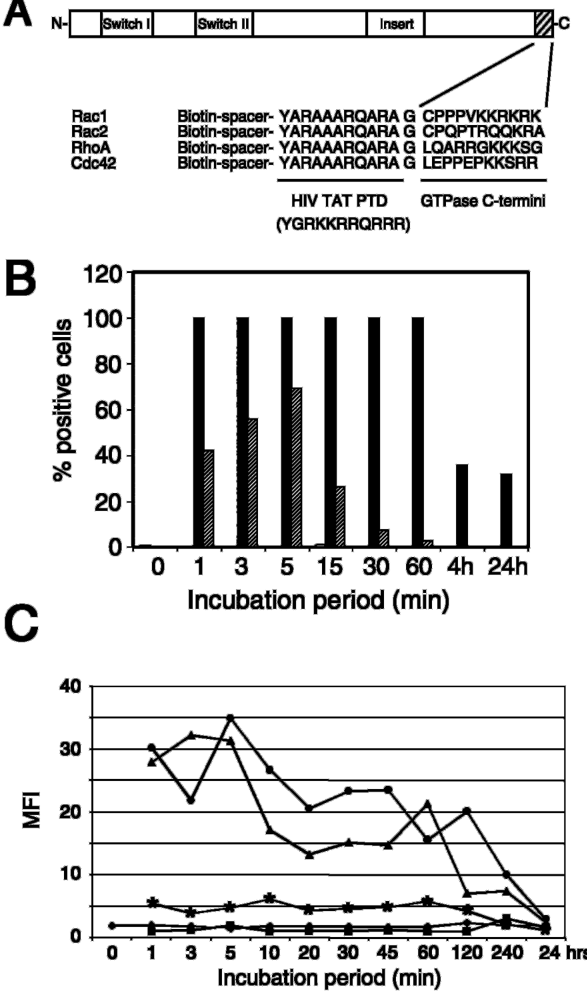
FIG. 1 Characterization of TAT Rho-like GTPase C-terminal peptides. A , schematic representation of the TAT Rho-like GTPase C-terminal peptides used in this study. B , efficiency of entry of biotinylated TAT-Rac1 C-terminal peptide in HL60 cells. Solid and hatched bars indicate percentage of positive cells obtained with 200 and 50 𝛍 g/ml, respectively. A representative example of three experiments is shown. C , kinetics of entry of the TAT C-terminal peptides (200 𝛍 g/ml) in HL60 cells. Diamonds , untreated; squares , TAT; triangles , TAT-Rac1; circles , TAT-Rac2; asterisks , TAT-Cdc42. A representative example of three experiments is shown.
FIG. 1 Characterization of TAT Rho-like GTPase C-terminal peptides. A , schematic representation of the TAT Rho-like GTPase C-terminal peptides used in this study. B , efficiency of entry of biotinylated TAT-Rac1 C-terminal peptide in HL60 cells. Solid and hatched bars indicate percentage of positive cells obtained with 200 and 50 𝛍 g/ml, respectively. A representative example of three experiments is shown. C , kinetics of entry of the TAT C-terminal peptides (200 𝛍 g/ml) in HL60 cells. Diamonds , untreated; squares , TAT; triangles , TAT-Rac1; circles , TAT-Rac2; asterisks , TAT-Cdc42. A representative example of three experiments is shown.CC-BY · From the PDF
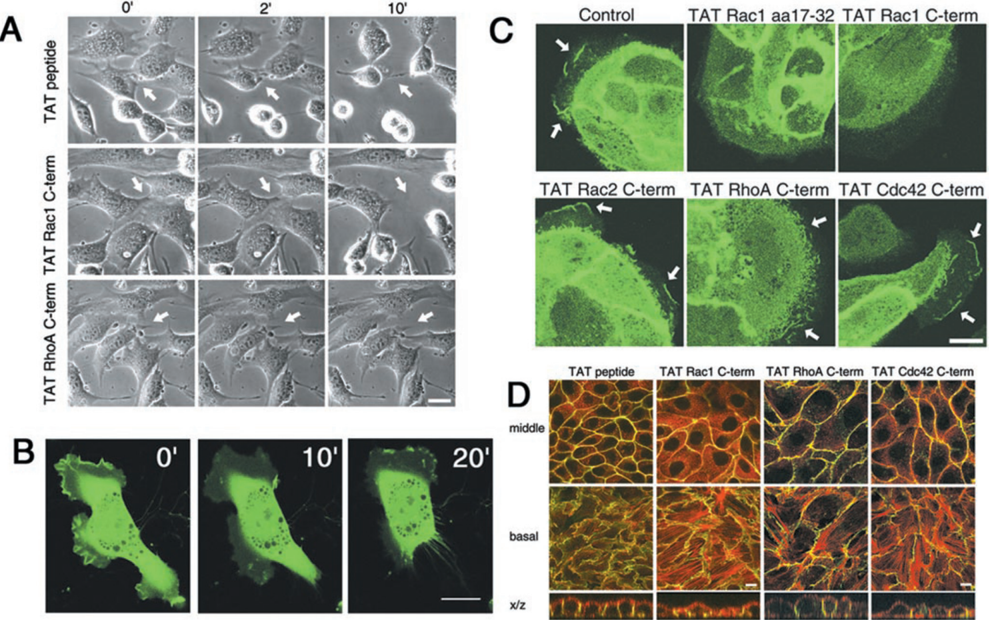
FIG. 2 Specificity of the C-terminal peptides. A , time-lapse analysis of serum-induced contraction of NIH3T3 fibroblasts. In the control situation, serum-starved NIH3T3 cells showed RhoA-mediated contraction ( arrow ) in response to 10% fetal calf serum in ∼ 10 min ( upper panels ). This response was not affected ( arrow in middle panels ) by a 30-min incubation of the cells with 400 𝛍 g/ml TAT-Rac1 but was inhibited ( arrow in lower panels ) by 400 𝛍 g/ml TAT-RhoA. Bar , 20 𝛍 m. B , spontaneous membrane ruffling in Rac1-GFP-transduced HUVEC was blocked by TAT-Rac1 and accompanied by a loss of GFP-Rac1 localization. Lamella spreading and contractility were not impaired. Bar , 20 𝛍 m. C , HGF-induced (10 ng/ml; 30 min) membrane ruffling in GFP-Rac1-transduced MDCKII cells was inhibited within 1 to 3 min by TAT-Rac1 or TAT-Rac1 17 -32 peptide but not by TAT-Rac2, TAT-RhoA, or TAT-Cdc42. Arrows indicate GFP-Rac1-positive membrane ruffles. Bar , 20 𝛍 m. D , effect of TAT Rho-like GTPase C-terminal peptides on epithelial cell-cell junctions. Confocal images of confluent MDCKII cells show E-cadherin in green and polymerized actin in red after incubation with different TAT C-terminal peptides (200 𝛍 g/ml for 18 h). The cells exposed to TAT-Rac1 or TAT-Cdc42 C-terminal peptide showed an increase in stress fibers and a decrease in E-cadherin-based cell-cell adhesion indicated by a reduction in polarized adherens junctions ( X / Z scan ). Bar , 20 𝛍 m.
FIG. 2 Specificity of the C-terminal peptides. A , time-lapse analysis of serum-induced contraction of NIH3T3 fibroblasts. In the control situation, serum-starved NIH3T3 cells showed RhoA-mediated contraction ( arrow ) in response to 10% fetal calf serum in ∼ 10 min ( upper panels ). This response was not affected ( arrow in middle panels ) by a 30-min incubation of the cells with 400 𝛍 g/ml TAT-Rac1 but was inhibited ( arrow in lower panels ) by 400 𝛍 g/ml TAT-RhoA. Bar , 20 𝛍 m. B , spontaneous membrane ruffling in Rac1-GFP-transduced HUVEC was blocked by TAT-Rac1 and accompanied by a loss of GFP-Rac1 localization. Lamella spreading and contractility were not impaired. Bar , 20 𝛍 m. C , HGF-induced (10 ng/ml; 30 min) membrane ruffling in GFP-Rac1-transduced MDCKII cells was inhibited within 1 to 3 min by TAT-Rac1 or TAT-Rac1 17 -32 peptide but not by TAT-Rac2, TAT-RhoA, or TAT-Cdc42. Arrows indicate GFP-Rac1-positive membrane ruffles. Bar , 20 𝛍 m. D , effect of TAT Rho-like GTPase C-terminal peptides on epithelial cell-cell junctions. Confocal images of confluent MDCKII cells show E-cadherin in green and polymerized actin in red after incubation with different TAT C-terminal peptides (200 𝛍 g/ml for 18 h). The cells exposed to TAT-Rac1 or TAT-Cdc42 C-terminal peptide showed an increase in stress fibers and a decrease in E-cadherin-based cell-cell adhesion indicated by a reduction in polarized adherens junctions ( X / Z scan ). Bar , 20 𝛍 m.CC-BY · From the PDF
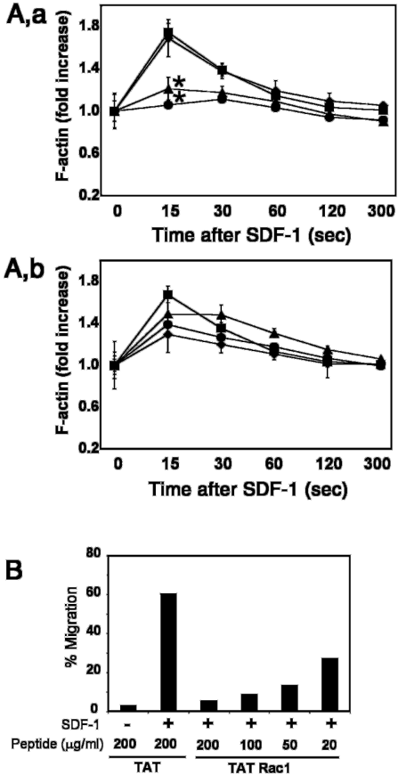
FIG. 3 Effect of C-terminal peptides on SDF-1-induced actin polymerization and migration in HL60 cells. A , a shows SDF-1induced actin polymerization (-fold increase of F-actin) in HL60 cells that were untreated ( squares ) or exposed to the PTD ( diamonds ), TATRac1 ( triangles ), or TAT-Rac1 17 -32 ( circles ) peptides. A , b shows the effect of treatment of HL60 cells with TAT-Rac2 ( diamonds ), TAT-RhoA ( triangles ), or TAT-Cdc42 ( circles ) C-terminal peptides on SDF-1-induced actin polymerization. Squares represent untreated cells. In conclusion, actin polymerization after 30 min of pre-incubation with 200 𝛍 g/ml of the C-terminal peptides was not significantly inhibited by the PTD ( a , diamonds ), TAT-RhoA ( b , triangles ), TAT-Rac2 ( b , diamonds ), or TAT-Cdc42 ( b , circles ). TAT-Rac1 ( a , triangles ) and TAT-Rac1 17 -32 ( a , circles ) did significantly inhibit the response at t = 15 s in comparison to untreated cells ( a and b , squares ). Data are depicted as mean -fold increase of F-actin ± S.E. ( n = 3 -6); *, p < 0.05. B , representative example ( n = 3) of SDF-1 (100 ng/ml)-induced migration after a 30-min pre-treatment with the TAT control or increasing concentrations of TAT-Rac1. Data are depicted as percentage migration.
FIG. 3 Effect of C-terminal peptides on SDF-1-induced actin polymerization and migration in HL60 cells. A , a shows SDF-1induced actin polymerization (-fold increase of F-actin) in HL60 cells that were untreated ( squares ) or exposed to the PTD ( diamonds ), TATRac1 ( triangles ), or TAT-Rac1 17 -32 ( circles ) peptides. A , b shows the effect of treatment of HL60 cells with TAT-Rac2 ( diamonds ), TAT-RhoA ( triangles ), or TAT-Cdc42 ( circles ) C-terminal peptides on SDF-1-induced actin polymerization. Squares represent untreated cells. In conclusion, actin polymerization after 30 min of pre-incubation with 200 𝛍 g/ml of the C-terminal peptides was not significantly inhibited by the PTD ( a , diamonds ), TAT-RhoA ( b , triangles ), TAT-Rac2 ( b , diamonds ), or TAT-Cdc42 ( b , circles ). TAT-Rac1 ( a , triangles ) and TAT-Rac1 17 -32 ( a , circles ) did significantly inhibit the response at t = 15 s in comparison to untreated cells ( a and b , squares ). Data are depicted as mean -fold increase of F-actin ± S.E. ( n = 3 -6); *, p < 0.05. B , representative example ( n = 3) of SDF-1 (100 ng/ml)-induced migration after a 30-min pre-treatment with the TAT control or increasing concentrations of TAT-Rac1. Data are depicted as percentage migration.CC-BY · From the PDF
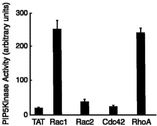
FIG. 4 Association of PIP5K with TAT, TAT-Rac1, TAT-Rac2, TAT-Cdc42, and TAT-RhoA peptides in HL60 cells. Cells were lysed prior to the addition of the various peptides, and peptide-protein complexes were collected using streptavidin-coated beads. Associated PIP5K activity was determined as described under ' Materials and Methods. ' PIP5K associated with TAT-Rac1 but not with the TAT control, TAT-Rac2, or TAT-Cdc42. TAT-RhoA also associated with PIP5K.
FIG. 4 Association of PIP5K with TAT, TAT-Rac1, TAT-Rac2, TAT-Cdc42, and TAT-RhoA peptides in HL60 cells. Cells were lysed prior to the addition of the various peptides, and peptide-protein complexes were collected using streptavidin-coated beads. Associated PIP5K activity was determined as described under ' Materials and Methods. ' PIP5K associated with TAT-Rac1 but not with the TAT control, TAT-Rac2, or TAT-Cdc42. TAT-RhoA also associated with PIP5K.CC-BY · From the PDF
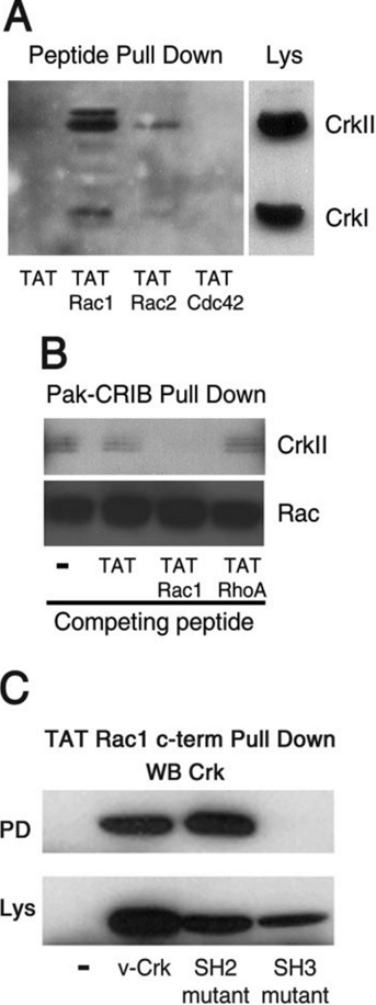
FIG. 5 Characterization of the Crk-Rac1 complex. A , the indicated peptides were added to cell lysates, and complexes were isolated using streptavidin-coated beads. Western blotting of the samples was used to analyze presence of associated Crk. Compared with TAT-Rac2, TAT-Cdc42, and TAT-RhoA, the TAT-Rac1 peptide associated most efficiently with endogenous Crk. B , endogenous Rac was isolated using the biotinylated peptide encoding the CRIB domain of PAK. Crk was also found associated with this complex. This association could be specifically disturbed by adding TAT-Rac1. The TAT-RhoA peptide did not compete for Crk binding to endogenous Rac1. C , peptide pull-down experiments with Cos7 cells, expressing versions of v-Crk, mutated in the SH2 or SH3 domain, showed that the Rac1 C-terminal peptide no longer bound to the Crk protein with a mutation in the SH3 domain, in contrast to the v-Crk version with a mutated SH2 domain.
FIG. 5 Characterization of the Crk-Rac1 complex. A , the indicated peptides were added to cell lysates, and complexes were isolated using streptavidin-coated beads. Western blotting of the samples was used to analyze presence of associated Crk. Compared with TAT-Rac2, TAT-Cdc42, and TAT-RhoA, the TAT-Rac1 peptide associated most efficiently with endogenous Crk. B , endogenous Rac was isolated using the biotinylated peptide encoding the CRIB domain of PAK. Crk was also found associated with this complex. This association could be specifically disturbed by adding TAT-Rac1. The TAT-RhoA peptide did not compete for Crk binding to endogenous Rac1. C , peptide pull-down experiments with Cos7 cells, expressing versions of v-Crk, mutated in the SH2 or SH3 domain, showed that the Rac1 C-terminal peptide no longer bound to the Crk protein with a mutation in the SH3 domain, in contrast to the v-Crk version with a mutated SH2 domain.CC-BY · From the PDF
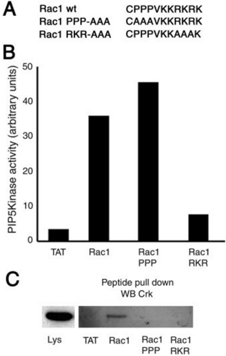
FIG. 6 Characterization of the Rac1 C-terminal protein binding motifs. A , amino acid sequence of the C-terminal TAT-Rac1 WT and the TAT-Rac1 PPP-AAA and TAT-Rac1 RKR-AAA mutant peptides. B , the Rac1 RKR-AAA C-terminal peptide no longer associated with PIP5K activity in the peptide pull-down assay, whereas the Rac1 PPP-AAA peptide did. C , conversely, the Rac1 PPP-AAA C-terminal peptide no longer associated with Crk in the pull-down assay, which shows that the three consecutive prolines in the Rac1 C terminus are required for the Rac1-Crk interaction. The Rac1 RKR-AAA peptide also did not associate to Crk. Representative examples of three experiments are shown.
FIG. 6 Characterization of the Rac1 C-terminal protein binding motifs. A , amino acid sequence of the C-terminal TAT-Rac1 WT and the TAT-Rac1 PPP-AAA and TAT-Rac1 RKR-AAA mutant peptides. B , the Rac1 RKR-AAA C-terminal peptide no longer associated with PIP5K activity in the peptide pull-down assay, whereas the Rac1 PPP-AAA peptide did. C , conversely, the Rac1 PPP-AAA C-terminal peptide no longer associated with Crk in the pull-down assay, which shows that the three consecutive prolines in the Rac1 C terminus are required for the Rac1-Crk interaction. The Rac1 RKR-AAA peptide also did not associate to Crk. Representative examples of three experiments are shown.CC-BY · From the PDF
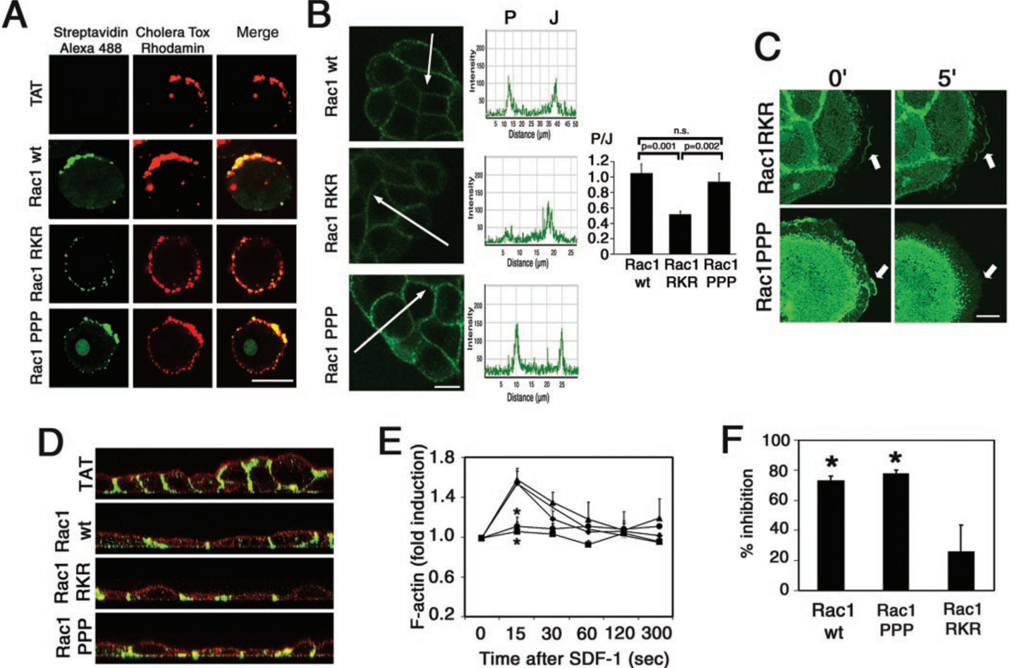
FIG. 7 Cellular localization and effects of Rac1 C-terminal mutant peptides in functional assays. A , co-localization of TAT-Rac1 C-terminal wild type and mutant peptides with lipid rafts in HL60 cells. Confocal images show peptides in green , lipid rafts (CTX) in red , and co-localization in yellow (merge). CTX and the Rac1 RKR-AAA mutant peptide co-localized to a lesser extent compared with co-localization of CTX with the Rac1 WT or the Rac1 PPP-AAA mutant peptide. Bar , 20 𝛍 m. B , localization of biotinylated TAT-Rac1 peptides in MDCKII cells. Confocal images show peptides in green . Rac1 WT and Rac1 PPP-AAA peptides localized to sites of cell-cell contact ( J ) and to the peripheral membrane ( P ). The TAT-Rac1 RKR-AAA peptide also accumulated at cell-cell junctions, whereas the peripheral membrane staining was strongly reduced. Bar , 20 𝛍 m. Pixel intensities along the indicated arrows are shown in the right panels . The difference in localization was significantly different as was shown by statistical analysis of the ratios of peripheral and junctional staining intensities for the three peptides used. Graph shows mean P/J ratio ( ± S.E.) of 10 intensity measurements for the Rac1 WT, Rac1 PPP-AAA, and Rac1 RKR-AAA peptides. Data acquisition was done using identical confocal settings for each peptide. n.s. , not significant. C , HGF-induced membrane ruffling of GFP-Rac1-expressing MDCKII cells was no longer blocked by the Rac1 RKR-AAA peptide, whereas the Rac1 PPP-AAA peptide still blocked membrane ruffling. D , the Rac1 WT and mutant peptides all affected MDCK cell polarity, as revealed by X/Z sectioning following 18 h of treatment with the peptides. E , SDF-1-induced action polymerization in HL60 cells was significantly blocked by the Rac1 PPP-AAA but not by the Rac1 RKR-AAA peptide at 15 s after addition of SDF-1. Diamonds , untreated; triangles , TAT peptide; circles , TAT-Rac1 WT; asterisks , TAT-Rac1 RKR; squares , TAT-Rac1 PPP. F , SDF-1 induced chemotaxis of HL60 cells was significantly blocked by the Rac1 PPP-AAA, similar to that by the WT peptide, but not by the Rac1 RKR-AAA peptide. *, p < 0.05 as compared with TAT-treated cells.
FIG. 7 Cellular localization and effects of Rac1 C-terminal mutant peptides in functional assays. A , co-localization of TAT-Rac1 C-terminal wild type and mutant peptides with lipid rafts in HL60 cells. Confocal images show peptides in green , lipid rafts (CTX) in red , and co-localization in yellow (merge). CTX and the Rac1 RKR-AAA mutant peptide co-localized to a lesser extent compared with co-localization of CTX with the Rac1 WT or the Rac1 PPP-AAA mutant peptide. Bar , 20 𝛍 m. B , localization of biotinylated TAT-Rac1 peptides in MDCKII cells. Confocal images show peptides in green . Rac1 WT and Rac1 PPP-AAA peptides localized to sites of cell-cell contact ( J ) and to the peripheral membrane ( P ). The TAT-Rac1 RKR-AAA peptide also accumulated at cell-cell junctions, whereas the peripheral membrane staining was strongly reduced. Bar , 20 𝛍 m. Pixel intensities along the indicated arrows are shown in the right panels . The difference in localization was significantly different as was shown by statistical analysis of the ratios of peripheral and junctional staining intensities for the three peptides used. Graph shows mean P/J ratio ( ± S.E.) of 10 intensity measurements for the Rac1 WT, Rac1 PPP-AAA, and Rac1 RKR-AAA peptides. Data acquisition was done using identical confocal settings for each peptide. n.s. , not significant. C , HGF-induced membrane ruffling of GFP-Rac1-expressing MDCKII cells was no longer blocked by the Rac1 RKR-AAA peptide, whereas the Rac1 PPP-AAA peptide still blocked membrane ruffling. D , the Rac1 WT and mutant peptides all affected MDCK cell polarity, as revealed by X/Z sectioning following 18 h of treatment with the peptides. E , SDF-1-induced action polymerization in HL60 cells was significantly blocked by the Rac1 PPP-AAA but not by the Rac1 RKR-AAA peptide at 15 s after addition of SDF-1. Diamonds , untreated; triangles , TAT peptide; circles , TAT-Rac1 WT; asterisks , TAT-Rac1 RKR; squares , TAT-Rac1 PPP. F , SDF-1 induced chemotaxis of HL60 cells was significantly blocked by the Rac1 PPP-AAA, similar to that by the WT peptide, but not by the Rac1 RKR-AAA peptide. *, p < 0.05 as compared with TAT-treated cells.CC-BY · From the PDF
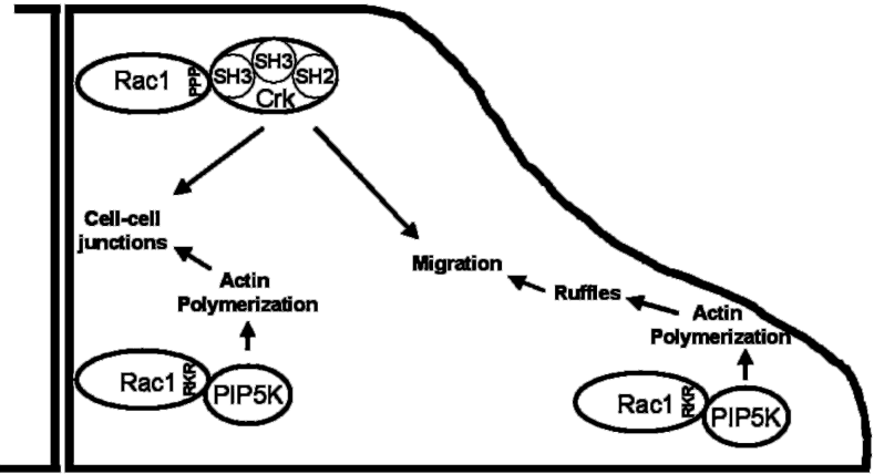
FIG. 8 Model of the interactions, mediated by the Rac1 C terminus, with different signaling proteins at cell-cell junctions and the cell periphery. See ' Discussion ' for details.
FIG. 8 Model of the interactions, mediated by the Rac1 C terminus, with different signaling proteins at cell-cell junctions and the cell periphery. See ' Discussion ' for details.CC-BY · From the PDF

Verification

Claims the paper makes

The claims and statistics of this paper haven’t been checked yet.

Run the check — free, no account needed. It reads the open-access text and checks the sources it cites.

A few free checks a day · papers already verified open instantly · results are public

Claims other papers make about this one

Each is checked against this paper’s own text: does it say what the citing paper says it does?

5 more claims about this paper

Create a free account to see every claim other papers make about this one.

See all claims — free account

Self-check

Percentages, sums and reported differences re-computed from the paper’s own numbers.

The self-check needs the paper’s full text.

Citations

Cites

No references could be resolved yet.

Research works printed but not linked: 44

  1. Hall, 1998, Science, 279, 509, 10.1126/science.279.5350.509 · DOI
  2. Hordijk, 1997, Science, 278, 1464, 10.1126/science.278.5342.1464 · DOI
  3. Braga, 1997, J. Cell Biol., 137, 1421, 10.1083/jcb.137.6.1421 · DOI
  4. Takaishi, 1997, J. Cell Biol., 139, 1047, 10.1083/jcb.139.4.1047 · DOI
  5. Tao, 2002, Blood, 100, 1679, 10.1182/blood.V100.5.1679.h81702001679_1679_1688 · DOI
  6. Kreck, 1996, Biochemistry, 35, 15683, 10.1021/bi962064l · DOI
  7. Knaus, 1998, J. Biol. Chem., 273, 21512, 10.1074/jbc.273.34.21512 · DOI · Paper page
  8. del Pozo, 2000, EMBO J., 19, 2008, 10.1093/emboj/19.9.2008 · DOI
  9. Michaelson, 2001, J. Cell Biol., 152, 111, 10.1083/jcb.152.1.111 · DOI · Paper page
  10. Green, 1988, Cell, 55, 1179, 10.1016/0092-8674(88)90262-0 · DOI
  11. Ho, 2001, Cancer Res., 61, 474
  12. van Wetering, 2002, J. Cell Sci., 115, 1837, 10.1242/jcs.115.9.1837 · DOI
  13. Voermans, 1999, Exp. Hematol., 27, 1806, 10.1016/S0301-472X(99)00113-7 · DOI
  14. Voermans, 2001, Exp. Hematol., 29, 1456, 10.1016/S0301-472X(01)00740-8 · DOI
  15. Sander, 1999, J. Cell Biol., 147, 1009, 10.1083/jcb.147.5.1009 · DOI
  16. Divecha, 2002, Curr. Biol., 12, 582, 10.1016/S0960-9822(02)00769-8 · DOI
  17. Royal, 2000, Mol. Biol. Cell, 11, 1709, 10.1091/mbc.11.5.1709 · DOI
  18. Vastrik, 1999, Curr. Biol., 9, 991, 10.1016/S0960-9822(99)80447-3 · DOI
  19. Kodama, 1999, Oncogene, 18, 3996, 10.1038/sj.onc.1202773 · DOI
  20. Zondag, 2000, J. Cell Biol., 149, 775, 10.1083/jcb.149.4.775 · DOI
  21. Olofsson, 1999, Cell Signal, 11, 545, 10.1016/S0898-6568(98)00063-1 · DOI
  22. Tolias, 1998, Mol. Cell. Biol., 18, 762, 10.1128/MCB.18.2.762 · DOI
  23. Divecha, 2000, EMBO J., 19, 5440, 10.1093/emboj/19.20.5440 · DOI
  24. Ren, 1996, Mol. Biol. Cell, 7, 435, 10.1091/mbc.7.3.435 · DOI
  25. Feller, 2001, Oncogene, 20, 6348, 10.1038/sj.onc.1204779 · DOI
  26. Li, 2001, Oncogene, 20, 6403, 10.1038/sj.onc.1204782 · DOI
  27. Nievers, 1997, J. Cell Sci., 110, 389, 10.1242/jcs.110.3.389 · DOI
  28. Tolias, 2000, Curr. Biol., 10, 153, 10.1016/S0960-9822(00)00315-8 · DOI
  29. Pike, 1998, J. Biol. Chem., 273, 22298, 10.1074/jbc.273.35.22298 · DOI · Paper page
  30. Rozelle, 2000, Curr. Biol., 10, 311, 10.1016/S0960-9822(00)00384-5 · DOI
  31. Heyworth, 1994, J. Biol. Chem., 269, 30749, 10.1016/S0021-9258(18)47341-3 · DOI
  32. Roberts, 1999, Immunity, 10, 183, 10.1016/S1074-7613(00)80019-9 · DOI · Paper page
  33. Croker, 2002, J. Immunol., 168, 3376, 10.4049/jimmunol.168.7.3376 · DOI
  34. Croker, 2002, Immunol. Cell Biol., 80, 231, 10.1046/j.1440-1711.2002.01077.x · DOI
  35. Worthylake, 2001, J. Cell Biol., 154, 147, 10.1083/jcb.200103048 · DOI
  36. Alblas, 2001, Mol. Biol. Cell, 12, 2137, 10.1091/mbc.12.7.2137 · DOI
  37. del Pozo, 1999, Eur. J. Immunol., 29, 3609, 10.1002/(SICI)1521-4141(199911)29:11<3609::AID-IMMU3609>3.0.CO;2-S · DOI
  38. Westwick, 1997, Mol. Cell. Biol., 17, 1324, 10.1128/MCB.17.3.1324 · DOI
  39. Insall, 2001, Dev. Cell, 1, 743, 10.1016/S1534-5807(01)00086-7 · DOI
  40. Mayer, 2001, J. Cell Sci., 114, 1253, 10.1242/jcs.114.7.1253 · DOI
  41. Manser, 1998, Mol. Cell, 1, 183, 10.1016/S1097-2765(00)80019-2 · DOI
  42. Wu, 1995, Structure, 3, 215, 10.1016/S0969-2126(01)00151-4 · DOI
  43. Kiyokawa, 1997, Crit. Rev. Oncog., 8, 329, 10.1615/CritRevOncog.v8.i4.30 · DOI
  44. Cho, 2002, J. Cell Biol., 156, 725, 10.1083/jcb.200111032 · DOI

Metadata

Authors
van Hennik, P. B., Klooster, J. P. T., Halstead, J. R., Voermans, C., Anthony, E. C., Divecha, N., & Hordijk, P. L.
Venue
Journal of Biological Chemistry
Publisher
Elsevier BV
Published
2003-10
Volume
278
Issue
40
Pages
39166-39175
ISSN
0021-9258
Cited by
90
References
0
Language
en

About these checks

Registry, retraction, Cites and statistics results are computed, not judged, with one exception: a printed reference that no index lists is read once by a model before it counts as a citation error. Claims are extracted by a model and kept only when their quote appears verbatim in the text; each cited source is then checked, with a model’s help, for whether it says what the claim relies on. Those verdicts can be wrong — read the claim’s quote and the cited source before relying on one.

Statistics and arithmetic are recomputed, not judged.

Confidence bands are shown only for fields calibrated on our annotated test set. None is calibrated yet, so no band is shown.

What counts as a claim ›How we measure confidence ›How we check this ›

Are you an author, or do you see a problem with this page? Request a review

How to cite

Cite CoChat for the checks on this page, and the paper for its own findings.

This page
CoChat. (2026). Citation check: The C-terminal Domain of Rac1 Contains Two Motifs That Control Targeting and Signaling Specificity. https://app.cochat.ai/papers/10.1074/jbc.m307001200
The paper
van Hennik, P. B., Klooster, J. P. T., Halstead, J. R., Voermans, C., Anthony, E. C., Divecha, N., & Hordijk, P. L. (2003). The C-terminal Domain of Rac1 Contains Two Motifs That Control Targeting and Signaling Specificity. Journal of Biological Chemistry. https://doi.org/10.1074/jbc.m307001200