Cited by 110

The DOI matches its Crossref record.

Acylation of CD44 and Its Association with Lipid Rafts Are Required for Receptor and Hyaluronan Endocytosis

Thankamony, Sai P., Knudson, Warren·2006·Journal of Biological Chemistry

Overview

The claims and statistics of this paper haven’t been checked yet.

Run the check — free, no account needed. It reads the open-access text and checks the sources it cites.

A few free checks a day · papers already verified open instantly · results are public

Integrity signals

Registry

Crossref · 10.1074/jbc.m601530200

Cites

Statistics

Figures

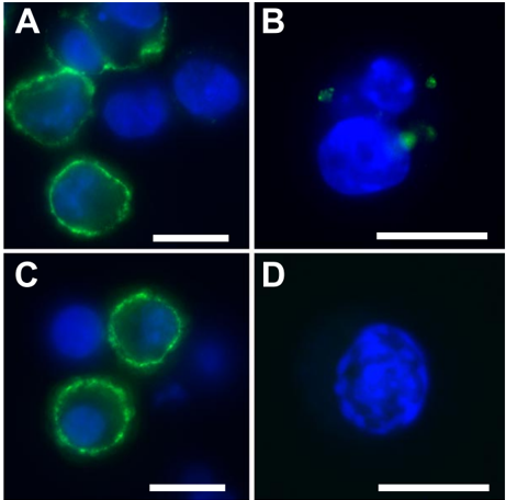
FIGURE1 MembranecholesterolisrequiredforCD44-mediatedinternalization of Fl-HA. COS-7 cell transfectants of CD44wt pre-treated without ( A and B ) or with ( C and D ) 10 mM M 𝛃 -CD, were released from monolayers with EDTA and incubated with complete medium containing 15 𝛍 g/ml Fl-HA. To visualize cell surface binding ( A and C ), the cells were incubated for 1 h at 4 °C with Fl-HA, washed, fixed, mounted onto glass slides with DAPI nuclear counterstain and visualized by fluorescence microscopy. To visualize internalized Fl-HA, a corresponding aliquot of cells from each group was incubated with Fl-HA for 3 h at 37 °C followed by extensive trypsinization to remove all extracellularly bound Fl-HA ( B and D ). Shown are digital overlay images of green and blue fluorescence channels. The bars shown in each panel represent 20 𝛍 m.
FIGURE1 MembranecholesterolisrequiredforCD44-mediatedinternalization of Fl-HA. COS-7 cell transfectants of CD44wt pre-treated without ( A and B ) or with ( C and D ) 10 mM M 𝛃 -CD, were released from monolayers with EDTA and incubated with complete medium containing 15 𝛍 g/ml Fl-HA. To visualize cell surface binding ( A and C ), the cells were incubated for 1 h at 4 °C with Fl-HA, washed, fixed, mounted onto glass slides with DAPI nuclear counterstain and visualized by fluorescence microscopy. To visualize internalized Fl-HA, a corresponding aliquot of cells from each group was incubated with Fl-HA for 3 h at 37 °C followed by extensive trypsinization to remove all extracellularly bound Fl-HA ( B and D ). Shown are digital overlay images of green and blue fluorescence channels. The bars shown in each panel represent 20 𝛍 m.CC-BY · From the PDF
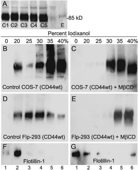
FIGURE 2 Association of CD44 with low buoyant density lipid raft membrane fractions. Panel A represents a typical Western blotting profile for CD44 expressed by five different selected clones of Flp-293 cells stably transfected with CD44wt. The clones are labeled as C1 through C5 , whereas E depicts endogenous levels of CD44 expressed by Flp-293 cells. COS-7 transfectants ( B ) and Flp-293 cells ( D ) expressing CD44wt were extracted in the presence of 1% Triton X-100 on ice and subjected to ultracentrifugation on step gradients of iodixanol. Equal volume fractions were collected and analyzed by Western blotting for CD44. The 0-20% iodixanol interface (predominately the 20% fraction) represents the low density, lipid raft fraction. In some instances, cells were cholesterol-depleted by pretreatment with 20 mM M 𝛃 -CDat37 °Cfor50minforCOS-7cells( C ) or 25 min for Flp-293 cells ( E ) prior to ultracentrifugation and Western blotting. The same fractions from the Flp293 stable transfectants shown in D and E were also assayed for the lipid raft marker flotillin-1 using the anti-flotillin-1 antibody, before ( F ) and after ( G ) cholesterol depletion. CD44 was shown to associate with a low buoyant density lipid raft fraction in both COS-7 and Flp-293 cells (verified by the migration of flotillin-1), and this association was dependent on the presence of cholesterol. CD44 was detected using either an anti-CD44 monoclonal antibody, BU-52 ( B and C ) or the epitope-tag anti-V5 monoclonal antibody ( D and E ).
FIGURE 2 Association of CD44 with low buoyant density lipid raft membrane fractions. Panel A represents a typical Western blotting profile for CD44 expressed by five different selected clones of Flp-293 cells stably transfected with CD44wt. The clones are labeled as C1 through C5 , whereas E depicts endogenous levels of CD44 expressed by Flp-293 cells. COS-7 transfectants ( B ) and Flp-293 cells ( D ) expressing CD44wt were extracted in the presence of 1% Triton X-100 on ice and subjected to ultracentrifugation on step gradients of iodixanol. Equal volume fractions were collected and analyzed by Western blotting for CD44. The 0-20% iodixanol interface (predominately the 20% fraction) represents the low density, lipid raft fraction. In some instances, cells were cholesterol-depleted by pretreatment with 20 mM M 𝛃 -CDat37 °Cfor50minforCOS-7cells( C ) or 25 min for Flp-293 cells ( E ) prior to ultracentrifugation and Western blotting. The same fractions from the Flp293 stable transfectants shown in D and E were also assayed for the lipid raft marker flotillin-1 using the anti-flotillin-1 antibody, before ( F ) and after ( G ) cholesterol depletion. CD44 was shown to associate with a low buoyant density lipid raft fraction in both COS-7 and Flp-293 cells (verified by the migration of flotillin-1), and this association was dependent on the presence of cholesterol. CD44 was detected using either an anti-CD44 monoclonal antibody, BU-52 ( B and C ) or the epitope-tag anti-V5 monoclonal antibody ( D and E ).CC-BY · From the PDF
Image 3
CC-BY · From the PDF
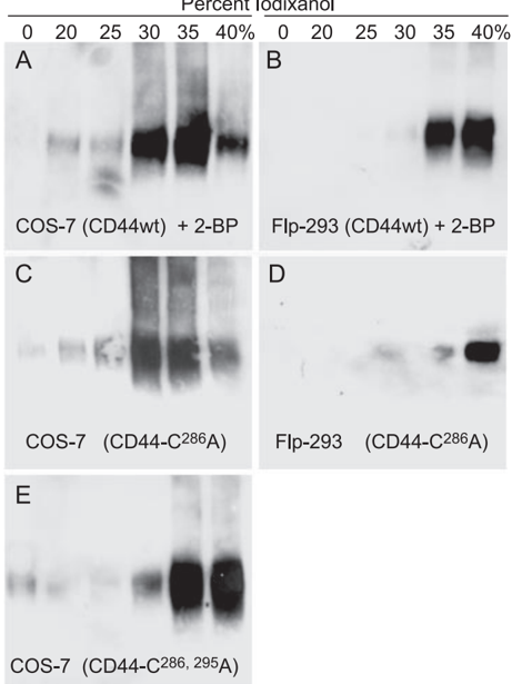
FIGURE 3 Palmitoylation is required for CD44 targeting into lipid rafts. 2-BP pre-treatment prevented CD44 association with low buoyant density iodixanol lipid rafts fractions in both COS-7 transient transfectants ( A ) and Flp-293 stable transfectants ( B ) of CD44wt. The palmitoylation mutants of CD44 (pCD44-C286A) expressed in COS-7 transfectants ( C ) and as Flp-293 stable transfectants ( D ) also exhibited significantly reduced association with the low density lipid rafts fraction. Similar results were obtained when the double mutation isoform of CD44, CD44-C286,295A, was expressed in COS-7 transfectants ( E ).
FIGURE 3 Palmitoylation is required for CD44 targeting into lipid rafts. 2-BP pre-treatment prevented CD44 association with low buoyant density iodixanol lipid rafts fractions in both COS-7 transient transfectants ( A ) and Flp-293 stable transfectants ( B ) of CD44wt. The palmitoylation mutants of CD44 (pCD44-C286A) expressed in COS-7 transfectants ( C ) and as Flp-293 stable transfectants ( D ) also exhibited significantly reduced association with the low density lipid rafts fraction. Similar results were obtained when the double mutation isoform of CD44, CD44-C286,295A, was expressed in COS-7 transfectants ( E ).CC-BY · From the PDF
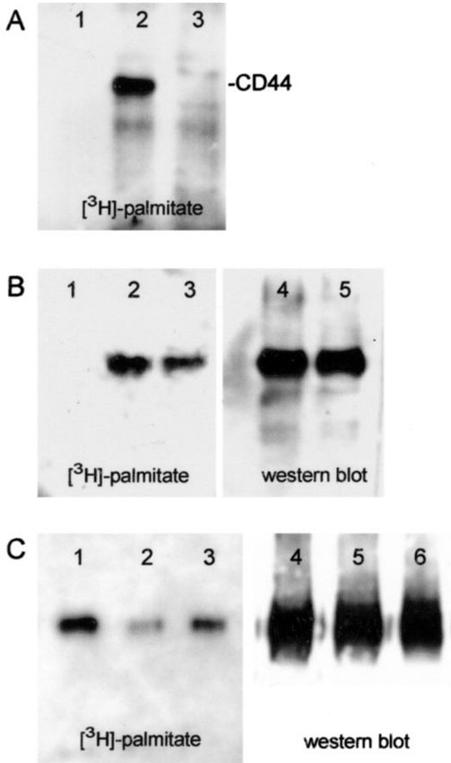
FIGURE 4 [ 3 H]Palmitate is incorporated into CD44 by acylation of membrane-proximalcysteines. A , [ 3 H]palmitateincorporation(250 𝛍 Ci/ml, 2 h at 37 °C) into Flp-293 stable transfectants of CD44wt, pretreated with or without 50 𝛍 M 2-BP ( lanes 1 and 2 , respectively). [ 3 H]Palmitate incorporation into Flp293 stable transfectants of Hyal-2 followed by immunoprecipitation using anti-V5 monoclonal antibody is shown in lane 3 . In B , the extent of [ 3 H]palmitate incorporation in parental Flp-293 cells ( lane 1 ) or Flp-293 cells expressing the wild-type CD44wt ( lane 2 ) were compared with the Flp-293 expressing the cysteine mutants CD44-C286A ( lane 3 ). Lanes 4 and 5 represent a Western blot (detection using an anti-V5 monoclonal antibody) of equivalent aliquots of lysates shown in lanes 2 and 3 , respectively. In C , incorporation of [ 3 H]palmitate into wild-type CD44wt ( lane 1 ), CD44-C286,295A ( lane 2 ), and CD44C286A ( lane 3 ) were compared side-by-side. Lanes 4-6 represent a Western blot (detection using an anti-V5 monoclonal antibody) of equivalent aliquots of lysates shown in lanes 1-3 , respectively. Changes in [ 3 H]palmitate incorporation were quantified by digital scanning of x-ray films, and the resultant data are expressed as the average ± S.D. ( n = 3) change in pixel density relative to control CD44 wild-type incorporation.
FIGURE 4 [ 3 H]Palmitate is incorporated into CD44 by acylation of membrane-proximalcysteines. A , [ 3 H]palmitateincorporation(250 𝛍 Ci/ml, 2 h at 37 °C) into Flp-293 stable transfectants of CD44wt, pretreated with or without 50 𝛍 M 2-BP ( lanes 1 and 2 , respectively). [ 3 H]Palmitate incorporation into Flp293 stable transfectants of Hyal-2 followed by immunoprecipitation using anti-V5 monoclonal antibody is shown in lane 3 . In B , the extent of [ 3 H]palmitate incorporation in parental Flp-293 cells ( lane 1 ) or Flp-293 cells expressing the wild-type CD44wt ( lane 2 ) were compared with the Flp-293 expressing the cysteine mutants CD44-C286A ( lane 3 ). Lanes 4 and 5 represent a Western blot (detection using an anti-V5 monoclonal antibody) of equivalent aliquots of lysates shown in lanes 2 and 3 , respectively. In C , incorporation of [ 3 H]palmitate into wild-type CD44wt ( lane 1 ), CD44-C286,295A ( lane 2 ), and CD44C286A ( lane 3 ) were compared side-by-side. Lanes 4-6 represent a Western blot (detection using an anti-V5 monoclonal antibody) of equivalent aliquots of lysates shown in lanes 1-3 , respectively. Changes in [ 3 H]palmitate incorporation were quantified by digital scanning of x-ray films, and the resultant data are expressed as the average ± S.D. ( n = 3) change in pixel density relative to control CD44 wild-type incorporation.CC-BY · From the PDF
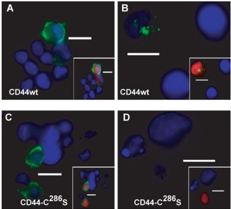
FIGURE 5 Palmitoylation of CD44 is required for HA internalization. COS-7 cells were co-transfected with red fluorescent protein and the wildtype CD44wt ( A and B ) or the CD44-C286S mutant isoform ( C and D ). The transfectants were released from monolayers with EDTA and incubated in complete medium containing 15 𝛍 g/ml Fl-HA. To visualize cell surface binding ( A and C ), the cells were incubated for 1 h at 4 °C with Fl-HA, washed, fixed, mounted onto glass slides with DAPI nuclear counterstain and visualized by fluorescence microscopy ( A and C ). To visualize internalized Fl-HA, a corresponding aliquot of cells from each group was incubated with Fl-HA for 3 h at 37 °C followed by extensive trypsinization to remove all extracellularlybound Fl-HA ( B and D ). Shown are digital overlay images of green and blue fluorescence channels. Insets depict three-channel digital overlays of green , red , and blue fluorescence signals. The bars represent 20 𝛍 m.
FIGURE 5 Palmitoylation of CD44 is required for HA internalization. COS-7 cells were co-transfected with red fluorescent protein and the wildtype CD44wt ( A and B ) or the CD44-C286S mutant isoform ( C and D ). The transfectants were released from monolayers with EDTA and incubated in complete medium containing 15 𝛍 g/ml Fl-HA. To visualize cell surface binding ( A and C ), the cells were incubated for 1 h at 4 °C with Fl-HA, washed, fixed, mounted onto glass slides with DAPI nuclear counterstain and visualized by fluorescence microscopy ( A and C ). To visualize internalized Fl-HA, a corresponding aliquot of cells from each group was incubated with Fl-HA for 3 h at 37 °C followed by extensive trypsinization to remove all extracellularlybound Fl-HA ( B and D ). Shown are digital overlay images of green and blue fluorescence channels. Insets depict three-channel digital overlays of green , red , and blue fluorescence signals. The bars represent 20 𝛍 m.CC-BY · From the PDF
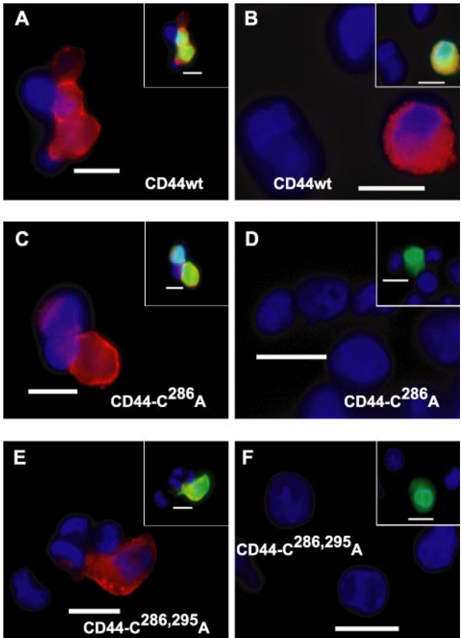
FIGURE 6 Binding and internalization of HA decorated with biotinylated HABP. COS-7 cells were co-transfected with green fluorescent protein and the wild-type CD44H (labeled CD44wt, A and B ), the CD44-C286A mutant isoform ( C and D ), or the CD44-C286,295A double mutant isoform ( E and F ). The transfectants were released from monolayers with EDTA and incubated in complete medium containing 15 𝛍 g/ml unlabeled HA pre-decorated with biotinylated HABP. To visualize cell surface binding ( A , C , and E ), the cells were incubated for 1 h at 4 °C with HA/HABP washed, fixed, mounted onto glass slides with DAPI nuclear counterstain, and visualized by using fluorescence microscopy. To visualize internalized HA/HABP, a corresponding aliquot of cells from each group was incubated with HA/HABP for 3 h at 37 °C followed byextensivetrypsinizationtoremoveallextracellularlyboundHA/HABP( B , D , and F ). Biotinylated HABP was detected using NeutrAvidin-Texas Red. Shown are digital overlay images of red and blue fluorescence channels. Insets depict three-channel digital overlays of green , red , and blue fluorescence signals. The bars represent 20 𝛍 m.
FIGURE 6 Binding and internalization of HA decorated with biotinylated HABP. COS-7 cells were co-transfected with green fluorescent protein and the wild-type CD44H (labeled CD44wt, A and B ), the CD44-C286A mutant isoform ( C and D ), or the CD44-C286,295A double mutant isoform ( E and F ). The transfectants were released from monolayers with EDTA and incubated in complete medium containing 15 𝛍 g/ml unlabeled HA pre-decorated with biotinylated HABP. To visualize cell surface binding ( A , C , and E ), the cells were incubated for 1 h at 4 °C with HA/HABP washed, fixed, mounted onto glass slides with DAPI nuclear counterstain, and visualized by using fluorescence microscopy. To visualize internalized HA/HABP, a corresponding aliquot of cells from each group was incubated with HA/HABP for 3 h at 37 °C followed byextensivetrypsinizationtoremoveallextracellularlyboundHA/HABP( B , D , and F ). Biotinylated HABP was detected using NeutrAvidin-Texas Red. Shown are digital overlay images of red and blue fluorescence channels. Insets depict three-channel digital overlays of green , red , and blue fluorescence signals. The bars represent 20 𝛍 m.CC-BY · From the PDF
Image 8
CC-BY · From the PDF
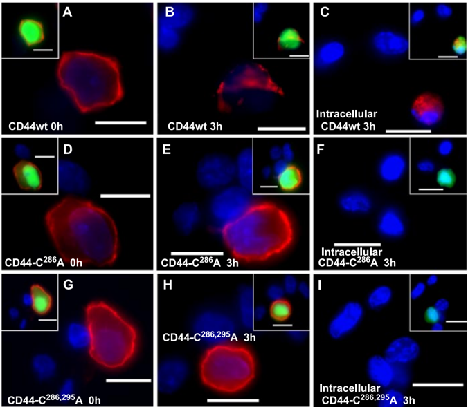
FIGURE 7 CD44 internalization in COS-7 transfectants is mediated by receptor palmitoylation. COS-7 cells were co-transfected with green fluorescent protein and the wild-type CD44H (labeled CD44wt, A-C ), the CD44-C286A mutant isoform ( D-F ), or the CD44-C286,295A double mutant isoform ( G-I ). The transfectants were then preincubated without ( A , D , and G ) or with 100 𝛍 g/ml cycloheximide for 3 h at 37 °C ( B , C , E , F , H , and I ) as labeled. To detect intracellular staining, the cells were co-incubated with cycloheximide and chloroquine; the latter was used to inhibit lysosomal degradation and enhance visualization of internalized receptor ( C , F , and I ). Cell surface CD44 was visualized by immunostaining of fixed cells with the anti-CD44 monoclonal antibody, BU-52, and anti-mouse cy-3-conjugated secondary antibody ( red fluorescence; A , B , D , E , G , and H ). To detect internalized CD44 ( C , F , and I ), cells were trypsinized, fixed, and permeabilized prior to immunostaining with the BU-52 antibody. Shown are digital overlay images of red and blue fluorescence channels. Insets depict three-channel digital overlays of green , red , and blue fluorescence signals. The bars represent 20 𝛍 m.
FIGURE 7 CD44 internalization in COS-7 transfectants is mediated by receptor palmitoylation. COS-7 cells were co-transfected with green fluorescent protein and the wild-type CD44H (labeled CD44wt, A-C ), the CD44-C286A mutant isoform ( D-F ), or the CD44-C286,295A double mutant isoform ( G-I ). The transfectants were then preincubated without ( A , D , and G ) or with 100 𝛍 g/ml cycloheximide for 3 h at 37 °C ( B , C , E , F , H , and I ) as labeled. To detect intracellular staining, the cells were co-incubated with cycloheximide and chloroquine; the latter was used to inhibit lysosomal degradation and enhance visualization of internalized receptor ( C , F , and I ). Cell surface CD44 was visualized by immunostaining of fixed cells with the anti-CD44 monoclonal antibody, BU-52, and anti-mouse cy-3-conjugated secondary antibody ( red fluorescence; A , B , D , E , G , and H ). To detect internalized CD44 ( C , F , and I ), cells were trypsinized, fixed, and permeabilized prior to immunostaining with the BU-52 antibody. Shown are digital overlay images of red and blue fluorescence channels. Insets depict three-channel digital overlays of green , red , and blue fluorescence signals. The bars represent 20 𝛍 m.CC-BY · From the PDF
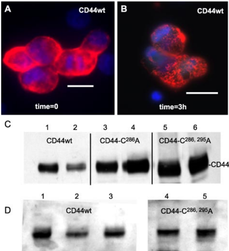
FIGURE8 CD44internalization is dependent on receptor palmitoylation. The two channel fluorescence overlay images of cells ( A and B ) represent another illustration of the loss of CD44 expression in COS-7 transfectants of CD44wt at time zero ( A ) as compared with 3 h following pre-treatment with cycloheximide ( B ). As in Fig. 7, the red fluorescence represents immunostaining of the cells with anti-CD44 monoclonal antibody, BU-52, followed by anti-mouse Cy3-conjugated secondary antibody. C depicts CD44 Western blotting of cell lysates prepared from COS-7 transfectants at time zero ( lanes 1 , 3 , and 5 ) or after 6 h of treatment with cycloheximide ( lanes 2 , 4 , and 6 ). Lysates of wild-type CD44 (CD44wt; lanes 1 and 2 ), CD44-C286A ( lanes 3 and 4 ), or CD44-C286,295A ( lanes 5 and 6 ) were examined. D represents a CD44 Western blot of another experiment that depicts transfectants of CD44H ( lanes 1-3 ) or CD44-C286,295A ( lanes 4 and 5 ) at time zero ( lanes 1 and 4 ), treated with cycloheximide for 3 h ( lanes 2 and 5 ) or treated with cycloheximide and chloroquine for 3 h ( lane 3 ). Changes in band intensity were quantified by digital scanning of x-ray films, and the resultant data are expressed as the average ± S.D. ( n = 3) change in pixel density relative to control CD44 wild-type expression.
FIGURE8 CD44internalization is dependent on receptor palmitoylation. The two channel fluorescence overlay images of cells ( A and B ) represent another illustration of the loss of CD44 expression in COS-7 transfectants of CD44wt at time zero ( A ) as compared with 3 h following pre-treatment with cycloheximide ( B ). As in Fig. 7, the red fluorescence represents immunostaining of the cells with anti-CD44 monoclonal antibody, BU-52, followed by anti-mouse Cy3-conjugated secondary antibody. C depicts CD44 Western blotting of cell lysates prepared from COS-7 transfectants at time zero ( lanes 1 , 3 , and 5 ) or after 6 h of treatment with cycloheximide ( lanes 2 , 4 , and 6 ). Lysates of wild-type CD44 (CD44wt; lanes 1 and 2 ), CD44-C286A ( lanes 3 and 4 ), or CD44-C286,295A ( lanes 5 and 6 ) were examined. D represents a CD44 Western blot of another experiment that depicts transfectants of CD44H ( lanes 1-3 ) or CD44-C286,295A ( lanes 4 and 5 ) at time zero ( lanes 1 and 4 ), treated with cycloheximide for 3 h ( lanes 2 and 5 ) or treated with cycloheximide and chloroquine for 3 h ( lane 3 ). Changes in band intensity were quantified by digital scanning of x-ray films, and the resultant data are expressed as the average ± S.D. ( n = 3) change in pixel density relative to control CD44 wild-type expression.CC-BY · From the PDF

Verification

Claims the paper makes

The claims and statistics of this paper haven’t been checked yet.

Run the check — free, no account needed. It reads the open-access text and checks the sources it cites.

A few free checks a day · papers already verified open instantly · results are public

Claims other papers make about this one

Each is checked against this paper’s own text: does it say what the citing paper says it does?

Self-check

Percentages, sums and reported differences re-computed from the paper’s own numbers.

The self-check needs the paper’s full text.

Citations

Cites

No references could be resolved yet.

Research works printed but not linked: 35

  1. Knudson, 1993, J. Cell Biol., 120, 825, 10.1083/jcb.120.3.825 · DOI
  2. Embry, 2003, Arthritis Rheum., 48, 3431, 10.1002/art.11323 · DOI
  3. Tammi, 2001, J. Biol. Chem., 276, 35111, 10.1074/jbc.M103481200 · DOI
  4. Underhill, 1993, Dev. Biol., 155, 324, 10.1006/dbio.1993.1032 · DOI
  5. Jiang, 2001, Arthritis Rheum., 44, 2599, 10.1002/1529-0131(200111)44:11<2599::AID-ART440>3.0.CO;2-Y · DOI
  6. Jiang, 2002, J. Biol. Chem., 277, 10531, 10.1074/jbc.M108654200 · DOI
  7. Knudson, 2002, Matrix Biol., 21, 15, 10.1016/S0945-053X(01)00186-X · DOI
  8. Knudson, 2004, Curr. Opin. Orthop., 15, 369, 10.1097/01.bco.0000134433.53684.04 · DOI
  9. Radeva, 2004, Biochem. J., 380, 219, 10.1042/bj20031348 · DOI
  10. Oliferenko, 1999, J. Cell Biol., 146, 843, 10.1083/jcb.146.4.843 · DOI
  11. Foger, 2000, Eur. J. Immunol., 30, 2888, 10.1002/1521-4141(200010)30:10<2888::AID-IMMU2888>3.0.CO;2-4 · DOI
  12. Bijlmakers, 2003, Trends Cell Biol., 13, 32, 10.1016/S0962-8924(02)00008-9 · DOI
  13. Liu, 1996, J. Exp. Med., 183, 1987, 10.1084/jem.183.5.1987 · DOI
  14. Roy, 2005, Mol. Cell. Biol., 25, 6722, 10.1128/MCB.25.15.6722-6733.2005 · DOI
  15. Percherancier, 2001, J. Biol. Chem., 276, 31936, 10.1074/jbc.M104013200 · DOI
  16. Bonner, 1974, Eur. J. Biochem., 46, 83, 10.1111/j.1432-1033.1974.tb03599.x · DOI
  17. Hua, 1993, J. Cell Sci., 106, 365, 10.1242/jcs.106.1.365 · DOI
  18. Aguiar, 1999, Exp. Cell Res., 252, 292, 10.1006/excr.1999.4641 · DOI
  19. Guo, 1993, Cell. Immunol., 152, 186, 10.1006/cimm.1993.1278 · DOI
  20. Guo, 1994, Int. Immunol., 6, 213, 10.1093/intimm/6.2.213 · DOI
  21. Bourguignon, 1991, J. Biol. Chem., 266, 11761, 10.1016/S0021-9258(18)99022-8 · DOI
  22. Stickney, 2004, Cancer Res., 64, 2717, 10.1158/0008-5472.CAN-03-3798 · DOI
  23. Bourguignon, 2004, J. Biol. Chem., 279, 26991, 10.1074/jbc.M311838200 · DOI
  24. Li, 2002, J. Virol., 76, 11845, 10.1128/JVI.76.23.11845-11852.2002 · DOI
  25. Abrami, 2006, J. Cell Biol., 172, 309, 10.1083/jcb.200507067 · DOI
  26. Munshi, 2005, Mol. Endocrinol., 19, 749, 10.1210/me.2004-0335 · DOI
  27. Hicke, 2001, Cell, 106, 527, 10.1016/S0092-8674(01)00485-8 · DOI
  28. Roth, 2000, J. Biol. Chem., 275, 8143, 10.1074/jbc.275.11.8143 · DOI
  29. Haglund, 2003, Trends Biochem. Sci., 28, 598, 10.1016/j.tibs.2003.09.005 · DOI · Paper page
  30. Marmor, 2004, Oncogene, 23, 2057, 10.1038/sj.onc.1207390 · DOI
  31. Knudson, 2005, 1, 213
  32. Knudson, 2004, Clin. Orthop., 427, 152, 10.1097/01.blo.0000143804.26638.82 · DOI
  33. Resh, 1999, Biochim. Biophys. Acta, 1451, 1, 10.1016/S0167-4889(99)00075-0 · DOI
  34. Yeh, 1999, J. Biol. Chem., 274, 33148, 10.1074/jbc.274.46.33148 · DOI
  35. Duncan, 1998, J. Biol. Chem., 273, 15830, 10.1074/jbc.273.25.15830 · DOI

Cited by

Sorted by citations

Metadata

Authors
Thankamony, S. P., & Knudson, W.
Venue
Journal of Biological Chemistry
Publisher
Elsevier BV
Published
2006-11
Volume
281
Issue
45
Pages
34601-34609
ISSN
0021-9258
Cited by
110
References
0
Language
en

About these checks

Registry, retraction, Cites and statistics results are computed, not judged, with one exception: a printed reference that no index lists is read once by a model before it counts as a citation error. Claims are extracted by a model and kept only when their quote appears verbatim in the text; each cited source is then checked, with a model’s help, for whether it says what the claim relies on. Those verdicts can be wrong — read the claim’s quote and the cited source before relying on one.

Statistics and arithmetic are recomputed, not judged.

Confidence bands are shown only for fields calibrated on our annotated test set. None is calibrated yet, so no band is shown.

What counts as a claim ›How we measure confidence ›How we check this ›

Are you an author, or do you see a problem with this page? Request a review