

The DOI matches its Crossref record; 42 of the 43 references it cites were found in a registry or an index.
Clonal identification and characterization of self-renewing pluripotent stem cells in the developing liver
Open accessAbstract
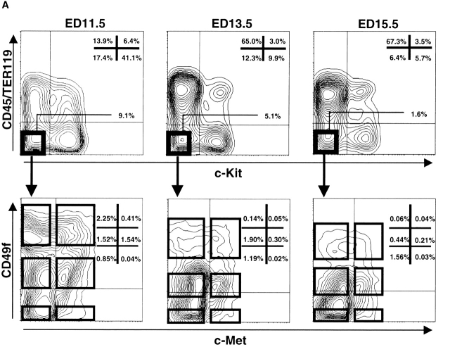
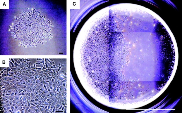
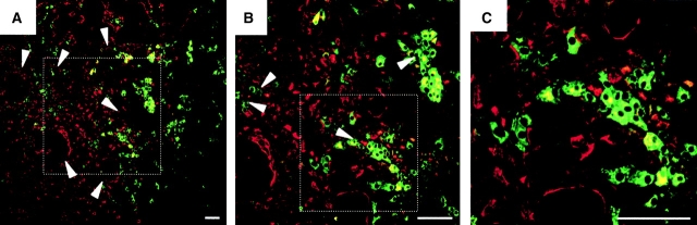
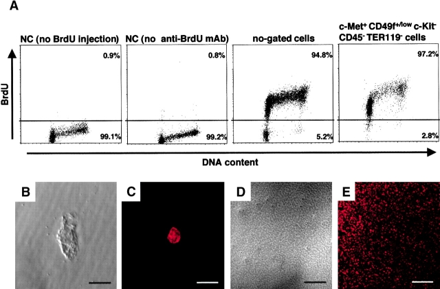
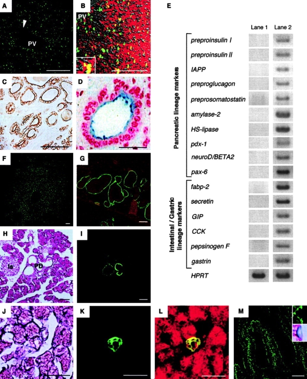
Using flow cytometry and single cell–based assays, we prospectively identified hepatic stem cells with multilineage differentiation potential and self-renewing capability. These cells could be clonally propagated in culture where they continuously produced hepatocytes and cholangiocytes as descendants while maintaining primitive stem cells. When cells that expanded in vitro were transplanted into recipient animals, they morphologically and functionally differentiated into hepatocytes and cholangiocytes with reconstitution of hepatocyte and bile duct structures. Furthermore, these cells differentiated into pancreatic ductal and acinar cells or intestinal epithelial cells when transplanted into pancreas or duodenal wall. These data indicate that self-renewing pluripotent stem cells persist in the developing mouse liver and that such cells can be induced to become cells of other organs of endodermal origin under appropriate microenvironment. Manipulation of hepatic stem cells may provide new insight into therapies for diseases of the digestive system.
Overview
The claims and statistics of this paper haven’t been checked yet.
Run the check — free, no account needed. It reads the open-access text and checks the sources it cites.
Integrity signals
Registry
Cites
Statistics
Figures
Verification
Claims the paper makes
The claims and statistics of this paper haven’t been checked yet.
Run the check — free, no account needed. It reads the open-access text and checks the sources it cites.
Claims other papers make about this one
Each is checked against this paper’s own text: does it say what the citing paper says it does?
The source backs part of the claim, not all of it — or backs it for a different population, condition or comparison.
In the citing paper
“Isolating c-Met + c-Kit - CD45 -Ter119 -(also known as Met, Kit, Ptprc and Ly76, respectively -Mouse Genome Informatics) cells achieved a much higher enrichment of hepatoblasts, and thus this method could facilitate the identification of a discrete set of transcription factors that are activated in this specific cell population.”
From Tbx3 controls the fate of hepatic progenitor cells in liver development by suppressing p19ARF expression (2008) · cites this paper as Suzuki et al., 2002
Experimental — accuracy not yet measuredThe source backs part of the claim, not all of it — or backs it for a different population, condition or comparison.
In the citing paper
“In particular, cells marked by the hepatocyte growth factor (Hgf) receptor c-Met displayed distinctive activities in response to Hgf stimulus, including self-renewing cell divisions and differentiation into both hepatocytes and cholangiocytes (Suzuki et al., 2002;Suzuki et al., 2003a).”
From Tbx3 controls the fate of hepatic progenitor cells in liver development by suppressing p19ARF expression (2008) · cites this paper as Suzuki et al., 2002
Experimental — accuracy not yet measured
Self-check
Percentages, sums and reported differences re-computed from the paper’s own numbers.
The self-check needs the paper’s full text.
Citations
Cites
A Direct Measurement of the Radiation Sensitivity of Normal Mouse Bone Marrow Cells1
Till, J. E., & McCulloch, E. A. (2010) · Radiation Research · doi:10.1667/rrxx28.1 · Cited by 4,036
Histopathologic Technic and Practical Histochemistry.
Small, J. A., & Lillie, R. D. (1954) · Bulletin of the Torrey Botanical Club · doi:10.2307/2481951 · Cited by 2,972
Michalopoulos, G. K., & DeFrances, M. C. (1997) · Science · doi:10.1126/science.276.5309.60 · Cited by 2,828
‘Green mice’ as a source of ubiquitous green cells
Okabe, M., Ikawa, M., Kominami, K., Nakanishi, T., & Nishimune, Y. (1997) · FEBS Letters · doi:10.1016/s0014-5793(97)00313-x · Cited by 2,581
Long-Term Lymphohematopoietic Reconstitution by a Single CD34-Low/Negative Hematopoietic Stem Cell
Osawa, M., Hanada, K., Hamada, H., & Nakauchi, H. (1996) · Science · doi:10.1126/science.273.5272.242 · Cited by 2,141
Self-citationScatter factor/hepatocyte growth factor is essential for liver development
Schmidt, C. S., Bladt, F., Gödecke, S., Brinkmann, V., Zschiesche, W., Sharpe, M. J., Gherardi, E., & Birchmeler, C. (1995) · Nature · doi:10.1038/373699a0 · Cited by 1,395
Control of endodermal endocrine development by Hes-1
Jensen, J. N., Pedersen, E. E., Galante, P. E., Hald, J., Heller, R. S., Ishibashi, M., Kageyama, R., Guillemot, F., Serup, P., & Madsen, O. D. (2000) · Nature Genetics · doi:10.1038/71657 · Cited by 1,155
Placental defect and embryonic lethality in mice lacking hepatocyte growth factor/scatter factor
Uehara, Y., Minowa, O., Mori, C., Shiota, K., Kuno, J., Noda, T., & Kitamura, N. (1995) · Nature · doi:10.1038/373702a0 · Cited by 1,041
Ory, D., Neugeboren, B. I., & Mulligan, R. C. (1996) · Proceedings of the National Academy of Sciences · doi:10.1073/pnas.93.21.11400 · Cited by 926
Zulewski, H., Abraham, E. J., Gerlach, M. J., Daniel, P. B., Moritz, W., Müller-Stich, B. P., Vallejo, M., Thomas, M. K., & Habener, J. F. (2001) · Diabetes · doi:10.2337/diabetes.50.3.521 · Cited by 785
Research works printed but not linked: 10
- 1995, Am. J. Pathol., 147, 707
- 1994, Lab. Invest., 70, 511
- 1993, Am. J. Pathol., 142, 1823
- 1999, Cell., 5, 737
- 1997, Am. J. Pathol., 151, 1273
- 1989, Am. J. Pathol., 134, 1069
- 1989, Am. J. Pathol., 134, 1347
- 1984, J. Embryol. Exp. Morphol., 79, 25
- 1991, Cancer Res., 51, 2611
- 1961, Radiat. Res., 14, 1419
Cited by
Sorted by citations
Items 301–356 of 356
Fetal Liver Cell Transplantation
Sumitran–Holgersson, S., Joshi, M. G., & Olausson, M. (2012) · Springer eBooks · doi:10.1007/978-1-4471-4171-6_17 · Cited by 2
Embryologic development of the liver, biliary tract, and pancreas
El‐Gohary, Y., Zhao, K., & Gittes, G. K. (2017) · Elsevier eBooks · doi:10.1016/b978-0-323-34062-5.00001-7 · Cited by 2
Stem Cells in the Gastrointestinal Tract
Preston, S. L., Wright, N. A., Direkze, N. C., & Brittan, M. (2004) · Elsevier eBooks · doi:10.1016/b978-012436643-5/50137-1 · Cited by 2
Itoh, T., TanakaTanaka, M., & Miyajima, A. (2010) · Springer eBooks · doi:10.1007/978-90-481-9075-1_14 · Cited by 1
Case History: Toxicology Biomarker Development Using Toxicogenomics
Watson, D. E., Ryan, T. P., & Stevens, J. L. (2007) · Springer eBooks · doi:10.1007/978-0-387-44961-6_12 · Cited by 1
Hosaka, K., Katsuda, T., Terai, S., & Ochiya, T. (2018) · Elsevier eBooks · doi:10.1016/b978-0-12-812301-0.00005-0 · Cited by 1
Grompe, M. (2012) · Elsevier eBooks · doi:10.1016/b978-0-12-385942-6.00072-x · Cited by 1
WNT AND BETA-CATENIN IN HEPATIC DIFFERENTIATION: WHICH WAY DOES THE WNT BLOW?
Lade, A. G. (2011) · D-Scholarship@Pitt (University of Pittsburgh) · Cited by 1
Afroze, S. H., Jensen, K. J., Rahal, K., Meng, F., Alpini, G. D., & Glaser, S. S. (2013) · Elsevier eBooks · doi:10.1016/b978-0-12-398523-1.00026-4 · Cited by 1
Manohar, R., & Lagasse, E. (2013) · Elsevier eBooks · doi:10.1016/b978-0-12-398358-9.00045-8 · Cited by 1
Goltsev, A. N., Дубрава, Т. Г., Gaevska, Y., Yampolskaya, E. E., & Ostankov, M. V. (2014) · Problems of Cryobiology and Cryomedicine · doi:10.15407/cryo24.02.118 · Cited by 1
Tauran, Y., Lereau, M., Ségard, B.-D., Danoy, M., Kimura, K., Shinohara, M., Brioude, A., Sakai, Y., de Jonge, H., Melnyk, O., et al. (2022) · Development Growth & Differentiation · doi:10.1111/dgd.12818 · Cited by 1
Elsheikh, E., El-Hady, E., Abd-Allah, S. H., Selem, A. A., & Konsowa, M. (2021) · Slovenian Veterinary Research · doi:10.26873/svr-1438-2021 · Cited by 1
Pancreatic Progenitor Cells in Injury and Regeneration
Afelik, S., & Jensen, J. (2008) · Springer eBooks · doi:10.1007/978-0-387-69252-4_7
THREE-DIMENSIONAL CULTURE OF FETAL LIVER CELLS
Qian, L. (2006) · eCommons (Cornell University)
Grompe, M. (2004) · Elsevier eBooks · doi:10.1016/b978-012436643-5/50134-6
Scatter Factors in Tumor Progression
Comoglio, P. M., & Boccaccio, C. (2006) · Springer eBooks · doi:10.1007/1-4020-4009-1_6
Role of Polycomb group proteins in the hepatic stem cell self-renewal
Ueno, Y., Naito, T., & Taniguchi, H. (2009) · SEIBUTSU BUTSURI KAGAKU · doi:10.2198/sbk.53.115
Self-citationHepatic Progenitors in Development and Transplantation
Shafritz, D. A., Oertel, M., & Dabeva, M. D. (2010) · Molecular pathology library · doi:10.1007/978-1-4419-7107-4_16
HEPATOCELLULAR CARCINOMA VIRAL ETIOLOGY AND CELLULAR MECHANISMS
Yildiz, E. (2002)
INVOLVEMENT OF SDF-1 IN STEM CELL-AIDED LIVER REGENERATION
Zheng, D. (2006)
Biología molecular y celular de las células troncales hepáticas
Fuenzalida, T., & Hugo, J. B. (2014)
Bone marrow stroma-derived adult stem cells and tissue regeneration
Satomura, K. (2009) · Japanese Journal of Oral & Maxillofacial Surgery · doi:10.5794/jjoms.55.474
Engineered cells for treatment of diabetes
Efrat, S. (2007) · Elsevier eBooks · doi:10.1533/9781845693077.4.388
Role of Stem Cells in the Gastrointestinal Tract and in the Development of Cancer
Huang, P., Li, B., & Zheng, Y. (2016) · Wiley eBooks · doi:10.1002/9781119135449.ch19 · Cited by 1
Self-citationChaudhary, K. R., & Baumgärtner, W. (2013) · RWTH Publications (RWTH Aachen)
Hepatic Progenitor Cell Transplantation
Shafritz, D. A. (2015) · Elsevier eBooks · doi:10.1016/b978-0-12-420128-6.00020-8
Chiba, T., Zheng, Y., Kita, K., Yokosuka, O., Saisho, H., Onodera, M., Miyoshi, H., Nakano, M., Zen, Y., Nakanuma, Y., et al. (2007)
Self-citationStem Cells in the Liver Cancer and the Organ Size Control
Tiyaboonchai, A., Su, Y., & Li, B. (2018) · Elsevier eBooks · doi:10.1016/b978-0-12-812301-0.00004-9
Plasticity of Liver Epithelial Cells in Healthy and Injured Livers
Tanimizu, N., & Mitaka, T. (2018) · Elsevier eBooks · doi:10.1016/b978-0-12-812301-0.00003-7
Long-Term Culture of Mouse Fetal Hepatic Stem/Progenitor Cells
Tsuchiya, A., & Terai, S. (2018) · Methods in molecular biology · doi:10.1007/978-1-4939-8961-4_1
Lagasse, E. (2007) · Elsevier eBooks · doi:10.1016/b978-012370615-7/50051-2
Stem Cells in the Gastrointestinal Tract
Preston, S. L., Wright, N. A., Direkze, N. C., & Brittan, M. (2012) · Elsevier eBooks · doi:10.1016/b978-0-12-385942-6.00067-6
The susceptibility of human hepatoma-derived oval-like cells to hepatitis B virus infection.
Sun, S., Li, Y., Liu, B., Zhang, B., Han, S., & Li, X. (2018) · Europe PMC (PubMed Central)
Stem cells and cancer in the liver
Taniguchia, H., & Chiba, T. (2014)
P-12 酸化モリブデン触媒を用いる脱水環化反応によるオキサゾリン、チアゾリンの効率的合成(ポスター発表の部)
坂倉彰, 近藤, 玲., 梅村, 周., & 石原, 一. (2005) · 天然有機化合物討論会講演要旨集
Alison, M. R., & Forbes, S. J. (2007) · Springer eBooks · doi:10.1007/1-4020-3420-2_11
Itoh, T., Takase, H. M., Tanaka, M., & Miyajima, A. (2013) · Springer eBooks · doi:10.1007/978-94-007-5690-8_13
Functional characterization of CD49f+ hepatic stem/progenitor cells in adult mice liver
Guo, Z., Pu, S., Li, Y., Wang, X., Hu, S., Zhao, H., Yang, C., & Zhou, Z. (2022) · Journal of Molecular Histology · doi:10.1007/s10735-022-10063-z
Alison, M. R. (2012) · Faculty Opinions – Post-Publication Peer Review of the Biomedical Literature · doi:10.3410/f.13437025.14811158
Jahagirdar, B., & Verfaillie, C. M. (2004) · CRC Press eBooks · doi:10.1201/b14440-3
Sharma, A. D., Iacob, R. A., Bock, M., Cantz, T., Manns, M. P., & Ott, M. (2010) · Springer eBooks · doi:10.1007/978-90-481-9075-1_33
N/A (2003) · World Chinese Journal of Digestology · doi:10.11569/wcjd.v11.i11.1735
Zheng, Y., & Ge, J.-Y. (2018) · Elsevier eBooks · doi:10.1016/b978-0-12-812301-0.00020-7
Self-citationSharma, A. D., Iacob, R. A., Cantz, T., Manns, M. P., & Ott, M. (2013) · Springer eBooks · doi:10.1007/978-94-007-5690-8_37
Auxiliary Liver Organ Formation by Implantation of Spleen-Encapsulated Hepatocytes
Nishio, R., Nakayama, M., Ikekita, M., & Watanabe, Y. (2006) · Tissue Engineering · doi:10.1089/ten.2006.12.ft-176
Faccioli, L. A. P., Dias, M. L., Martins-Santos, R., Paredes, B. D., Takiya, C. M., & Goldenberg, R. C. D. S. (2023) · Elsevier eBooks · doi:10.1016/b978-0-443-15289-4.00015-9
Ge, J.-Y., 鄭, 允., Tsuchida, T., Furuya, K., Isoda, H., Taniguchi, H., Ohkohchi, N., & Oda, T. (2020) · Figshare · doi:10.6084/m9.figshare.13023899
Self-citationGe, J.-Y., 鄭, 允., Tsuchida, T., Furuya, K., Isoda, H., Taniguchi, H., Ohkohchi, N., & Oda, T. (2020) · Figshare · doi:10.6084/m9.figshare.13023902
Self-citationGe, J.-Y., 鄭, 允., Tsuchida, T., Furuya, K., Isoda, H., Taniguchi, H., Ohkohchi, N., & Oda, T. (2020) · Figshare · doi:10.6084/m9.figshare.13023896
Self-citationHabib, R. S., Zaman, B. A., Khanamir, R. A., Hassan, S. M. A., & Merza, M. A. (2025) · Anatomia Histologia Embryologia · doi:10.1111/ahe.70080
Cell polarity in hepatic cells physiopathology
Akkari, L. (2010) · theses.fr (ABES) · doi:10.70675/50d0b5fez8356z488fzb2b0z029ae5e17d3a
Rôle du microenvironnement cellulaire de la mégacaryopoïèse
Jost, C. (2019) · theses.fr (ABES) · doi:10.70675/02f08865z4d42z4d02za666zccd4c74db835
Insulin-Producing Cells Generated from Nonpancreatic Tissues
Efrat, S. (2006) · Springer eBooks · doi:10.1007/1-84628-142-3_21
Regenerative Therapies for Liver Diseases
Sharma, A. D., Rittelmeyer, I., Cantz, T., & Ott, M. (2012) · Humana Press eBooks · doi:10.1007/978-1-62703-098-4_10
Metadata
- Authors
- Suzuki, A., Zheng, Y.-W., Kaneko, S., Onodera, M., Fukao, K., Nakauchi, H., & Taniguchi, H.
- Affiliations
- University of Tsukuba (JP)
- Venue
- The Journal of Cell Biology
- Publisher
- Rockefeller University Press
- Published
- 2002-01-07
- Volume
- 156
- Issue
- 1
- Pages
- 173-184
- ISSN
- 0021-9525 (ISSN-L)
- 1540-8140
- PMC
- PMC2173576
- OpenAlex
- W2121054813
- Cited by
- 356
- References
- 43
- Citations by year
- 2025: 2 · 2024: 3 · 2023: 4 · 2022: 3 · 2021: 1 · 2020: 7 · 2019: 8 · 2018: 10 · 2017: 9 · 2016: 5 · 2015: 4 · 2014: 14 · 2013: 19 · 2012: 15
- FWCI
- 20.45
- Type
- journal article
- Language
- en
- Open access
- green
- www.ncbi.nlm.nih.gov
- Licence
- cc-by-nc-sa
- Locations
- The Journal of Cell Biology (publishedVersion)
- Europe PMC (PubMed Central) (publishedVersion, cc-by-nc-sa)
- PubMed Central (publishedVersion, cc-by-nc-sa)
- PubMed (publishedVersion)
- Topics
- Health Sciences › Medicine › Hepatology › Liver physiology and pathology
- Life Sciences › Biochemistry, Genetics and Molecular Biology › Cell Biology › Mesenchymal stem cell research
- Health Sciences › Medicine › Hematology › Hematopoietic Stem Cell Transplantation
- Keywords
- hepatic stem cells, pluripotent stem cells, self-renewal, clonal propagation, liver development, hepatocyte differentiation, cholangiocyte differentiation, multilineage differentiation, pancreatic differentiation, intestinal epithelial differentiation, cell transplantation
- MeSH terms
- Animals, Bile Ducts, Cell Differentiation, Cells, Cultured, Clone Cells, Epithelial Cells, Fetus, Flow Cytometry, Intestines, Liver, Liver Regeneration, Microscopy, Electron, Pancreas, Stem Cells, Biomarkers, Cell Transplantation, Proto-Oncogene Proteins c-kit, Cell Lineage, Proto-Oncogene Proteins c-met, Reverse Transcriptase Polymerase Chain Reaction, Hepatocytes, Mice
About these checks
Registry, retraction, Cites and statistics results are computed, not judged, with one exception: a printed reference that no index lists is read once by a model before it counts as a citation error. Claims are extracted by a model and kept only when their quote appears verbatim in the text; each cited source is then checked, with a model’s help, for whether it says what the claim relies on. Those verdicts can be wrong — read the claim’s quote and the cited source before relying on one.
Statistics and arithmetic are recomputed, not judged.
Confidence bands are shown only for fields calibrated on our annotated test set. None is calibrated yet, so no band is shown.
What counts as a claim ›How we measure confidence ›How we check this ›
Are you an author, or do you see a problem with this page? Request a review
