

The DOI matches its Crossref record; 4 of the 4 references it cites were found in a registry or an index.
A novel in vitro assay reveals SNARE topology and the role of Ykt6 in autophagosome fusion with vacuoles
Abstract
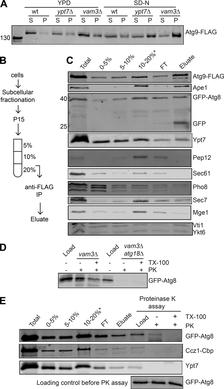
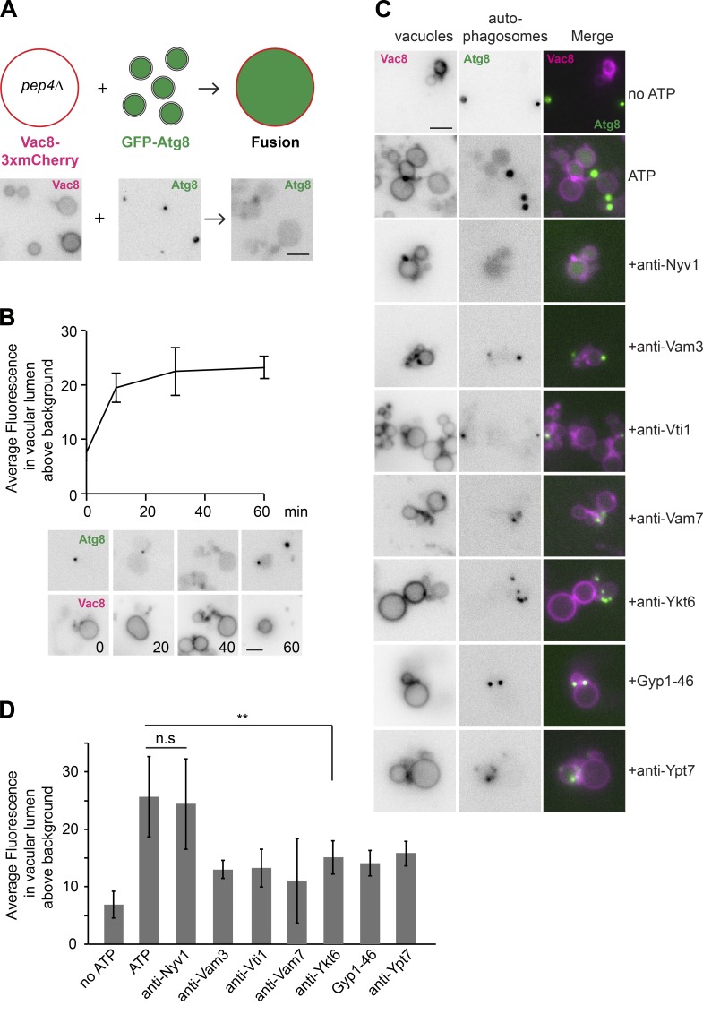
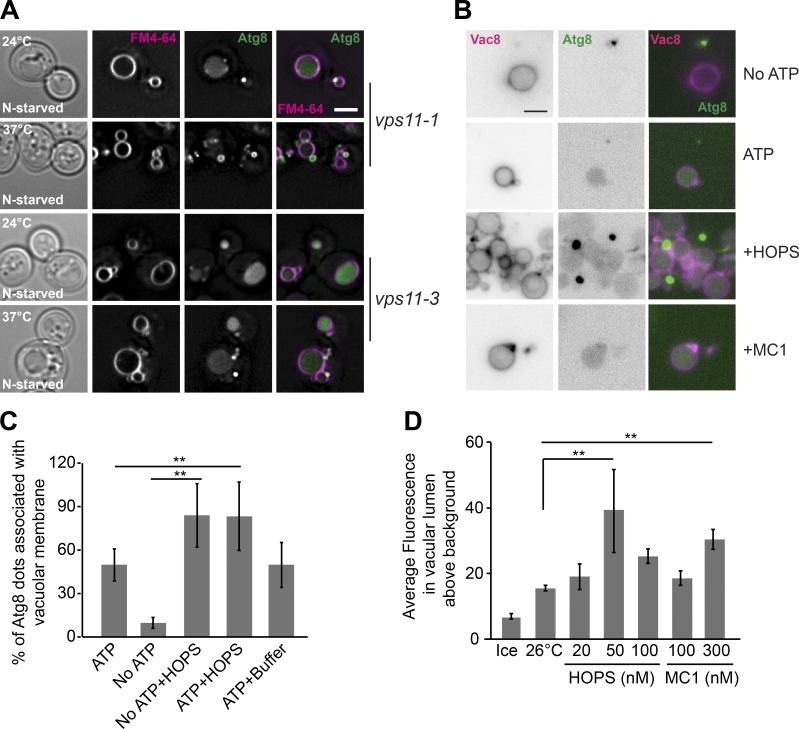
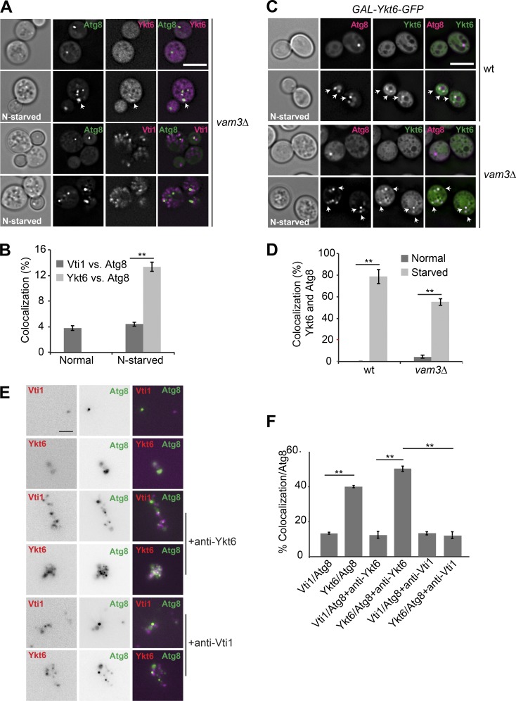
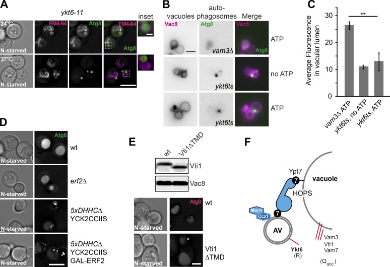
Autophagy is a catabolic pathway that delivers intracellular material to the mammalian lysosomes or the yeast and plant vacuoles. The final step in this process is the fusion of autophagosomes with vacuoles, which requires SNARE proteins, the homotypic vacuole fusion and protein sorting tethering complex, the RAB7-like Ypt7 GTPase, and its guanine nucleotide exchange factor, Mon1-Ccz1. Where these different components are located and function during fusion, however, remains to be fully understood. Here, we present a novel in vitro assay to monitor fusion of intact and functional autophagosomes with vacuoles. This process requires ATP, physiological temperature, and the entire fusion machinery to tether and fuse autophagosomes with vacuoles. Importantly, we uncover Ykt6 as the autophagosomal SNARE. Our assay and findings thus provide the tools to dissect autophagosome completion and fusion in a test tube.
Overview
The claims and statistics of this paper haven’t been checked yet.
Run the check — free, no account needed. It reads the open-access text and checks the sources it cites.
Integrity signals
Registry
Cites
Statistics
Figures
Verification
Claims the paper makes
The claims and statistics of this paper haven’t been checked yet.
Run the check — free, no account needed. It reads the open-access text and checks the sources it cites.
Claims other papers make about this one
Each is checked against this paper’s own text: does it say what the citing paper says it does?
The source backs part of the claim, not all of it — or backs it for a different population, condition or comparison.
In the citing paper
“In vitro reconstitutions of autophagosome-vacuole and autophagosome-lysosome fusion have partially overcome this problem and made it possible to identify relevant proteins and their functions in this specific step of autophagy.”
From Guidelines for the use and interpretation of assays for monitoring autophagy (4th edition) 1 (2021) · cites this paper as [507]
Experimental — accuracy not yet measuredIn the citing paper
“Both autophagosome-vacuole fusion in yeast and autophagosome-lysosome fusion in mammals has been recently reconstituted, using partially purified fractions of autophagosomes, vacuoles/lysosomes and cytosol [506-508].”
From Guidelines for the use and interpretation of assays for monitoring autophagy (4th edition) 1 (2021) · cites this paper as [507]
Self-check
Percentages, sums and reported differences re-computed from the paper’s own numbers.
The self-check needs the paper’s full text.
Citations
Cites
Guidelines for the use and interpretation of assays for monitoring autophagy (3rd edition).
Klionsky, D. J., Abdelmohsen, K., Abe, A., Abedin, M. J., Abeliovich, H., Acevedo Arozena, A., Adachi, H., Adams, C. M., Adams, P. D., Adeli, K., et al. (2016) · Autophagy · doi:10.1080/15548627.2015.1100356 · Cited by 5,894
Self-citationFormation Process of Autophagosome Is Traced with Apg8/Aut7p in Yeast
Kirisako, T., Baba, M., Ishihara, N., Miyazawa, K., Ohsumi, M., Yoshimori, T., Noda, T., & Ohsumi, Y. (1999) · The Journal of Cell Biology · doi:10.1083/jcb.147.2.435 · Cited by 881
Wurmser, A. E., Sato, T. K., & Emr, S. D. (2000) · The Journal of Cell Biology · doi:10.1083/jcb.151.3.551 · Cited by 425
Guan, J., Stromhaug, P. E., George, M. D., Habibzadegah-Tari, P., Bevan, A., Dunn, W. A., & Klionsky, D. J. (2001) · Molecular Biology of the Cell · doi:10.1091/mbc.12.12.3821 · Cited by 204
Research works printed but not linked: 78
- Arasaki, 2015, A role for the ancient SNARE syntaxin 17 in regulating mitochondrial division, Dev. Cell., 32, 304, 10.1016/j.devcel.2014.12.011 · DOI
- Baba, 1994, Ultrastructural analysis of the autophagic process in yeast: detection of autophagosomes and their characterization, J. Cell Biol., 124, 903, 10.1083/jcb.124.6.903 · DOI
- Baker, 2015, A direct role for the Sec1/Munc18-family protein Vps33 as a template for SNARE assembly, Science., 349, 1111, 10.1126/science.aac7906 · DOI
- Balderhaar, 2013, CORVET and HOPS tethering complexes - coordinators of endosome and lysosome fusion, J. Cell Sci., 126, 1307, 10.1242/jcs.107805 · DOI
- Barth, 2001, Autophagy and the cytoplasm to vacuole targeting pathway both require Aut10p, FEBS Lett., 508, 23, 10.1016/S0014-5793(01)03016-2 · DOI · Paper page
- Bas, 2018, Reconstitution reveals Ykt6 as the autophagosomal SNARE in autophagosome–vacuole fusion, J. Cell Biol., 10.1083/jcb.201804028 · DOI · Paper page
- Brett, 2008, Efficient termination of vacuolar Rab GTPase signaling requires coordinated action by a GAP and a protein kinase, J. Cell Biol., 182, 1141, 10.1083/jcb.200801001 · DOI
- Bröcker, 2012, Molecular architecture of the multisubunit homotypic fusion and vacuole protein sorting (HOPS) tethering complex, Proc. Natl. Acad. Sci. USA., 109, 1991, 10.1073/pnas.1117797109 · DOI
- Cebollero, 2012, Phosphatidylinositol-3-phosphate clearance plays a key role in autophagosome completion, Curr. Biol., 22, 1545, 10.1016/j.cub.2012.06.029 · DOI
- Cheever, 2001, Phox domain interaction with PtdIns(3)P targets the Vam7 t-SNARE to vacuole membranes, Nat. Cell Biol., 3, 613, 10.1038/35083000 · DOI · Paper page
- Chen, 2016, Multiple ER-Golgi SNARE transmembrane domains are dispensable for trafficking but required for SNARE recycling, Mol. Biol. Cell., 27, 2633, 10.1091/mbc.e16-05-0277 · DOI
- D’Agostino, 2017, A tethering complex drives the terminal stage of SNARE-dependent membrane fusion, Nature., 551, 634, 10.1038/nature24469 · DOI
- Darsow, 1997, A multispecificity syntaxin homologue, Vam3p, essential for autophagic and biosynthetic protein transport to the vacuole, J. Cell Biol., 138, 517, 10.1083/jcb.138.3.517 · DOI
- Darsow, 1998, Acidic di-leucine motif essential for AP-3-dependent sorting and restriction of the functional specificity of the Vam3p vacuolar t-SNARE, J. Cell Biol., 142, 913, 10.1083/jcb.142.4.913 · DOI
- Dietrich, 2005, ATP-independent control of Vac8 palmitoylation by a SNARE subcomplex on yeast vacuoles, J. Biol. Chem., 280, 15348, 10.1074/jbc.M410582200 · DOI
- Dilcher, 2001, Genetic interactions with the yeast Q-SNARE VTI1 reveal novel functions for the R-SNARE YKT6, J. Biol. Chem., 276, 34537, 10.1074/jbc.M101551200 · DOI
- Fischer von Mollard, 1999, The Saccharomyces cerevisiae v-SNARE Vti1p is required for multiple membrane transport pathways to the vacuole, Mol. Biol. Cell., 10, 1719, 10.1091/mbc.10.6.1719 · DOI
- Fukasawa, 2004, Localization and activity of the SNARE Ykt6 determined by its regulatory domain and palmitoylation, Proc. Natl. Acad. Sci. USA., 101, 4815, 10.1073/pnas.0401183101 · DOI
- Gao, 2018, Molecular mechanism to target the endosomal Mon1-Ccz1 GEF complex to the pre-autophagosomal structure, eLife., 7, 765, 10.7554/eLife.31145 · DOI
- Ge, 2014, The protein-vesicle network of autophagy, Curr. Opin. Cell Biol., 29, 18, 10.1016/j.ceb.2014.02.005 · DOI
- Goody, 2017, Mechanisms of action of Rab proteins, key regulators of intracellular vesicular transport, Biol. Chem., 398, 565, 10.1515/hsz-2016-0274 · DOI
- Haas, 1995, A quantitative assay to measure homotypic vacuole fusion in vitro, Methods Cell Sci., 17, 283, 10.1007/BF00986234 · DOI
- Haas, 1994, G-protein ligands inhibit in vitro reactions of vacuole inheritance, J. Cell Biol., 126, 87, 10.1083/jcb.126.1.87 · DOI
- Hasegawa, 2003, Mammalian ykt6 is a neuronal SNARE targeted to a specialized compartment by its profilin-like amino terminal domain, Mol. Biol. Cell., 14, 698, 10.1091/mbc.e02-09-0556 · DOI
- Hegedüs, 2016, The Ccz1-Mon1-Rab7 module and Rab5 control distinct steps of autophagy, Mol. Biol. Cell., 27, 3132, 10.1091/mbc.e16-03-0205 · DOI
- Hickey, 2010, HOPS initiates vacuole docking by tethering membranes before trans-SNARE complex assembly, Mol. Biol. Cell., 21, 2297, 10.1091/mbc.e10-01-0044 · DOI
- Ho, 2015, The HOPS/class C Vps complex tethers membranes by binding to one Rab GTPase in each apposed membrane, Mol. Biol. Cell., 26, 2655, 10.1091/mbc.e14-04-0922 · DOI
- Ishihara, 2001, Autophagosome requires specific early Sec proteins for its formation and NSF/SNARE for vacuolar fusion, Mol. Biol. Cell., 12, 3690, 10.1091/mbc.12.11.3690 · DOI
- Itakura, 2012, The hairpin-type tail-anchored SNARE syntaxin 17 targets to autophagosomes for fusion with endosomes/lysosomes, Cell., 151, 1256, 10.1016/j.cell.2012.11.001 · DOI
- Janke, 2004, A versatile toolbox for PCR-based tagging of yeast genes: new fluorescent proteins, more markers and promoter substitution cassettes, Yeast., 21, 947, 10.1002/yea.1142 · DOI
- Karim, 2018, The Na+(K+)/H+ exchanger Nhx1 controls multivesicular body-vacuolar lysosome fusion, Mol. Biol. Cell., 29, 317, 10.1091/mbc.e17-08-0496 · DOI
- Karim, 2018, Distinct features of multivesicular body-lysosome fusion revealed by a new cell-free content-mixing assay, Traffic., 19, 138, 10.1111/tra.12543 · DOI
- Kumar, 2018, Mechanism of Stx17 recruitment to autophagosomes via IRGM and mammalian Atg8 proteins, J. Cell Biol., 217, 997, 10.1083/jcb.201708039 · DOI
- Kweon, 2003, Ykt6p is a multifunctional yeast R-SNARE that is required for multiple membrane transport pathways to the vacuole, Mol. Biol. Cell., 14, 1868, 10.1091/mbc.e02-10-0687 · DOI
- Lai, 2017, Molecular Mechanisms of Synaptic Vesicle Priming by Munc13 and Munc18, Neuron., 95, 591, 10.1016/j.neuron.2017.07.004 · DOI
- Lamb, 2013, Endocytosis and autophagy: Shared machinery for degradation, BioEssays., 35, 34, 10.1002/bies.201200130 · DOI
- Lamb, 2013, The autophagosome: origins unknown, biogenesis complex, Nat. Rev. Mol. Cell Biol., 14, 759, 10.1038/nrm3696 · DOI
- Langemeyer, 2018, A guanine nucleotide exchange factor (GEF) limits Rab GTPase-driven membrane fusion, J. Biol. Chem., 293, 731, 10.1074/jbc.M117.812941 · DOI
- Linder, 2013, Mechanism and function of DHHC S-acyltransferases, Biochem. Soc. Trans., 41, 29, 10.1042/BST20120328 · DOI
- Lobingier, 2012, Sec1/Munc18 protein Vps33 binds to SNARE domains and the quaternary SNARE complex, Mol. Biol. Cell, 23, 4611, 10.1091/mbc.e12-05-0343 · DOI
- Lürick, 2017, Multivalent Rab interactions determine tether-mediated membrane fusion, Mol. Biol. Cell., 28, 322, 10.1091/mbc.e16-11-0764 · DOI
- Mari, 2010, Atg9 reservoirs, a new organelle of the yeast endomembrane system?, Autophagy., 6, 1221, 10.4161/auto.6.8.13792 · DOI
- Matsui, 2018, Autophagosomal YKT6 is required for fusion with lysosomes independently of syntaxin 17, J. Cell Biol., 217, 2633, 10.1083/jcb.201712058 · DOI · Paper page
- McNew, 1997, Ykt6p, a prenylated SNARE essential for endoplasmic reticulum-Golgi transport, J. Biol. Chem., 272, 17776, 10.1074/jbc.272.28.17776 · DOI
- McNew, 2000, Compartmental specificity of cellular membrane fusion encoded in SNARE proteins, Nature., 407, 153, 10.1038/35025000 · DOI
- Meiringer, 2008, Depalmitoylation of Ykt6 prevents its entry into the multivesicular body pathway, Traffic, 9, 1510, 10.1111/j.1600-0854.2008.00778.x · DOI
- Meiringer, 2011, The Dsl1 protein tethering complex is a resident endoplasmic reticulum complex, which interacts with five soluble NSF (N-ethylmaleimide-sensitive factor) attachment protein receptors (SNAREs): implications for fusion and fusion regulation, J. Biol. Chem., 286, 25039, 10.1074/jbc.M110.215327 · DOI
- Mitchell, 2006, Protein palmitoylation by a family of DHHC protein S-acyltransferases, J. Lipid Res., 47, 1118, 10.1194/jlr.R600007-JLR200 · DOI
- Nair, 2011, SNARE proteins are required for macroautophagy, Cell., 146, 290, 10.1016/j.cell.2011.06.022 · DOI
- Noda, 1995, Novel system for monitoring autophagy in the yeast Saccharomyces cerevisiae, Biochem. Biophys. Res. Commun., 210, 126, 10.1006/bbrc.1995.1636 · DOI
- Nordmann, 2010, The Mon1-Ccz1 complex is the GEF of the late endosomal Rab7 homolog Ypt7, Curr. Biol., 20, 1654, 10.1016/j.cub.2010.08.002 · DOI
- Ohashi, 2010, Membrane delivery to the yeast autophagosome from the Golgi-endosomal system, Mol. Biol. Cell., 21, 3998, 10.1091/mbc.e10-05-0457 · DOI
- Orr, 2017, HOPS catalyzes the interdependent assembly of each vacuolar SNARE into a SNARE complex, Mol. Biol. Cell., 28, 975, 10.1091/mbc.e16-10-0743 · DOI
- Peterson, 2001, The class C Vps complex functions at multiple stages of the vacuolar transport pathway, Traffic., 2, 476, 10.1034/j.1600-0854.2001.20705.x · DOI
- Puig, 1998, New constructs and strategies for efficient PCR-based gene manipulations in yeast, Yeast., 14, 1139, 10.1002/(SICI)1097-0061(19980915)14:12<1139::AID-YEA306>3.0.CO;2-B · DOI
- Reggiori, 2017, Autophagosome Maturation and Fusion, J. Mol. Biol., 429, 486, 10.1016/j.jmb.2017.01.002 · DOI
- Rieter, 2013, Atg18 function in autophagy is regulated by specific sites within its β-propeller, J. Cell Sci., 126, 593, 10.1242/jcs.115725 · DOI
- Rohde, 2003, The transmembrane domain of Vam3 affects the composition of cis- and trans-SNARE complexes to promote homotypic vacuole fusion, J. Biol. Chem., 278, 1656, 10.1074/jbc.M209522200 · DOI
- Roth, 2006, Global analysis of protein palmitoylation in yeast, Cell, 125, 1003, 10.1016/j.cell.2006.03.042 · DOI
- Seals, 2000, A Ypt/Rab effector complex containing the Sec1 homolog Vps33p is required for homotypic vacuole fusion, Proc. Natl. Acad. Sci. USA., 97, 9402, 10.1073/pnas.97.17.9402 · DOI
- Shibutani, 2014, A current perspective of autophagosome biogenesis, Cell Res., 24, 58, 10.1038/cr.2013.159 · DOI
- Takáts, 2013, Autophagosomal Syntaxin17-dependent lysosomal degradation maintains neuronal function in Drosophila, J. Cell Biol., 201, 531, 10.1083/jcb.201211160 · DOI
- Takáts, 2014, Interaction of the HOPS complex with Syntaxin 17 mediates autophagosome clearance in Drosophila, Mol. Biol. Cell., 25, 1338, 10.1091/mbc.e13-08-0449 · DOI
- Takáts, 2018, Non-canonical role of the SNARE protein Ykt6 in autophagosome-lysosome fusion, PLoS Genet., 14, e1007359, 10.1371/journal.pgen.1007359 · DOI
- Torggler, 2017, Assays to Monitor Autophagy in Saccharomyces cerevisiae, Cells., 6, 23, 10.3390/cells6030023 · DOI
- Tsui, 2000, Yeast Golgi SNARE interactions are promiscuous, J. Cell Sci., 113, 145, 10.1242/jcs.113.1.145 · DOI
- Ungermann, 1998, Vam7p, a vacuolar SNAP-25 homolog, is required for SNARE complex integrity and vacuole docking and fusion, EMBO J., 17, 3269, 10.1093/emboj/17.12.3269 · DOI
- Ungermann, 1998, A vacuolar v-t-SNARE complex, the predominant form in vivo and on isolated vacuoles, is disassembled and activated for docking and fusion, J. Cell Biol., 140, 61, 10.1083/jcb.140.1.61 · DOI
- Ungermann, 1999, Three v-SNAREs and two t-SNAREs, present in a pentameric cis-SNARE complex on isolated vacuoles, are essential for homotypic fusion, J. Cell Biol., 145, 1435, 10.1083/jcb.145.7.1435 · DOI
- Veit, 2004, The human SNARE protein Ykt6 mediates its own palmitoylation at C-terminal cysteine residues, Biochem. J., 384, 233, 10.1042/BJ20041474 · DOI
- von Mollard, 1997, The yeast v-SNARE Vti1p mediates two vesicle transport pathways through interactions with the t-SNAREs Sed5p and Pep12p, J. Cell Biol., 137, 1511, 10.1083/jcb.137.7.1511 · DOI
- Wickner, 2010, Membrane fusion: five lipids, four SNAREs, three chaperones, two nucleotides, and a Rab, all dancing in a ring on yeast vacuoles, Annu. Rev. Cell Dev. Biol., 26, 115, 10.1146/annurev-cellbio-100109-104131 · DOI
- Wickner, 2017, A cascade of multiple proteins and lipids catalyzes membrane fusion, Mol. Biol. Cell., 28, 707, 10.1091/mbc.e16-07-0517 · DOI
- Xu, 2011, A lipid-anchored SNARE supports membrane fusion, Proc. Natl. Acad. Sci. USA., 108, 17325, 10.1073/pnas.1113888108 · DOI
- Yamamoto, 2012, Atg9 vesicles are an important membrane source during early steps of autophagosome formation, J. Cell Biol., 198, 219, 10.1083/jcb.201202061 · DOI · Paper page
- Zick, 2012, Phosphorylation of the effector complex HOPS by the vacuolar kinase Yck3p confers Rab nucleotide specificity for vacuole docking and fusion, Mol. Biol. Cell., 23, 3429, 10.1091/mbc.e12-04-0279 · DOI
- Zick, 2013, The tethering complex HOPS catalyzes assembly of the soluble SNARE Vam7 into fusogenic trans-SNARE complexes, Mol. Biol. Cell., 24, 3746, 10.1091/mbc.e13-07-0419 · DOI
- Zick, 2016, Improved reconstitution of yeast vacuole fusion with physiological SNARE concentrations reveals an asymmetric Rab(GTP) requirement, Mol. Biol. Cell., 27, 2590, 10.1091/mbc.e16-04-0230 · DOI
Cited by
Sorted by citations
Guidelines for the use and interpretation of assays for monitoring autophagy (4th edition) 1
Klionsky, D. J., Abdel-Aziz, A. K., Abdelfatah, S., Abdellatif, M., Abdoli, A., Abel, S., Abeliovich, H., Abildgaard, M. H., Abudu, Y. P., Acevedo-Arozena, A., et al. (2021) · Autophagy · doi:10.1080/15548627.2020.1797280 · Cited by 2,897
Self-citationLőrincz, P., & Juhász, G. (2020) · Journal of Molecular Biology · doi:10.1016/j.jmb.2019.10.028 · Cited by 328
Metadata
- Authors
- Gao, J., Reggiori, F., & Ungermann, C.
- Affiliations
- Biochemistry Section, Department of Biology/Chemistry, University of Osnabrück, Osnabrück, Germany 1
- Department of Cell Biology, University of Groningen, University Medical Center Groningen, Groningen, Netherlands 2
- Venue
- Journal of Cell Biology
- Publisher
- Rockefeller University Press
- Published
- 2018-08-10
- Volume
- 217
- Issue
- 10
- Pages
- 3670-3682
- ISSN
- 0021-9525
- 1540-8140
- PMC
- PMC6168247
- Cited by
- 90
- References
- 4
- Language
- en
- Funders
- ALW (822.02.014)
- Deutsche Forschungsgemeinschaft (UN111/7-3)
- Netherlands Organisation for Scientific Research (DN82-303)
- Swiss National Science Foundation (CRSII3_154421)
- Marie Skłodowska-Curie (713660)
- ZonMW (016.130.606)
- Sonderforschungsbereich (Project P11)
About these checks
Registry, retraction, Cites and statistics results are computed, not judged, with one exception: a printed reference that no index lists is read once by a model before it counts as a citation error. Claims are extracted by a model and kept only when their quote appears verbatim in the text; each cited source is then checked, with a model’s help, for whether it says what the claim relies on. Those verdicts can be wrong — read the claim’s quote and the cited source before relying on one.
Statistics and arithmetic are recomputed, not judged.
Confidence bands are shown only for fields calibrated on our annotated test set. None is calibrated yet, so no band is shown.
What counts as a claim ›How we measure confidence ›How we check this ›
Are you an author, or do you see a problem with this page? Request a review
