Cited by 168T-cell and B-cell ImmunologySystemic Lupus Erythematosus ResearchImmune Response and Inflammation

The DOI matches its Crossref record; 63 of the 63 references it cites were found in a registry or an index.

Sustained Activation of Lyn Tyrosine Kinase In Vivo Leads to Autoimmunity

Open access

Abstract

Genetic ablation of the Lyn tyrosine kinase has revealed unique inhibitory roles in B lymphocyte signaling. We now report the consequences of sustained activation of Lyn in vivo using a targeted gain-of-function mutation (Lynup/up mice). Lynup/up mice have reduced numbers of conventional B lymphocytes, down-regulated surface immunoglobulin M and costimulatory molecules, and elevated numbers of B1a B cells. Lynup/up B cells are characterized by the constitutive phosphorylation of negative regulators of B cell antigen receptor (BCR) signaling including CD22, SHP-1, and SHIP-1, and display attributes of lymphocytes rendered tolerant by constitutive engagement of the antigen receptor. However, exaggerated positive signaling is also apparent as evidenced by the constitutive phosphorylation of Syk and phospholipase Cγ2 in resting Lynup/up B cells. Similarly, Lynup/up B cells show a heightened calcium flux in response to BCR stimulation. Surprisingly, Lynup/up mice develop circulating autoreactive antibodies and lethal autoimmune glomerulonephritis, suggesting that enhanced positive signaling eventually overrides constitutive negative signaling. These studies highlight the difficulty in maintaining tolerance in the face of chronic stimulation and emphasize the pivotal role of Lyn in B cell signaling.

Overview

The claims and statistics of this paper haven’t been checked yet.

Run the check — free, no account needed. It reads the open-access text and checks the sources it cites.

A few free checks a day · papers already verified open instantly · results are public

Integrity signals

Registry

Crossref · 10.1084/jem.20020515

Cites

63 of 63 references verified0 citation errors · 0 not checkable

Statistics

Figures

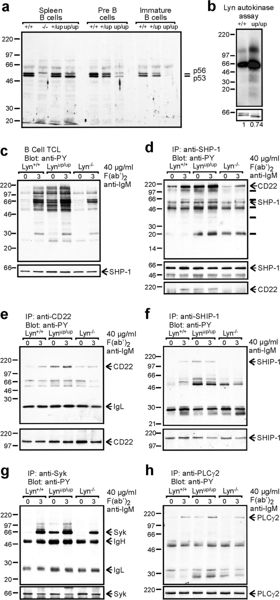
Figure 1. Altered Lyn protein levels and signaling in primary B cells from Lyn mutant mice. (a) Mature B cells were purified from the spleen (Spleen B cells) and Pre-B and immature B cells were purified from the BM of the indicated mice (+/+, Lyn+/+; −/−, Lyn−/−; +/up, Lyn+/up; up/up, Lynup/up) by FACS® sorting. Total cell lysates (TCL; 5 × 10⁶ cell equivalents for mature and Pre-B cells and 2.5 × 10⁶ cell equivalents for immature B cells) were immunoblotted with anti-Lyn Abs. The p56 and p53 isoforms of Lyn are indicated. (b) Lyn kinase activity. Equalized levels of Lyn protein (bottom, including densitometric measurements) immunoprecipitated from Lyn+/+ (+/+) and Lynup/up (up/up) B cells were subjected to autokinase reactions. (c) Primary B cells from Lyn+/+, Lynup/up, and Lyn−/− mice were stimulated for 0 or 3 min with 40 μg/ml F(ab′)2 anti-IgM. 25 μg TCLs were immunoblotted with anti-PY. The blot was stripped and reprobed with anti–SHP-1 as a protein loading control. (d) SHP-1 was immunoprecipitated from the indicated B cell lysates and immunoblotted with anti-PY. The blot was stripped and reprobed with anti–SHP-1 (middle) and an antiserum to CD22 (bottom) to demonstrate coassociation of SHP-1 and phospho-CD22. CD22 (e), SHIP-1 (f), Syk (g), and PLCγ2 (h) were immunoprecipitated from the indicated B cell lysates and immunoblotted with anti-PY. Blots were stripped and re-probed with the indicated antibodies to ensure equal protein loading.
Figure 1. Altered Lyn protein levels and signaling in primary B cells from Lyn mutant mice. (a) Mature B cells were purified from the spleen (Spleen B cells) and Pre-B and immature B cells were purified from the BM of the indicated mice (+/+, Lyn+/+; −/−, Lyn−/−; +/up, Lyn+/up; up/up, Lynup/up) by FACS® sorting. Total cell lysates (TCL; 5 × 10⁶ cell equivalents for mature and Pre-B cells and 2.5 × 10⁶ cell equivalents for immature B cells) were immunoblotted with anti-Lyn Abs. The p56 and p53 isoforms of Lyn are indicated. (b) Lyn kinase activity. Equalized levels of Lyn protein (bottom, including densitometric measurements) immunoprecipitated from Lyn+/+ (+/+) and Lynup/up (up/up) B cells were subjected to autokinase reactions. (c) Primary B cells from Lyn+/+, Lynup/up, and Lyn−/− mice were stimulated for 0 or 3 min with 40 μg/ml F(ab′)2 anti-IgM. 25 μg TCLs were immunoblotted with anti-PY. The blot was stripped and reprobed with anti–SHP-1 as a protein loading control. (d) SHP-1 was immunoprecipitated from the indicated B cell lysates and immunoblotted with anti-PY. The blot was stripped and reprobed with anti–SHP-1 (middle) and an antiserum to CD22 (bottom) to demonstrate coassociation of SHP-1 and phospho-CD22. CD22 (e), SHIP-1 (f), Syk (g), and PLCγ2 (h) were immunoprecipitated from the indicated B cell lysates and immunoblotted with anti-PY. Blots were stripped and re-probed with the indicated antibodies to ensure equal protein loading.CC BY-NC-SA · PubMed Central
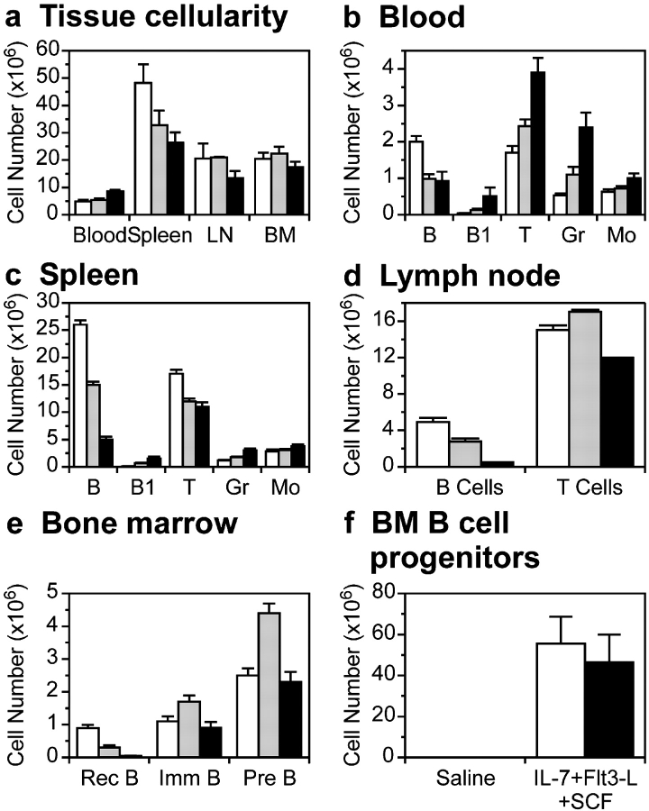
Figure 2. Cellularity and composition of lymphoid tissue in 8-wk-old Lyn+/+, Lyn+/up, and Lynup/up mice. (a) Nucleated cells in lymphoid tissue in Lyn+/+, Lyn+/up, and Lynup/up mice. Numbers represent per ml blood, per whole spleen, per mesenteric lymph nodes, and per femur. (b) Numbers of conventional B cells, B1 cells, T cells, granulocytes (Gr), and monocytes (Mo) in blood and (c) spleen of Lyn+/+, Lyn+/up, and Lynup/up mice. (d) Numbers of B and T cells in mesenteric lymph nodes of Lyn+/+, Lyn+/up, and Lynup/up mice. (e) Numbers of recirculating B cells (Rec B), immature B cells (Imm B), and Pre-B cells in the BM of Lyn+/+, Lyn+/up, and Lynup/up mice. Data in a–e is compiled from nine mice in three experiments, except B cell and B1 cell numbers in blood and spleen, which were compiled from six mice in two experiments. Data is mean ± standard error. (f) BM B cell progenitors in Lyn+/+ and Lynup/up mice. Numbers are the mean ± standard deviation for five mice of each genotype. For a–f, white bars, Lyn+/+; gray bars, Lyn+/up; black bars, Lynup/up.
Figure 2. Cellularity and composition of lymphoid tissue in 8-wk-old Lyn+/+, Lyn+/up, and Lynup/up mice. (a) Nucleated cells in lymphoid tissue in Lyn+/+, Lyn+/up, and Lynup/up mice. Numbers represent per ml blood, per whole spleen, per mesenteric lymph nodes, and per femur. (b) Numbers of conventional B cells, B1 cells, T cells, granulocytes (Gr), and monocytes (Mo) in blood and (c) spleen of Lyn+/+, Lyn+/up, and Lynup/up mice. (d) Numbers of B and T cells in mesenteric lymph nodes of Lyn+/+, Lyn+/up, and Lynup/up mice. (e) Numbers of recirculating B cells (Rec B), immature B cells (Imm B), and Pre-B cells in the BM of Lyn+/+, Lyn+/up, and Lynup/up mice. Data in a–e is compiled from nine mice in three experiments, except B cell and B1 cell numbers in blood and spleen, which were compiled from six mice in two experiments. Data is mean ± standard error. (f) BM B cell progenitors in Lyn+/+ and Lynup/up mice. Numbers are the mean ± standard deviation for five mice of each genotype. For a–f, white bars, Lyn+/+; gray bars, Lyn+/up; black bars, Lynup/up.CC BY-NC-SA · PubMed Central
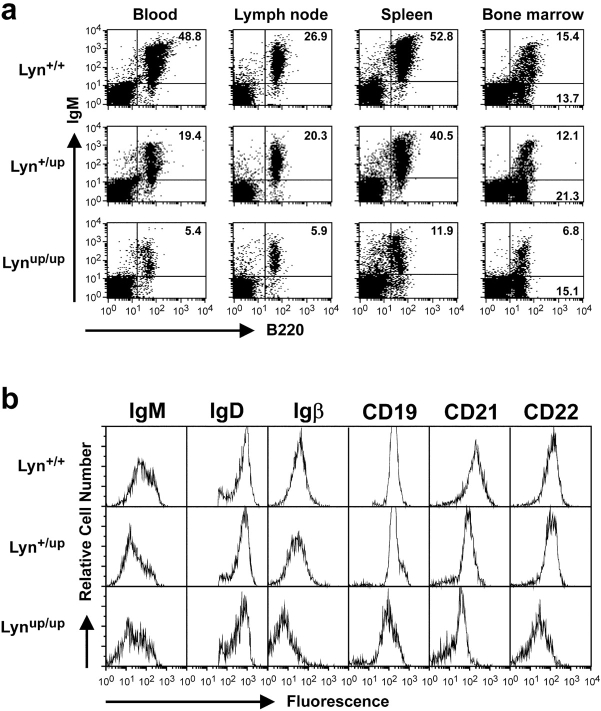
Figure 3. Lynup/up mice have reduced levels of recirculating B cells. (a) Representative two-color fluorescence analysis of lymphoid tissues from 8-wk-old Lyn+/+, Lyn+/up, and Lynup/up mice. Staining was performed with Abs against B220 and IgM to determine proportions of B cells and levels of surface IgM. The mean fluorescence intensity of IgM on peripheral blood B cells from Lyn+/+, Lyn+/up, and Lynup/up mice is 163, 101, and 103, respectively, on lymph node B cells is 300, 163, and 213, respectively, and on splenic B cells is 196, 122, and 133, respectively. (b) Representative single-color FACS® analysis of splenic B220+ cells from 8-wk-old Lyn+/+, Lyn+/up, and Lynup/up mice. Staining was performed with Abs against the indicated cell surface molecules to assess expression level.
Figure 3. Lynup/up mice have reduced levels of recirculating B cells. (a) Representative two-color fluorescence analysis of lymphoid tissues from 8-wk-old Lyn+/+, Lyn+/up, and Lynup/up mice. Staining was performed with Abs against B220 and IgM to determine proportions of B cells and levels of surface IgM. The mean fluorescence intensity of IgM on peripheral blood B cells from Lyn+/+, Lyn+/up, and Lynup/up mice is 163, 101, and 103, respectively, on lymph node B cells is 300, 163, and 213, respectively, and on splenic B cells is 196, 122, and 133, respectively. (b) Representative single-color FACS® analysis of splenic B220+ cells from 8-wk-old Lyn+/+, Lyn+/up, and Lynup/up mice. Staining was performed with Abs against the indicated cell surface molecules to assess expression level.CC BY-NC-SA · PubMed Central
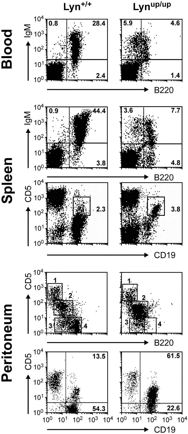
Figure 4. Lynup/up mice have increased numbers of B1 cells. Two-color fluorescence analysis of blood, spleen, and peritoneal lymphoid cells from 8-wk-old Lyn+/+ and Lynup/up mice. Blood and spleen cells were stained with B220 and IgM to identify IgM+ B220lo B1 cells. Spleen cells were also stained with CD19 and CD5 Abs to show CD19hi CD5+ B1a cells in Lynup/up spleen (boxed region). Peritoneal cells were stained using Abs to B220 and CD5 to identify: Region 1, T cells (Lyn+/+, 14.4%; Lynup/up, 13.0%), Region 2, B1a B cells (Lyn+/+, 34.6%; Lynup/up, 76.1%), Region 3, B1b B cells (Lyn+/+, 9.4%; Lynup/up, 4.3%), and Region 4, B2 cells (Lyn+/+, 31.7%; Lynup/up, 1.8%). Alternatively, peritoneal B1a cells were revealed with Abs to CD19 and CD5.
Figure 4. Lynup/up mice have increased numbers of B1 cells. Two-color fluorescence analysis of blood, spleen, and peritoneal lymphoid cells from 8-wk-old Lyn+/+ and Lynup/up mice. Blood and spleen cells were stained with B220 and IgM to identify IgM+ B220lo B1 cells. Spleen cells were also stained with CD19 and CD5 Abs to show CD19hi CD5+ B1a cells in Lynup/up spleen (boxed region). Peritoneal cells were stained using Abs to B220 and CD5 to identify: Region 1, T cells (Lyn+/+, 14.4%; Lynup/up, 13.0%), Region 2, B1a B cells (Lyn+/+, 34.6%; Lynup/up, 76.1%), Region 3, B1b B cells (Lyn+/+, 9.4%; Lynup/up, 4.3%), and Region 4, B2 cells (Lyn+/+, 31.7%; Lynup/up, 1.8%). Alternatively, peritoneal B1a cells were revealed with Abs to CD19 and CD5.CC BY-NC-SA · PubMed Central
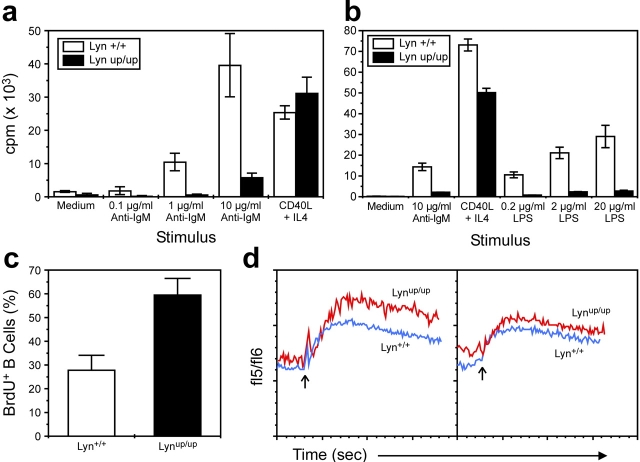
Figure 5. Proliferation, turnover, and calcium responses of Lyn+/+ and Lynup/up B lymphocytes. 5 × 10⁴ FACS®-sorted splenic B cells from Lyn+/+ mice (open bars) and Lynup/up mice (solid bars) were cultured in the presence of (a) the indicated concentrations of anti-IgM or CD40 ligand plus interleukin-4 for 2 d, and in (b) the indicated concentrations of LPS, 10 μg/ml anti-IgM, or CD40 ligand plus interleukin-4 for 3 d. (c) Turnover of peripheral B lymphocytes by incorporation of BrdU. Spleen cells from BrdU-treated Lyn+/+ mice (open bars) and Lynup/up mice (solid bars) were stained for surface CD19 and intranuclear BrdU to determine the percentage of BrdU+ B cells. Data is representative of two experiments in which five mice of each genotype were analyzed. (d) Changes in [Ca2+]i induced by cross-linking surface IgM on Indo-1–loaded spleen cells from Lyn+/+ and Lynup/up mice. The fluorescence ratio of the cells (fl5/fl6) was measured by flow cytometry and B cells were identified by counter-staining with anti-B220. Cross-linking reagent was added at 60 s (arrow) and the measurement continued for 300 s Two representative examples from three experiments involving six mice of each genotype are shown with the response of Lyn+/+ B cells depicted in blue and Lynup/up B cells in red.
Figure 5. Proliferation, turnover, and calcium responses of Lyn+/+ and Lynup/up B lymphocytes. 5 × 10⁴ FACS®-sorted splenic B cells from Lyn+/+ mice (open bars) and Lynup/up mice (solid bars) were cultured in the presence of (a) the indicated concentrations of anti-IgM or CD40 ligand plus interleukin-4 for 2 d, and in (b) the indicated concentrations of LPS, 10 μg/ml anti-IgM, or CD40 ligand plus interleukin-4 for 3 d. (c) Turnover of peripheral B lymphocytes by incorporation of BrdU. Spleen cells from BrdU-treated Lyn+/+ mice (open bars) and Lynup/up mice (solid bars) were stained for surface CD19 and intranuclear BrdU to determine the percentage of BrdU+ B cells. Data is representative of two experiments in which five mice of each genotype were analyzed. (d) Changes in [Ca2+]i induced by cross-linking surface IgM on Indo-1–loaded spleen cells from Lyn+/+ and Lynup/up mice. The fluorescence ratio of the cells (fl5/fl6) was measured by flow cytometry and B cells were identified by counter-staining with anti-B220. Cross-linking reagent was added at 60 s (arrow) and the measurement continued for 300 s Two representative examples from three experiments involving six mice of each genotype are shown with the response of Lyn+/+ B cells depicted in blue and Lynup/up B cells in red.CC BY-NC-SA · PubMed Central
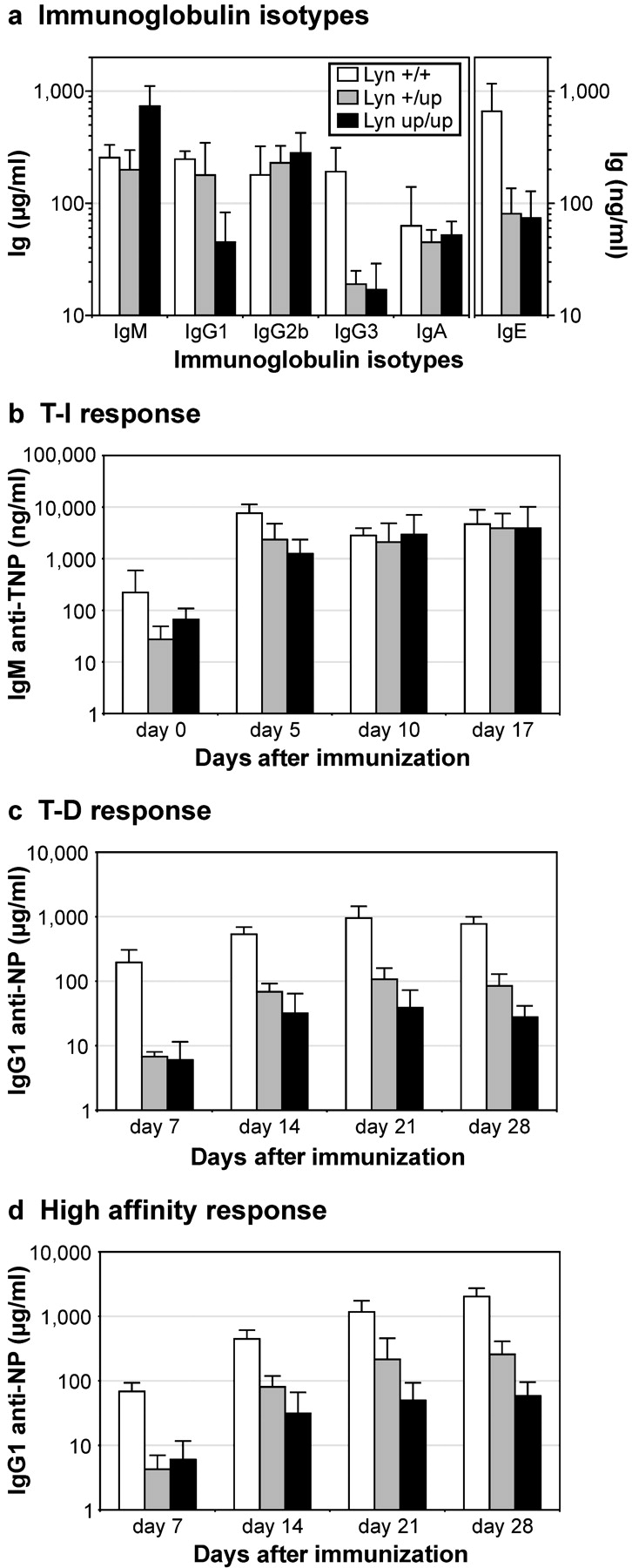
Figure 6. Immunoglobulin levels and immune responses. (a) Levels of immunoglobulin isotypes in the serum of 8–20 unchallenged Lyn+/+, Lyn+/up, and Lynup/up mice. (b) Specific IgM response of a group of six Lyn+/+, Lyn+/up, and Lynup/up mice at the indicated times after immunization with 10 μg DNP-dextran. Data is representative of two experiments. (c) Specific IgG1 response of a group of six Lyn+/+, Lyn+/up, and Lynup/up mice at the indicated times after immunization with 100 μg NP-KLH. Data is representative of two experiments. (d) High affinity IgG1 response of the group of mice indicated in c. Data is representative of two experiments. Data are mean ± standard deviation. For a–d, white bars, Lyn+/+; gray bars, Lyn+/up; black bars, Lynup/up.
Figure 6. Immunoglobulin levels and immune responses. (a) Levels of immunoglobulin isotypes in the serum of 8–20 unchallenged Lyn+/+, Lyn+/up, and Lynup/up mice. (b) Specific IgM response of a group of six Lyn+/+, Lyn+/up, and Lynup/up mice at the indicated times after immunization with 10 μg DNP-dextran. Data is representative of two experiments. (c) Specific IgG1 response of a group of six Lyn+/+, Lyn+/up, and Lynup/up mice at the indicated times after immunization with 100 μg NP-KLH. Data is representative of two experiments. (d) High affinity IgG1 response of the group of mice indicated in c. Data is representative of two experiments. Data are mean ± standard deviation. For a–d, white bars, Lyn+/+; gray bars, Lyn+/up; black bars, Lynup/up.CC BY-NC-SA · PubMed Central
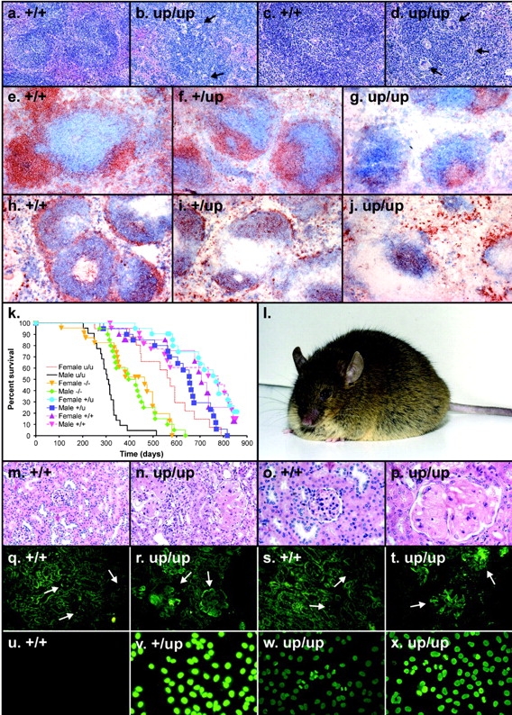
Figure 7. Histology, immunohistochemistry, survival, autoantibodies, and immunofluorescence analysis of Lyn+/+ and Lyn mutant mice. (a) Spleen from a Lyn+/+ mouse showing lymphoid follicles with germinal centers. (b) Spleen from a Lynup/up mouse showing lymphoid follicles with numerous multinucleate giant cells (arrows). (c) Higher power view of Lyn+/+ spleen. (d) Higher power view of Lynup/up spleen depicting multinucleate giant cells within a lymphoid follicle (arrows). Spleen sections from (e) Lyn+/+, (f) Lyn+/up, and (g) Lynup/up mice stained with B220 and CD3 to detect B (brown) and T cells (blue). Spleen sections from (h) Lyn+/+, (i) Lyn+/up, and (j) Lynup/up mice stained with IgM (brown) and IgD (blue) to detect marginal zones (IgMhi). (k) Kaplan-Meier survival curve showing the percent survival of a cohort of 20 male and 20 female Lyn+/+, Lyn+/up, Lynup/up, and Lyn−/− mice. (l) Male Lynup/up mouse of 10 mo of age showing extensive edema. (m) Low power view of a renal cortex from a Lyn+/+ mouse showing several normal glomeruli. (n) Low power view of Lynup/up renal cortex showing two very enlarged sclerotic glomeruli. (o) High power view of Lyn+/+ renal cortex showing a normal glomerulus. (p) High power view of Lynup/up renal cortex showing a severely damaged glomerulus with lobularity and sclerosis. View of the renal cortex from (q) Lyn+/+ and (r) Lynup/up mice stained with anti-Ig. Glomeruli are indicated with arrows. View of the renal cortex from (s) Lyn+/+ and (t) Lynup/up mice stained with anti-IgG. Glomeruli are indicated with arrows. Immunofluorescence analysis of HEp-2 cells stained with antisera from (u) female Lyn+/+, (v) male Lyn+/up, (w) female Lynup/up, and (x) male Lynup/up mice.
Figure 7. Histology, immunohistochemistry, survival, autoantibodies, and immunofluorescence analysis of Lyn+/+ and Lyn mutant mice. (a) Spleen from a Lyn+/+ mouse showing lymphoid follicles with germinal centers. (b) Spleen from a Lynup/up mouse showing lymphoid follicles with numerous multinucleate giant cells (arrows). (c) Higher power view of Lyn+/+ spleen. (d) Higher power view of Lynup/up spleen depicting multinucleate giant cells within a lymphoid follicle (arrows). Spleen sections from (e) Lyn+/+, (f) Lyn+/up, and (g) Lynup/up mice stained with B220 and CD3 to detect B (brown) and T cells (blue). Spleen sections from (h) Lyn+/+, (i) Lyn+/up, and (j) Lynup/up mice stained with IgM (brown) and IgD (blue) to detect marginal zones (IgMhi). (k) Kaplan-Meier survival curve showing the percent survival of a cohort of 20 male and 20 female Lyn+/+, Lyn+/up, Lynup/up, and Lyn−/− mice. (l) Male Lynup/up mouse of 10 mo of age showing extensive edema. (m) Low power view of a renal cortex from a Lyn+/+ mouse showing several normal glomeruli. (n) Low power view of Lynup/up renal cortex showing two very enlarged sclerotic glomeruli. (o) High power view of Lyn+/+ renal cortex showing a normal glomerulus. (p) High power view of Lynup/up renal cortex showing a severely damaged glomerulus with lobularity and sclerosis. View of the renal cortex from (q) Lyn+/+ and (r) Lynup/up mice stained with anti-Ig. Glomeruli are indicated with arrows. View of the renal cortex from (s) Lyn+/+ and (t) Lynup/up mice stained with anti-IgG. Glomeruli are indicated with arrows. Immunofluorescence analysis of HEp-2 cells stained with antisera from (u) female Lyn+/+, (v) male Lyn+/up, (w) female Lynup/up, and (x) male Lynup/up mice.CC BY-NC-SA · PubMed Central

Verification

Claims the paper makes

The claims and statistics of this paper haven’t been checked yet.

Run the check — free, no account needed. It reads the open-access text and checks the sources it cites.

A few free checks a day · papers already verified open instantly · results are public

Claims other papers make about this one

Each is checked against this paper’s own text: does it say what the citing paper says it does?

Self-check

Percentages, sums and reported differences re-computed from the paper’s own numbers.

The self-check needs the paper’s full text.

Citations

Cites

Show all 63 references

Research works printed but not linked: 1

  1. 1999, Curr. Top. Microbiol. Immunol., 244, 57

Cited by

Sorted by citations

See all 168

Metadata

Authors
Hibbs, M. L., Harder, K. W., Armes, J., Kountouri, N., Quilici, C., Casagranda, F., Dunn, A. R., & Tarlinton, D. M.
Affiliations
  • Ludwig Cancer Research (AU)
  • Royal Women's Hospital (AU)
  • The Royal Melbourne Hospital (AU)
Corresponding author
Hibbs, Margaret L.
Venue
The Journal of Experimental Medicine
Published
2002-12-16
Volume
196
Issue
12
Pages
1593-1604
ISSN
  • 0022-1007 (ISSN-L)
  • 1540-9538
OpenAlex
W2168601978
Cited by
168
References
63
Citations by year
2026: 2 · 2025: 8 · 2024: 4 · 2023: 10 · 2022: 5 · 2021: 4 · 2020: 5 · 2019: 2 · 2018: 5 · 2017: 3 · 2016: 5 · 2015: 5 · 2014: 12 · 2013: 13 · 2012: 13
FWCI
2.41
Type
journal article
Language
en
Open access
Locations
  • The Journal of Experimental Medicine (publishedVersion)
  • PubMed Central (publishedVersion, cc-by-nc-sa)
  • PubMed (publishedVersion)
  • Minerva Access (University of Melbourne) (submittedVersion, cc-by-nc-sa)
Topics
  • Life Sciences › Immunology and Microbiology › Immunology › T-cell and B-cell Immunology
  • Health Sciences › Medicine › Rheumatology › Systemic Lupus Erythematosus Research
  • Life Sciences › Immunology and Microbiology › Immunology › Immune Response and Inflammation
Keywords
Lyn kinase, B cell signaling, autoimmunity, B cell tolerance, B cell receptor signaling, CD22, SHP-1, SHIP1, Syk, PLCγ2, calcium flux, autoantibodies, glomerulonephritis
MeSH terms
Animals, Antigen-Antibody Complex, Autoantibodies, Autoimmune Diseases, B-Lymphocytes, Calcium, Cell Separation, Cells, Cultured, Enzyme Activation, Female, Flow Cytometry, Glomerulonephritis, Humans, Immunoglobulins, Kidney, Lymph Nodes, Male, Mice, Inbred C57BL, Mice, Transgenic, Phenotype, Spleen, Signal Transduction, Autoimmunity, Antigens, CD, Up-Regulation, Point Mutation, src-Family Kinases, Mice

About these checks

Registry, retraction, Cites and statistics results are computed, not judged, with one exception: a printed reference that no index lists is read once by a model before it counts as a citation error. Claims are extracted by a model and kept only when their quote appears verbatim in the text; each cited source is then checked, with a model’s help, for whether it says what the claim relies on. Those verdicts can be wrong — read the claim’s quote and the cited source before relying on one.

Statistics and arithmetic are recomputed, not judged.

Confidence bands are shown only for fields calibrated on our annotated test set. None is calibrated yet, so no band is shown.

What counts as a claim ›How we measure confidence ›How we check this ›

Are you an author, or do you see a problem with this page? Request a review