

The DOI matches its Crossref record; 22 of the 22 references it cites were found in a registry or an index.
Man against machine: diagnostic performance of a deep learning convolutional neural network for dermoscopic melanoma recognition in comparison to 58 dermatologists
Open accessAbstract
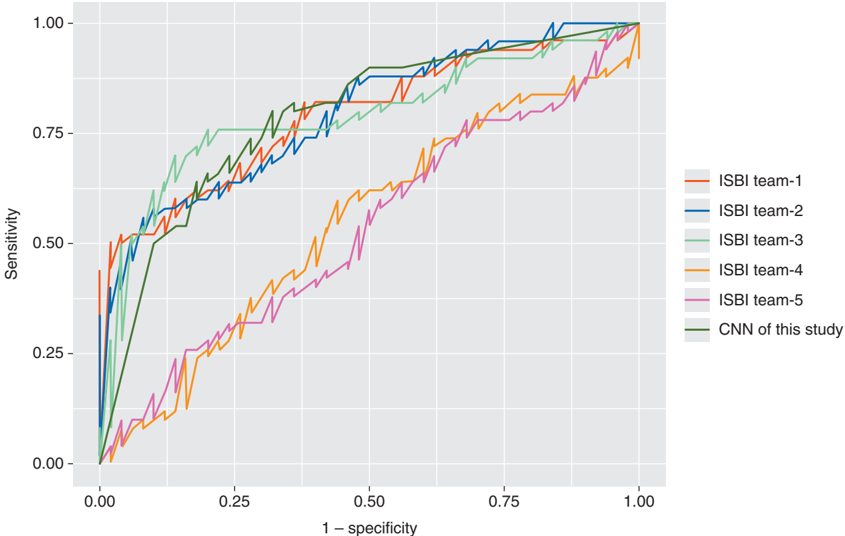
Background Deep learning convolutional neural networks (CNN) may facilitate melanoma detection, but data comparing a CNN's diagnostic performance to larger groups of dermatologists are lacking. Methods Google's Inception v4 CNN architecture was trained and validated using dermoscopic images and corresponding diagnoses. In a comparative cross-sectional reader study a 100-image test-set was used (level-I: dermoscopy only; level-II: dermoscopy plus clinical information and images). Main outcome measures were sensitivity, specificity and area under the curve (AUC) of receiver operating characteristics (ROC) for diagnostic classification (dichotomous) of lesions by the CNN versus an international group of 58 dermatologists during level-I or -II of the reader study. Secondary end points included the dermatologists' diagnostic performance in their management decisions and differences in the diagnostic performance of dermatologists during level-I and -II of the reader study. Additionally, the CNN's performance was compared with the top-five algorithms of the 2016 International Symposium on Biomedical Imaging (ISBI) challenge. Results In level-I dermatologists achieved a mean (±standard deviation) sensitivity and specificity for lesion classification of 86.6% (±9.3%) and 71.3% (±11.2%), respectively. More clinical information (level-II) improved the sensitivity to 88.9% (±9.6%, P = 0.19) and specificity to 75.7% (±11.7%, P < 0.05). The CNN ROC curve revealed a higher specificity of 82.5% when compared with dermatologists in level-I (71.3%, P < 0.01) and level-II (75.7%, P < 0.01) at their sensitivities of 86.6% and 88.9%, respectively. The CNN ROC AUC was greater than the mean ROC area of dermatologists (0.86 versus 0.79, P < 0.01). The CNN scored results close to the top three algorithms of the ISBI 2016 challenge. Conclusions For the first time we compared a CNN's diagnostic performance with a large international group of 58 dermatologists, including 30 experts. Most dermatologists were outperformed by the CNN. Irrespective of any physicians' experience, they may benefit from assistance by a CNN's image classification. Clinical trial number This study was registered at the German Clinical Trial Register (DRKS-Study-ID: DRKS00013570; https://www.drks.de/drks_web/).
Overview
The claims and statistics of this paper haven’t been checked yet.
Run the check — free, no account needed. It reads the open-access text and checks the sources it cites.
Integrity signals
Registry
Cites
Statistics
Figures





Verification
Claims the paper makes
The claims and statistics of this paper haven’t been checked yet.
Run the check — free, no account needed. It reads the open-access text and checks the sources it cites.
Claims other papers make about this one
Each is checked against this paper’s own text: does it say what the citing paper says it does?
The source backs part of the claim, not all of it — or backs it for a different population, condition or comparison.
In the citing paper
“A series of works have demonstrated that CNNs can match the performance of board-certified dermatologists at classifying malignant skin lesions from benign ones 7,93,94 .”
From Deep learning-enabled medical computer vision (2021) · cites this paper as [93]
Experimental — accuracy not yet measuredThe source backs part of the claim, not all of it — or backs it for a different population, condition or comparison.
In the citing paper
“For instance, AI is shown to diagnose skin cancer more efficiently than dermatologists [54].”
From Use of AI-based tools for healthcare purposes: a survey study from consumers’ perspectives (2020) · cites this paper as [54]
Experimental — accuracy not yet measuredThe cited source says what the claim attributes to it.
In the citing paper
“Several algorithms incorporating artificial intelligence (AI) have been developed to help interpret both clinical11,12,13,14,15 and dermoscopic16,17,18,19,20,21,22,23 images for a variety of skin conditions”
In the citing paper
“These studies have sequentially tested increasing numbers of dermatologists (25-7 57-93 , 157-94 ), consistently demonstrating a sensitivity and specificity in classification that matches or even exceeds physician levels.”
From Deep learning-enabled medical computer vision (2021) · cites this paper as [93]
Self-check
Percentages, sums and reported differences re-computed from the paper’s own numbers.
The self-check needs the paper’s full text.
Citations
Cites
The publisher hasn’t deposited this paper’s reference list.
Cited by
High-performance medicine: the convergence of human and artificial intelligence
Topol, E. J. (2019) · Nature Medicine · doi:10.1038/s41591-018-0300-7 · Cited by 10,450
A guide to deep learning in healthcare
Esteva, A., Robicquet, A., Ramsundar, B., Kuleshov, V., DePristo, M., Chou, K., Cui, C., Corrado, G., Thrun, S., & Dean, J. (2019) · Nature Medicine · doi:10.1038/s41591-018-0316-z · Cited by 5,312
Revolutionizing healthcare: the role of artificial intelligence in clinical practice
Alowais, S. A., Alghamdi, S. S., Alsuhebany, N., Alqahtani, T., Alshaya, A. I., Almohareb, S. N., Aldairem, A., Alrashed, M., Bin Saleh, K., Badreldin, H. A., et al. (2023) · BMC Medical Education · doi:10.1186/s12909-023-04698-z · Cited by 3,146
Key challenges for delivering clinical impact with artificial intelligence
Kelly, C. J., Karthikesalingam, A., Suleyman, M., Corrado, G., & King, D. (2019) · BMC Medicine · doi:10.1186/s12916-019-1426-2 · Cited by 3,136
Artificial intelligence in healthcare
Yu, K.-H., Beam, A. L., & Kohane, I. S. (2018) · Nature Biomedical Engineering · doi:10.1038/s41551-018-0305-z · Cited by 2,817
Metadata
- Authors
- Haenssle, H. A., Fink, C., Schneiderbauer, R., Toberer, F., Buhl, T., Blum, A., Kalloo, A., Hassen, A. B. H., Thomas, L., Enk, A., Uhlmann, L., Alt, C., Arenbergerova, M., Bakos, R., Baltzer, A., Bertlich, I., Blum, A., Bokor-Billmann, T., Bowling, J., Braghiroli, N., Braun, R., Buder-Bakhaya, K., Buhl, T., Cabo, H., Cabrijan, L., Cevic, N., Classen, A., Deltgen, D., Fink, C., Georgieva, I., Hakim-Meibodi, L.-E., Hanner, S., Hartmann, F., Hartmann, J., Haus, G., Hoxha, E., Karls, R., Koga, H., Kreusch, J., Lallas, A., Majenka, P., Marghoob, A., Massone, C., Mekokishvili, L., Mestel, D., Meyer, V., Neuberger, A., Nielsen, K., Oliviero, M., Pampena, R., Paoli, J., Pawlik, E., Rao, B., Rendon, A., Russo, T., Sadek, A., Samhaber, K., Schneiderbauer, R., Schweizer, A., Toberer, F., Trennheuser, L., Vlahova, L., Wald, A., Winkler, J., Wölbing, P., & Zalaudek, I.
- Affiliations
- Heidelberg University (DE)
- Memorial Sloan Kettering Cancer Center (US)
- University of Passau (DE)
- Lyon 1 Université (FR)
- Corresponding author
- Haenssle, H.A.
- ORCID
- Venue
- Annals of Oncology
- Publisher
- Elsevier BV
- Published
- 2018-08
- Volume
- 29
- Issue
- 8
- Pages
- 1836-1842
- ISSN
- 0923-7534 (ISSN-L)
- OpenAlex
- W2806853752
- Cited by
- 1,571
- References
- 0
- Citations by year
- 2026: 117 · 2025: 202 · 2024: 224 · 2023: 217 · 2022: 185 · 2021: 215 · 2020: 187 · 2019: 185 · 2018: 36
- FWCI
- 58.54
- Type
- journal article
- Language
- en
- Open access
- hybrid
- doi.org
- Licence
- cc-by-nc-nd
- www.elsevier.com/tdm/userlicense/1.0/ (tdm)
- www.elsevier.com/open-access/userlicense/1.0/ (vor, 450 d)
- Locations
- Annals of Oncology (publishedVersion, cc-by-nc-nd)
- PubMed (publishedVersion)
- ArTS Archivio della ricerca di Trieste (University of Trieste https://www.units.it/) (submittedVersion, other-oa)
- Lund University Publications (Lund University) (submittedVersion)
- GoeScholar The Publication Server of the Georg-August-Universität Göttingen (Georg-August-Universität Göttingen) (submittedVersion, other-oa)
- Topics
- Health Sciences › Medicine › Oncology › Cutaneous Melanoma Detection and Management
- Physical Sciences › Computer Science › Artificial Intelligence › AI in cancer detection
- Health Sciences › Health Professions › Health Information Management › Artificial Intelligence in Healthcare
- Keywords
- melanoma, dermoscopy, convolutional neural networks, Inception-v4, computer-aided diagnosis, skin lesion classification, sensitivity and specificity, clinical decision support, artificial intelligence in dermatology
- MeSH terms
- Dermatologists, Deep Learning, Clinical Competence, Cross-Sectional Studies, Humans, Image Processing, Computer-Assisted, International Cooperation, Melanoma, Retrospective Studies, ROC Curve, Skin, Skin Neoplasms, Dermoscopy
About these checks
Registry, retraction, Cites and statistics results are computed, not judged, with one exception: a printed reference that no index lists is read once by a model before it counts as a citation error. Claims are extracted by a model and kept only when their quote appears verbatim in the text; each cited source is then checked, with a model’s help, for whether it says what the claim relies on. Those verdicts can be wrong — read the claim’s quote and the cited source before relying on one.
Statistics and arithmetic are recomputed, not judged.
Confidence bands are shown only for fields calibrated on our annotated test set. None is calibrated yet, so no band is shown.
What counts as a claim ›How we measure confidence ›How we check this ›
Are you an author, or do you see a problem with this page? Request a review
How to cite
Cite CoChat for the checks on this page, and the paper for its own findings.
- This page
- CoChat. (2026). Citation check: Man against machine: diagnostic performance of a deep learning convolutional neural network for dermoscopic melanoma recognition in comparison to 58 dermatologists. https://app.cochat.ai/papers/10.1093/annonc/mdy166
- The paper
- Haenssle, H. A., Fink, C., Schneiderbauer, R., Toberer, F., Buhl, T., Blum, A., Kalloo, A., Hassen, A. B. H., Thomas, L., Enk, A., Uhlmann, L., Alt, C., Arenbergerova, M., Bakos, R., Baltzer, A., Bertlich, I., Blum, A., Bokor-Billmann, T., Bowling, J., . . . Zalaudek, I. (2018). Man against machine: diagnostic performance of a deep learning convolutional neural network for dermoscopic melanoma recognition in comparison to 58 dermatologists. Annals of Oncology. https://doi.org/10.1093/annonc/mdy166
Data sources
Bibliographic data: Semantic Scholar, OpenAlex, Crossref.
Contains information from the Semantic Scholar Academic Graph Datasets, made available under the ODC Attribution License (ODC-BY 1.0).
Semantic Scholar’s data is described in: Kinney, R., et al. (2023). The Semantic Scholar Open Data Platform. arXiv:2301.10140.
