Student or educator? Sign up with your school email and get 6 months of Pro free.
CoChat

The DOI matches its Crossref record; the references it cites are not checked yet.

Proline: an efficient and user-friendly software suite for large-scale proteomics

Open access

Abstract

Motivation: The proteomics field requires the production and publication of reliable mass spectrometry-based identification and quantification results. Although many tools or algorithms exist, very few consider the importance of combining, in a unique software environment, efficient processing algorithms and a data management system to process and curate hundreds of datasets associated with a single proteomics study. Results: Here, we present Proline, a robust software suite for analysis of MS-based proteomics data, which collects, processes and allows visualization and publication of proteomics datasets. We illustrate its ease of use for various steps in the validation and quantification workflow, its data curation capabilities and its computational efficiency. The DDA label-free quantification workflow efficiency was assessed by comparing results obtained with Proline to those obtained with a widely used software using a spiked-in sample. This assessment demonstrated Proline’s ability to provide high quantification accuracy in a user-friendly interface for datasets of any size. Availability and implementation: Proline is available for Windows and Linux under CECILL open-source license. It can be deployed in client–server mode or in standalone mode at http://proline.profiproteomics.fr/#downloads. Supplementary information: Supplementary data are available at Bioinformatics online.

Overview

The claims and statistics of this paper haven’t been checked yet.

Free: it reads the open-access text and checks the first 5 claims that cite a source we can check. Create a free account to check every claim.

A few free checks a day · papers already verified open instantly · results are public

Integrity signals

Registry

Crossref · 10.1093/bioinformatics/btaa118

Cites

Statistics

Figures

Fig. 1. Schematic representation of the scope of the application: input and output data are represented by gray boxes; tasks which are steps in the data analysis process are represented in blue. Proline provides a set of predefined tasks (dark blue) that can be executed and the paths linking the tasks defines analysis workflows
Fig. 1. Schematic representation of the scope of the application: input and output data are represented by gray boxes; tasks which are steps in the data analysis process are represented in blue. Proline provides a set of predefined tasks (dark blue) that can be executed and the paths linking the tasks defines analysis workflowsCC BY-NC · PubMed Central
Fig. 2. Missing values and CV distributions of yeast ions. (A) Proportions of MVs were represented as percentages of ions matching a yeast protein for which an abundance value was defined in more than n samples. The proportion of ions quantified in the 40 runs was different for Proline (57%) and MaxQuant (45%). (B) CV distribution of yeast ions before applying the cross-assignment procedure. Vertical lines indicate median CV values, 15.2% and 14.6%, respectively, for MaxQuant and Proline. (C) CV distribution of yeast ions after cross-assignment. Median CV values were increased to 17.6% and 16% for MaxQuant and Proline, respectively. In both (B) and (C), solid lines represent CV values calculated from raw intensities, whereas dashed lines represent CV values after median normalization
Fig. 2. Missing values and CV distributions of yeast ions. (A) Proportions of MVs were represented as percentages of ions matching a yeast protein for which an abundance value was defined in more than n samples. The proportion of ions quantified in the 40 runs was different for Proline (57%) and MaxQuant (45%). (B) CV distribution of yeast ions before applying the cross-assignment procedure. Vertical lines indicate median CV values, 15.2% and 14.6%, respectively, for MaxQuant and Proline. (C) CV distribution of yeast ions after cross-assignment. Median CV values were increased to 17.6% and 16% for MaxQuant and Proline, respectively. In both (B) and (C), solid lines represent CV values calculated from raw intensities, whereas dashed lines represent CV values after median normalizationCC BY-NC · PubMed Central
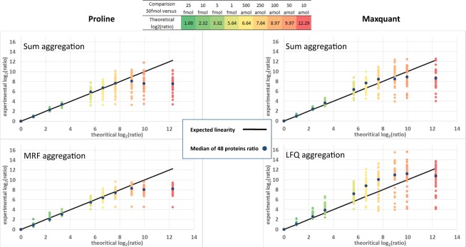
Fig. 3. Estimated versus expected ratios for UPS1 proteins. The abundances of the 48 UPS1 proteins were extracted by Proline (left panel) and MaxQuant (right panel) in each sample from the standard dataset, using either a sum aggregation (upper panels) or ratio-fitting algorithms (bottom panels). The ratios determined, calculated relative to the 50 fmol/µg concentration, were plotted against the expected ratios for the UPS1 proteins across the 10 different concentration points. Both Proline and MaxQuant accurately estimated the ratios calculated for concentration spikes down to 1 fmol/µg (expected ratio 5:6) for most UPS1 proteins. For lower concentration points, when some peptides fell below their limit of detection, the two software behaved differently, with a trend for overestimation of the ratio for MaxQuant, while Proline ratios were still well-fitted down to 250 amol/µg (expected ratio 7:6). When the ratios were calculated using MRF in Proline (bottom left), ratio variability around expected values was reduced compared with the sum method; variability was increased when MaxQuant MaxLFQ was applied (bottom right)
Fig. 3. Estimated versus expected ratios for UPS1 proteins. The abundances of the 48 UPS1 proteins were extracted by Proline (left panel) and MaxQuant (right panel) in each sample from the standard dataset, using either a sum aggregation (upper panels) or ratio-fitting algorithms (bottom panels). The ratios determined, calculated relative to the 50 fmol/µg concentration, were plotted against the expected ratios for the UPS1 proteins across the 10 different concentration points. Both Proline and MaxQuant accurately estimated the ratios calculated for concentration spikes down to 1 fmol/µg (expected ratio 5:6) for most UPS1 proteins. For lower concentration points, when some peptides fell below their limit of detection, the two software behaved differently, with a trend for overestimation of the ratio for MaxQuant, while Proline ratios were still well-fitted down to 250 amol/µg (expected ratio 7:6). When the ratios were calculated using MRF in Proline (bottom left), ratio variability around expected values was reduced compared with the sum method; variability was increased when MaxQuant MaxLFQ was applied (bottom right)CC BY-NC · PubMed Central
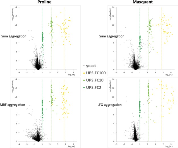
Fig. 4. Volcano plots of the mixed dataset differential analysis. Each protein in the mixed dataset obtained from the quantitative output of three different pairwise comparisons was plotted in a cartesian coordinate defined by the fold change (FC, in log2) on the horizontal axis and the inverse of the P-value (log10) on the vertical axis. The graphs illustrate the quantitative results for the UPS1 proteins quantified in each binary comparison (dark green: comparison of 25 versus 50 fmol/µg, theoretical fold change of 2; light green: comparison of 5 versus 50 fmol/µg, theoretical fold change of 10; yellow: comparison of 500 amol/µg versus 50 fmol/µg, theoretical fold change of 100). Black circles correspond to yeast proteins. The expected ratios of the different concentration points are represented by the dashed vertical lines. For each software, two different peptide-to-protein aggregation methods were implemented: the simplest one consists in an aggregation of non-shared peptides abundances by a sum function (upper part), whereas the second one determines the protein abundances by fitting protein ratios to all observed peptide ratios (MRF or MaxLFQ methods, lower part)
Fig. 4. Volcano plots of the mixed dataset differential analysis. Each protein in the mixed dataset obtained from the quantitative output of three different pairwise comparisons was plotted in a cartesian coordinate defined by the fold change (FC, in log2) on the horizontal axis and the inverse of the P-value (log10) on the vertical axis. The graphs illustrate the quantitative results for the UPS1 proteins quantified in each binary comparison (dark green: comparison of 25 versus 50 fmol/µg, theoretical fold change of 2; light green: comparison of 5 versus 50 fmol/µg, theoretical fold change of 10; yellow: comparison of 500 amol/µg versus 50 fmol/µg, theoretical fold change of 100). Black circles correspond to yeast proteins. The expected ratios of the different concentration points are represented by the dashed vertical lines. For each software, two different peptide-to-protein aggregation methods were implemented: the simplest one consists in an aggregation of non-shared peptides abundances by a sum function (upper part), whereas the second one determines the protein abundances by fitting protein ratios to all observed peptide ratios (MRF or MaxLFQ methods, lower part)CC BY-NC · PubMed Central
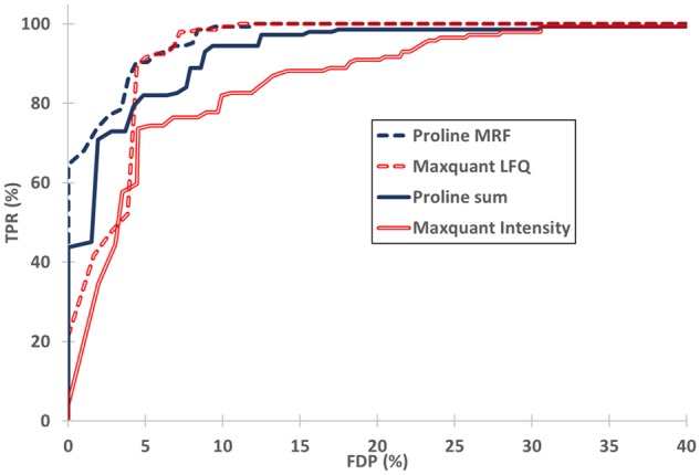
Fig. 5. Differential analysis results in terms of sensitivity and FDP. For each software, proteins from the mixed dataset were classified as variant through the application of q-value thresholding. Sensitivity (TPR = TP/144, TP UPS1 proteins) was plotted as a function of FDP [FDP = FP/(TP+ FP), FP yeast proteins]
Fig. 5. Differential analysis results in terms of sensitivity and FDP. For each software, proteins from the mixed dataset were classified as variant through the application of q-value thresholding. Sensitivity (TPR = TP/144, TP UPS1 proteins) was plotted as a function of FDP [FDP = FP/(TP+ FP), FP yeast proteins]CC BY-NC · PubMed Central
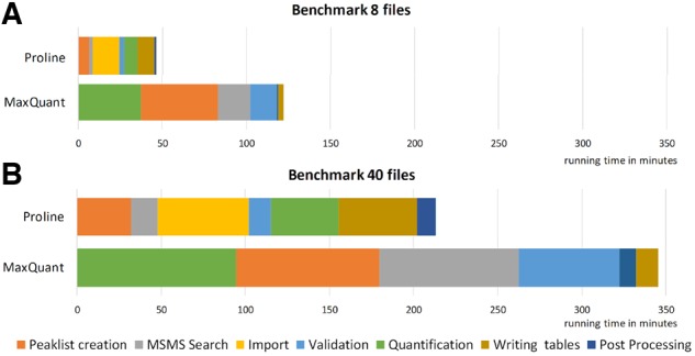
Fig. 6. Workflows computation time. Performance of the Mascot–Proline and Andromeda–MaxQuant label-free workflows were compared on two datasets of different sizes. The main steps of the compared workflows are shown in the same color when possible. Time values were taken from the ‘runningTimes.txt’ output file for MaxQuant and from the log files for Proline. (A) Performance observed for a dataset containing eight UPS1-Yeast LC-MS/MS runs: Total processing time was 122 min for MaxQuant and 46 min for Proline (average time per file 15.26 and 5.79 min, respectively). (B) Performance observed for the whole UPS1-Yeast dataset (40 LC-MS/MS runs): Total processing time 346 min for MaxQuant and 214 min for Proline (average time per file: 8.63 and 5.34 min, respectively)
Fig. 6. Workflows computation time. Performance of the Mascot–Proline and Andromeda–MaxQuant label-free workflows were compared on two datasets of different sizes. The main steps of the compared workflows are shown in the same color when possible. Time values were taken from the ‘runningTimes.txt’ output file for MaxQuant and from the log files for Proline. (A) Performance observed for a dataset containing eight UPS1-Yeast LC-MS/MS runs: Total processing time was 122 min for MaxQuant and 46 min for Proline (average time per file 15.26 and 5.79 min, respectively). (B) Performance observed for the whole UPS1-Yeast dataset (40 LC-MS/MS runs): Total processing time 346 min for MaxQuant and 214 min for Proline (average time per file: 8.63 and 5.34 min, respectively)CC BY-NC · PubMed Central

Verification

Claims the paper makes

The claims and statistics of this paper haven’t been checked yet.

Free: it reads the open-access text and checks the first 5 claims that cite a source we can check. Create a free account to check every claim.

A few free checks a day · papers already verified open instantly · results are public

Claims other papers make about this one

Each is checked against this paper’s own text: does it say what the citing paper says it does?

Self-check

Percentages, sums and reported differences re-computed from the paper’s own numbers.

The self-check needs the paper’s full text.

Citations

The citation lists are queued

They are fetched later and appear here then.

References 24 · Cited by 300

Metadata

Authors
Bouyssié, D., Hesse, A.-M., Mouton-Barbosa, E., Rompais, M., Macron, C., Carapito, C., Gonzalez de Peredo, A., Couté, Y., Dupierris, V., Burel, A., Menetrey, J.-P., Kalaitzakis, A., Poisat, J., Romdhani, A., Burlet-Schiltz, O., Cianférani, S., Garin, J., & Bruley, C.
Affiliations
  • Centre National de la Recherche Scientifique (FR)
  • Inserm (FR)
  • Institut de Pharmacologie et de Biologie Structurale (IPBS) , Université de Toulouse, CNRS, UPS, Toulouse, France
  • Université Grenoble Alpes , Inserm, CEA, IRIG, BGE, Grenoble 38000, France
Corresponding author
Bruley, Christophe
Venue
Bioinformatics
Published
2020-02-25
Volume
36
Issue
10
Pages
3148-3155
ISSN
  • 1367-4803 (ISSN-L)
  • 1367-4811
OpenAlex
W3006945494
Cited by
300
References
24
Citations by year
2026: 30 · 2025: 47 · 2024: 54 · 2023: 46 · 2022: 54 · 2021: 47 · 2020: 21
FWCI
16.12
Type
journal article
Language
en
Open access
Locations
  • Bioinformatics (publishedVersion, cc-by-nc)
  • PubMed Central (publishedVersion, cc-by-nc)
  • PubMed (publishedVersion)
  • HAL (submittedVersion, other-oa)
Funders
  • French National Agency for Research
  • ANR (ANR-10-INBS-08)
  • ProFI project
  • Infrastructures Nationales en Biologie et Santé
Topics
  • Physical Sciences › Chemistry › Spectroscopy › Advanced Proteomics Techniques and Applications
  • Physical Sciences › Chemistry › Spectroscopy › Mass Spectrometry Techniques and Applications
  • Life Sciences › Biochemistry, Genetics and Molecular Biology › Molecular Biology › Bioinformatics and Genomic Networks
Keywords
proteomics data analysis, mass spectrometry data analysis, label-free quantification, data curation, proteomics data management, data visualization, open source software
MeSH terms
Algorithms, Proline, Software, Mass Spectrometry, Proteomics

About these checks

Registry, retraction, Cites and statistics results are computed, not judged, with one exception: a printed reference that no index lists is read once by a model before it counts as a citation error. Claims are extracted by a model and kept only when their quote appears verbatim in the text; each cited source is then checked, with a model’s help, for whether it says what the claim relies on. Those verdicts can be wrong — read the claim’s quote and the cited source before relying on one.

Statistics and arithmetic are recomputed, not judged.

Confidence bands are shown only for fields calibrated on our annotated test set. None is calibrated yet, so no band is shown.

What counts as a claim ›How we measure confidence ›How we check this ›

Are you an author, or do you see a problem with this page? Request a review

How to cite

Cite CoChat for the checks on this page, and the paper for its own findings.

This page
CoChat. (2026). Citation check: Proline: an efficient and user-friendly software suite for large-scale proteomics. https://app.cochat.ai/papers/10.1093/bioinformatics/btaa118
The paper
Bouyssié, D., Hesse, A.-M., Mouton-Barbosa, E., Rompais, M., Macron, C., Carapito, C., Gonzalez de Peredo, A., Couté, Y., Dupierris, V., Burel, A., Menetrey, J.-P., Kalaitzakis, A., Poisat, J., Romdhani, A., Burlet-Schiltz, O., Cianférani, S., Garin, J., & Bruley, C. (2020). Proline: an efficient and user-friendly software suite for large-scale proteomics. Bioinformatics. https://doi.org/10.1093/bioinformatics/btaa118

Data sources

Bibliographic data: Semantic Scholar, OpenAlex, Crossref.

Contains information from the Semantic Scholar Academic Graph Datasets, made available under the ODC Attribution License (ODC-BY 1.0).

Semantic Scholar’s data is described in: Kinney, R., et al. (2023). The Semantic Scholar Open Data Platform. arXiv:2301.10140.