

The DOI matches its Crossref record; 55 of the 55 references it cites were found in a registry or an index.
The Ancestral Shape of the Access Proton Path of Mitochondrial ATP Synthases Revealed by a Split Subunit-a
Open accessPublished version of a preprint
Abstract
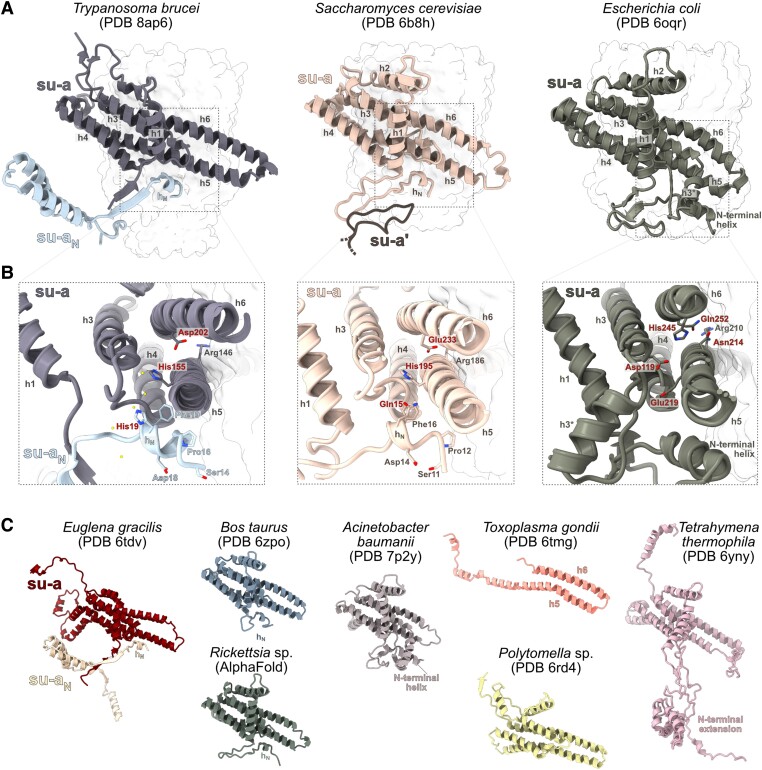
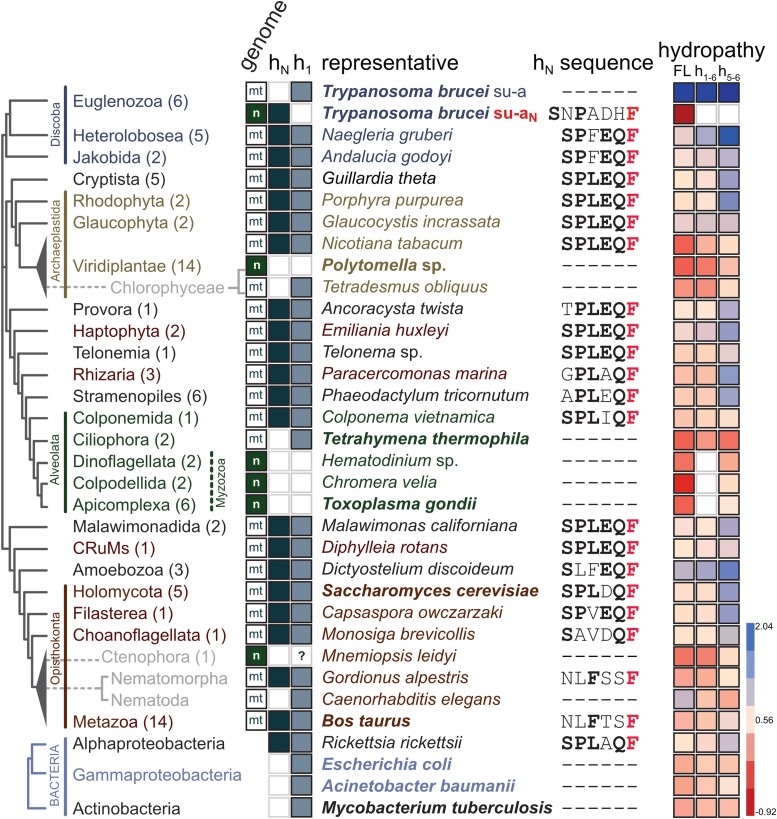
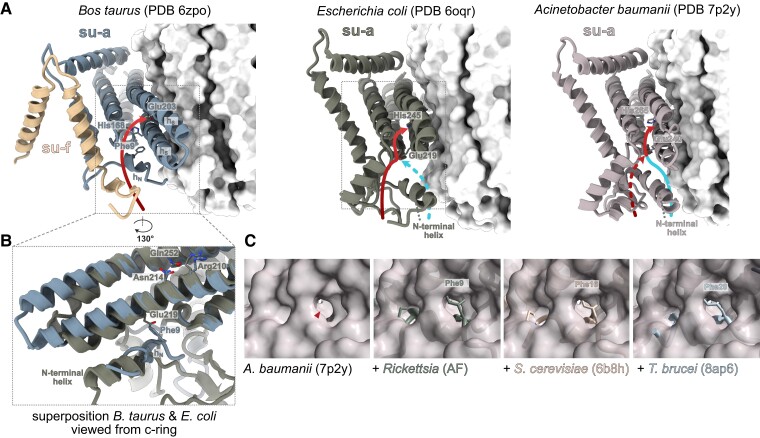
The passage of protons across membranes through F1Fo-ATP synthases spins their rotors and drives the synthesis of ATP. While the principle of torque generation by proton transfer is known, the mechanisms and routes of proton access and release and their evolution are not fully understood. Here, we show that the entry site and path of protons in the lumenal half channel of mitochondrial ATP synthases are largely defined by a short N-terminal α-helix of subunit-a. In Trypanosoma brucei and other Euglenozoa, the α-helix is part of another polypeptide chain that is a product of subunit-a gene fragmentation. This α-helix and other elements forming the proton pathway are widely conserved across eukaryotes and in Alphaproteobacteria, the closest extant relatives of mitochondria, but not in other bacteria. The α-helix blocks one of two proton routes found in Escherichia coli, resulting in a single proton entry site in mitochondrial and alphaproteobacterial ATP synthases. Thus, the shape of the access half channel predates eukaryotes and originated in the lineage from which mitochondria evolved by endosymbiosis.
Overview
The claims and statistics of this paper haven’t been checked yet.
Run the check — free, no account needed. It reads the open-access text and checks the sources it cites.
Integrity signals
Registry
Cites
Statistics
Figures
Verification
Claims the paper makes
The claims and statistics of this paper haven’t been checked yet.
Run the check — free, no account needed. It reads the open-access text and checks the sources it cites.
Self-check
Percentages, sums and reported differences re-computed from the paper’s own numbers.
The self-check needs the paper’s full text.
Citations
Cites
MUSCLE: multiple sequence alignment with high accuracy and high throughput
Edgar, R. C. (2004) · Nucleic Acids Research · doi:10.1093/nar/gkh340 · Cited by 44,662
Highly accurate protein structure prediction with AlphaFold
Jumper, J., Evans, R., Pritzel, A., Green, T., Figurnov, M., Ronneberger, O., Tunyasuvunakool, K., Bates, R., Žídek, A., Potapenko, A., et al. (2021) · Nature · doi:10.1038/s41586-021-03819-2 · Cited by 43,764
A simple method for displaying the hydropathic character of a protein
Kyte, J., & Doolittle, R. F. (1982) · Journal of Molecular Biology · doi:10.1016/0022-2836(82)90515-0 · Cited by 22,843
UCSF ChimeraX: Meeting modern challenges in visualization and analysis
Goddard, T. D., Huang, C. C., Meng, E. C., Pettersen, E. F., Couch, G. S., Morris, J. H., & Ferrin, T. E. (2018) · Protein Science · doi:10.1002/pro.3235 · Cited by 5,267
Potter, S. C., Luciani, A., Eddy, S. R., Park, Y., Lopez, R., & Finn, R. D. (2018) · Nucleic Acids Research · doi:10.1093/nar/gky448 · Cited by 2,607
The HMMTOP transmembrane topology prediction server
Tusnády, G. E., & Simon, I. (2001) · Bioinformatics · doi:10.1093/bioinformatics/17.9.849 · Cited by 1,907
Burki, F., Roger, A. J., Brown, M. W., & Simpson, A. G. B. (2020) · Trends in Ecology & Evolution · doi:10.1016/j.tree.2019.08.008 · Cited by 913
Jones, D. T., Taylor, W. R., & Thornton, J. M. (1994) · Biochemistry · doi:10.1021/bi00176a037 · Cited by 778
Structure and Mechanisms of F-Type ATP Synthases
Kühlbrandt, W. (2019) · Annual Review of Biochemistry · doi:10.1146/annurev-biochem-013118-110903 · Cited by 444
Atomic model for the dimeric F O region of mitochondrial ATP synthase
Guo, H., Bueler, S. A., & Rubinstein, J. L. (2017) · Science · doi:10.1126/science.aao4815 · Cited by 225
Structure of the dimeric ATP synthase from bovine mitochondria
Spikes, T. E., Montgomery, M. G., & Walker, J. E. (2020) · Proceedings of the National Academy of Sciences · doi:10.1073/pnas.2013998117 · Cited by 211
Rotary substates of mitochondrial ATP synthase reveal the basis of flexible F 1 -F o coupling
Murphy, B. J., Klusch, N., Langer, J. D., Mills, D. J., Yıldız, Ö., & Kühlbrandt, W. (2019) · Science · doi:10.1126/science.aaw9128 · Cited by 209
Cryo-EM structure of the entire mammalian F-type ATP synthase
Pinke, G., Zhou, L., & Sazanov, L. A. (2020) · Nature Structural & Molecular Biology · doi:10.1038/s41594-020-0503-8 · Cited by 207
Structure of mycobacterial ATP synthase bound to the tuberculosis drug bedaquiline
Guo, H., Courbon, G. M., Bueler, S. A., Mai, J., Liu, J., & Rubinstein, J. L. (2021) · Nature · doi:10.1038/s41586-020-3004-3 · Cited by 200
Proton Translocation by the F1FOATPase of Escherichia coli
Cain, B. D., & Simoni, R. (1989) · Journal of Biological Chemistry · doi:10.1016/s0021-9258(18)94065-2 · Cited by 159
Structural basis of mitochondrial membrane bending by the I–II–III2–IV2 supercomplex
Mühleip, A., Flygaard, R. K., Baradaran, R., Haapanen, O., Gruhl, T., Tobiasson, V., Maréchal, A., Sharma, V., & Amunts, A. (2023) · Nature · doi:10.1038/s41586-023-05817-y · Cited by 136
Interaction between Glu-219 and His-245 within the a subunit of F1F0-ATPase in Escherichia coli.
Cain, B. D., & Simoni, R. (1988) · Journal of Biological Chemistry · doi:10.1016/s0021-9258(18)68684-3 · Cited by 129
ATP synthase hexamer assemblies shape cristae of Toxoplasma mitochondria
Mühleip, A., Flygaard, R. K., Ovciarikova, J., Lacombe, A., Fernandes, P., Sheiner, L., & Amunts, A. (2021) · Nature Communications · doi:10.1038/s41467-020-20381-z · Cited by 124
Cryo-EM structures provide insight into how E. coli F1Fo ATP synthase accommodates symmetry mismatch
Sobti, M., Walshe, J. L., Wu, D., Ishmukhametov, R. R., Zeng, Y. C., Robinson, C. V., Berry, R. M., & Stewart, A. G. (2020) · Nature Communications · doi:10.1038/s41467-020-16387-2 · Cited by 116
Muñoz-Gómez, S. A., Susko, E., Williamson, K., Eme, L., Slamovits, C. H., Moreira, D., López-García, P., & Roger, A. J. (2022) · Nature Ecology & Evolution · doi:10.1038/s41559-021-01638-2 · Cited by 111
The Proton-Driven Rotor of ATP Synthase: Ohmic Conductance (10 fS), and Absence of Voltage Gating
Feniouk, B. A., Kozlova, M. A., Кнорре, Д. А., Cherepanov, D. A., Mulkidjanian, A. Y., & Junge, W. (2004) · Biophysical Journal · doi:10.1529/biophysj.103.036962 · Cited by 108
Structure of a mitochondrial ATP synthase with bound native cardiolipin
Mühleip, A., McComas, S. E., & Amunts, A. (2019) · eLife · doi:10.7554/elife.51179 · Cited by 108
Pérez-Martínez, X., Antaramián, A., Vázquez‐Acevedo, M., Funes, S., Tolkunova, E. N., d’Alayer, J., Claros, M. G., Davidson, E., King, M. P., & González‐Halphen, D. (2001) · Journal of Biological Chemistry · doi:10.1074/jbc.m010244200 · Cited by 104
Structures of Tetrahymena ’s respiratory chain reveal the diversity of eukaryotic core metabolism
Zhou, L., Maldonado, M., Padavannil, A., Guo, F., & Letts, J. A. (2022) · Science · doi:10.1126/science.abn7747 · Cited by 95
Hatch, L. P., Cox, G. B., & Howitt, S. M. (1995) · Journal of Biological Chemistry · doi:10.1074/jbc.270.49.29407 · Cited by 84
Microbial predators form a new supergroup of eukaryotes
Tikhonenkov, D. V., Mikhailov, K. V., Gawryluk, R. M. R., Belyaev, A. O., Mathur, V., Karpov, S. A., Zagumyonnyi, D. G., Borodina, A. S., Prokina, K. I., Mylnikov, A. P., et al. (2022) · Nature · doi:10.1038/s41586-022-05511-5 · Cited by 84
Identification of cryptic subunits from an apicomplexan ATP synthase
Huet, D., Rajendran, E., van Dooren, G. G., & Lourido, S. (2018) · eLife · doi:10.7554/elife.38097 · Cited by 78
Structural basis of proton translocation and force generation in mitochondrial ATP synthase
Klusch, N., Murphy, B. J., Mills, D. J., Yıldız, Ö., & Kühlbrandt, W. (2017) · eLife · doi:10.7554/elife.33274 · Cited by 75
Lightowlers, R. N., Howitt, S. M., Hatch, L. P., et al. (1988) · Biochimica et Biophysica Acta (BBA) - Bioenergetics · doi:10.1016/0005-2728(88)90031-x · Cited by 74
Vik, S. B., Cain, B. D., Chun, K. T., & Simoni, R. (1988) · Journal of Biological Chemistry · doi:10.1016/s0021-9258(18)68683-1 · Cited by 65
Hartzog, P. E., & Cain, B. D. (1994) · Journal of Biological Chemistry · doi:10.1016/s0021-9258(18)31637-5 · Cited by 59
The Oxytricha trifallax Mitochondrial Genome
Swart, E. C., Nowacki, M., Shum, J., Stiles, H., Higgins, B. P., Doak, T. G., Schotanus, K., Magrini, V. J., Minx, P. J., Mardis, E. R., et al. (2011) · Genome Biology and Evolution · doi:10.1093/gbe/evr136 · Cited by 59
Cryo-EM reveals distinct conformations of E. coli ATP synthase on exposure to ATP
Sobti, M., Ishmukhametov, R. R., Bouwer, J. C., Ayer, A., Suarna, C., Smith, N. J., Christie, M., Stocker, R., Duncan, T. M., & Stewart, A. G. (2019) · eLife · doi:10.7554/elife.43864 · Cited by 55
Role of the carboxyl terminal region of H+-ATPase (F0F1a subunit from Escherichia coli
Seiji, E. Y. A., Maeda, M., & Futai, M. (1991) · Archives of Biochemistry and Biophysics · doi:10.1016/0003-9861(91)90265-k · Cited by 53
Structure of ATP synthase under strain during catalysis
Guo, H., & Rubinstein, J. L. (2022) · Nature Communications · doi:10.1038/s41467-022-29893-2 · Cited by 51
Ciliate mitoribosome illuminates evolutionary steps of mitochondrial translation
Tobiasson, V., & Amunts, A. (2020) · eLife · doi:10.7554/elife.59264 · Cited by 49
Montgomery, M. G., Petri, J., Spikes, T. E., & Walker, J. E. (2021) · Proceedings of the National Academy of Sciences · doi:10.1073/pnas.2111899118 · Cited by 49
Alveolate and chlorophycean mitochondrial cox2 genes split twice independently
Waller, R. F., & Keeling, P. J. (2006) · Gene · doi:10.1016/j.gene.2006.07.003 · Cited by 45
Flygaard, R. K., Mühleip, A., Tobiasson, V., & Amunts, A. (2020) · Nature Communications · doi:10.1038/s41467-020-18993-6 · Cited by 45
Structure of ATP synthase from ESKAPE pathogen Acinetobacter baumannii
Demmer, J. K., Phillips, B. P., Uhrig, O. L., Filloux, A., Allsopp, L. P., Bublitz, M., & Meier, T. W. (2022) · Science Advances · doi:10.1126/sciadv.abl5966 · Cited by 32
An ancestral interaction module promotes oligomerization in divergent mitochondrial ATP synthases
Gahura, O., Mühleip, A., Hierro-Yap, C., Panicucci, B., Jain, M. S., Hollaus, D., Slapničková, M., Zı́ková, A., & Amunts, A. (2022) · Nature Communications · doi:10.1038/s41467-022-33588-z · Cited by 32
Self-citationFu, C.-J., Sheikh, S., Miao, W., Andersson, S. G. E., & Baldauf, S. L. (2014) · Genome Biology and Evolution · doi:10.1093/gbe/evu180 · Cited by 31
Mutational analysis of the function of the a-subunit of the F0F1-ATPase of Escherichia coli
Howitt, S. M., Lightowlers, R. N., Gibson, F., et al. (1990) · Biochimica et Biophysica Acta (BBA) - Bioenergetics · doi:10.1016/0005-2728(90)90030-8 · Cited by 28
Montgomery, M. G., Gahura, O., Leslie, A. G. W., Zı́ková, A., & Walker, J. E. (2018) · Proceedings of the National Academy of Sciences · doi:10.1073/pnas.1720940115 · Cited by 28
Self-citationPredatory colponemids are the sister group to all other alveolates
Tikhonenkov, D. V., Strassert, J. F. H., Janouškovec, J., Mylnikov, A. P., Aleoshin, V. V., Burki, F., & Keeling, P. J. (2020) · Molecular Phylogenetics and Evolution · doi:10.1016/j.ympev.2020.106839 · Cited by 27
Rodríguez-Salinas, E., Riveros-Rosas, H., Li, Z.-K., Fučíková, K., Brand, J., Lewis, L., & González-Halphen, D. (2012) · Molecular Phylogenetics and Evolution · doi:10.1016/j.ympev.2012.03.014 · Cited by 24
Tobiasson, V., Berzina, I., & Amunts, A. (2022) · Nature Communications · doi:10.1038/s41467-022-33582-5 · Cited by 24
The F 1 ‐ ATP ase from Trypanosoma brucei is elaborated by three copies of an additional p18‐subunit
Gahura, O., Šubrtová, K., Váchová, H., Panicucci, B., Fearnley, I. M., Harbour, M. E., Walker, J. E., & Zı́ková, A. (2017) · FEBS Journal · doi:10.1111/febs.14364 · Cited by 23
Self-citationRedesigned and reversed: architectural and functional oddities of the trypanosomal ATP synthase
Gahura, O., Hierro-Yap, C., & Zíková, A. (2021) · Parasitology · doi:10.1017/s0031182021000202 · Cited by 23
Self-citationpH-dependent 11° F1FO ATP synthase sub-steps reveal insight into the FO torque generating mechanism
Yanagisawa, S., & Frasch, W. D. (2021) · eLife · doi:10.7554/elife.70016 · Cited by 20
Structure, Mechanism and Regulation of ATP Synthases
Walker, J. E. (2017) · Royal Society of Chemistry eBooks · doi:10.1039/9781788010405-00338 · Cited by 19
The persistent homology of mitochondrial ATP synthases
Sinha, S. D., & Wideman, J. G. (2023) · iScience · doi:10.1016/j.isci.2023.106700 · Cited by 18
Szafrański, P. (2017) · BMC Genomics · doi:10.1186/s12864-017-3626-5 · Cited by 16
Intercompartmental Piecewise Gene Transfer
Szafrański, P. (2017) · Genes · doi:10.3390/genes8100260 · Cited by 9
Ivontsin, L. A., Mashkovtseva, E. V., & Nartsissov, Y. R. (2022) · Archives of Biochemistry and Biophysics · doi:10.1016/j.abb.2022.109135 · Cited by 9
Metadata
- Authors
- Wong, J. E., Zíková, A., & Gahura, O.
- Affiliations
- Czech Academy of Sciences (CZ)
- Institute of Parasitology, Biology Centre, Czech Academy of Sciences , České Budějovice , Czech Republic
- Corresponding author
- Gahura, Ondřej
- ORCID
- Gahura, Ondřej
- Venue
- Molecular Biology and Evolution
- Publisher
- Oxford University Press (OUP)
- Published
- 2023-06-01
- Volume
- 40
- Issue
- 6
- ISSN
- 0737-4038 (ISSN-L)
- 1537-1719
- PMC
- PMC10306403
- OpenAlex
- W4381309015
- Cited by
- 3
- References
- 55
- Citations by year
- 2026: 1 · 2025: 1 · 2023: 1
- FWCI
- 0.27
- Type
- journal article
- Language
- en
- Open access
- gold
- doi.org
- Licence
- Locations
- Molecular Biology and Evolution (publishedVersion, cc-by-nc)
- bioRxiv (Cold Spring Harbor Laboratory) (acceptedVersion, cc-by-nc-nd)
- PubMed Central (publishedVersion, cc-by-nc)
- PubMed (publishedVersion)
- Funders
- European Commission (CZ.02.1.01, HORIZON2020)
- Ministerstvo Školství, Mládeže a Tělovýchovy (CZ.02.1.01/0.0/0.0/16_019, 0000759, 16_019/0000759, CZ.02.1.01, CZ.02.1.01/0.0/0.0/16 019/0000759)
- Grantová Agentura České Republiky (CZ.02.1.01/0.0, GJ20-04150Y, CZ.02.1.01/0.0/0.0/ 16_019/0000759, CZ.02.1.01)
- European Regional Development Fund (02.1.01/0.0/0.0, CZ.02, CZ.02.1.01/0.0/0.0/16_019/0000759, CZ.02.1.01, CZ.02.1.01/0.0/0.0, 16_019/0000759, 02.1.01/0.0/0.0/16_019/0000759, /16_019)
- HORIZON EUROPE European Research Council (101044951)
- Topics
- Life Sciences › Biochemistry, Genetics and Molecular Biology › Molecular Biology › ATP Synthase and ATPases Research
- Life Sciences › Biochemistry, Genetics and Molecular Biology › Structural Biology › Enzyme Structure and Function
- Life Sciences › Biochemistry, Genetics and Molecular Biology › Molecular Biology › Mitochondrial Function and Pathology
- Keywords
- F1FO-ATP synthase, mitochondrial ATP synthase, subunit a, N-terminal alpha-helix, gene fragmentation, Trypanosoma brucei, Euglenozoa, Alphaproteobacteria, endosymbiotic origin of mitochondria, Escherichia coli
- MeSH terms
- Adenosine Triphosphate, Escherichia coli, Proton-Translocating ATPases, Protons, Mitochondrial Proton-Translocating ATPases, Eukaryota
About these checks
Registry, retraction, Cites and statistics results are computed, not judged, with one exception: a printed reference that no index lists is read once by a model before it counts as a citation error. Claims are extracted by a model and kept only when their quote appears verbatim in the text; each cited source is then checked, with a model’s help, for whether it says what the claim relies on. Those verdicts can be wrong — read the claim’s quote and the cited source before relying on one.
Statistics and arithmetic are recomputed, not judged.
Confidence bands are shown only for fields calibrated on our annotated test set. None is calibrated yet, so no band is shown.
What counts as a claim ›How we measure confidence ›How we check this ›
Are you an author, or do you see a problem with this page? Request a review
Data sources
Bibliographic data: Semantic Scholar, OpenAlex, Crossref.
Contains information from the Semantic Scholar Academic Graph Datasets, made available under the ODC Attribution License (ODC-BY 1.0).
Semantic Scholar’s data is described in: Kinney, R., et al. (2023). The Semantic Scholar Open Data Platform. arXiv:2301.10140.
