Cited by 13

The DOI matches its Crossref record.

The RdgC protein employs a novel mechanism involving a finger domain to bind to circular DNA

Briggs, G. S., Yu, J., Mahdi, A. A., Lloyd, R. G.·2010·Nucleic Acids Research

Overview

The claims and statistics of this paper haven’t been checked yet.

Run the check — free, no account needed. It reads the open-access text and checks the sources it cites.

A few free checks a day · papers already verified open instantly · results are public

Integrity signals

Registry

Crossref · 10.1093/nar/gkq509

Cites

Statistics

Figures

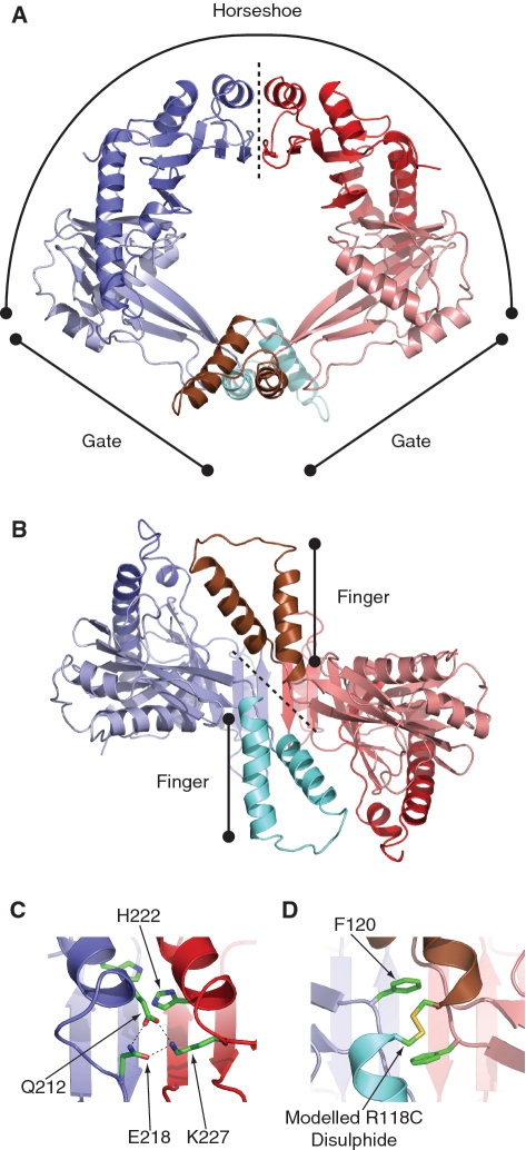
Figure 1. Structure of RdgC. (PDB co-ordinates 2OWL). (A) A ribbon diagram showing the ring structure of RdgC and identifying the horseshoe and gate regions. The dotted line indicates the horseshoe domain interface. (B) A rotated ribbon diagram illustrating projection of the finger domains. The dotted line indicates the gate domain interface. (C) Detail of one of the hydrogen bond networks at the horseshoe interface between the Q212 and E218 residues on one dimer and K227 on the other. The conserved H222 residues are also shown. (D) Detail of the gate interface interactions, showing the conserved F120 residues and the modelled disulphide formed in the R118C derivative.
Figure 1. Structure of RdgC. (PDB co-ordinates 2OWL). (A) A ribbon diagram showing the ring structure of RdgC and identifying the horseshoe and gate regions. The dotted line indicates the horseshoe domain interface. (B) A rotated ribbon diagram illustrating projection of the finger domains. The dotted line indicates the gate domain interface. (C) Detail of one of the hydrogen bond networks at the horseshoe interface between the Q212 and E218 residues on one dimer and K227 on the other. The conserved H222 residues are also shown. (D) Detail of the gate interface interactions, showing the conserved F120 residues and the modelled disulphide formed in the R118C derivative.CC BY-NC · PubMed Central
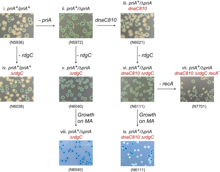
Figure 2. The effect of ΔrdgC on the viability of ΔpriA and ΔpriA dnaC810 strains. Photographs of plates illustrating the results of synthetic lethality assays. All constructs contained a priA+ derivative of the pRC7 plasmid, and white colonies indicate the segregation of plasmid-free cells. Cells were plated on either LB agar (rich medium) or MA (56/2 minimal agar medium) as indicated.
Figure 2. The effect of ΔrdgC on the viability of ΔpriA and ΔpriA dnaC810 strains. Photographs of plates illustrating the results of synthetic lethality assays. All constructs contained a priA+ derivative of the pRC7 plasmid, and white colonies indicate the segregation of plasmid-free cells. Cells were plated on either LB agar (rich medium) or MA (56/2 minimal agar medium) as indicated.CC BY-NC · PubMed Central
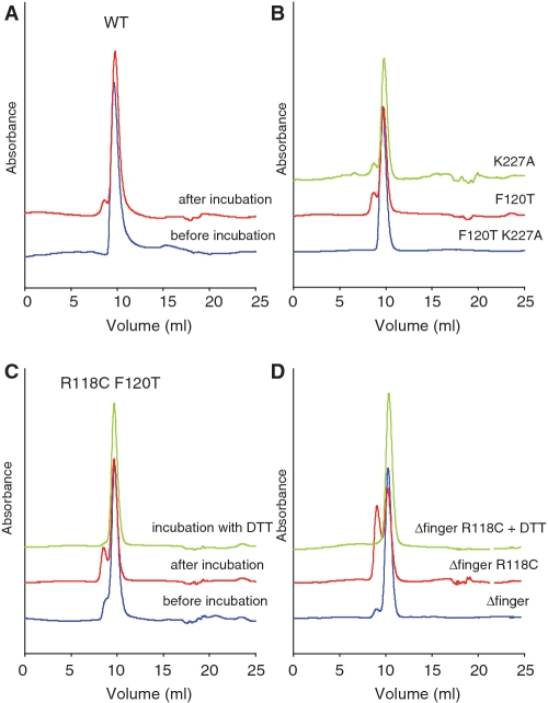
Figure 3. Analytical gel filtration analysis of RdgC proteins. (A) Gel filtration of purified WT RdgC protein. Protein was diluted to a final concentration of 10 μM (dimer) in gel filtration buffer and either analysed immediately or incubated overnight at 4°C before analysis on a Superdex 75 10/300 column (GE Healthcare). (B) Gel filtration of purified RdgC proteins. Protein was diluted to a final concentration of 10 μM (dimer) in gel filtration buffer and incubated overnight at 4°C before analysis on a Superdex 75 10/300 column (GE Healthcare). (C) Gel filtration of purified RdgC R118C F120T protein. Protein was diluted to a final concentration of 10 μM (dimer) in gel filtration buffer and either analysed immediately or incubated overnight at 4°C before analysis on a Superdex 75 10/300 column (GE Healthcare). An additional sample was incubated and analysed in gel filtration buffer containing 5 mM DTT. (D) Gel filtration of purified RdgC Δfinger proteins. Proteins were diluted to a final concentration of 10 μM (dimer) in gel filtration buffer and incubated overnight at 4°C before analysis on a Superdex 75 10/300 column (GE Healthcare). An additional sample of Δfinger R118C was incubated and analysed in gel filtration buffer containing 5 mM DTT.
Figure 3. Analytical gel filtration analysis of RdgC proteins. (A) Gel filtration of purified WT RdgC protein. Protein was diluted to a final concentration of 10 μM (dimer) in gel filtration buffer and either analysed immediately or incubated overnight at 4°C before analysis on a Superdex 75 10/300 column (GE Healthcare). (B) Gel filtration of purified RdgC proteins. Protein was diluted to a final concentration of 10 μM (dimer) in gel filtration buffer and incubated overnight at 4°C before analysis on a Superdex 75 10/300 column (GE Healthcare). (C) Gel filtration of purified RdgC R118C F120T protein. Protein was diluted to a final concentration of 10 μM (dimer) in gel filtration buffer and either analysed immediately or incubated overnight at 4°C before analysis on a Superdex 75 10/300 column (GE Healthcare). An additional sample was incubated and analysed in gel filtration buffer containing 5 mM DTT. (D) Gel filtration of purified RdgC Δfinger proteins. Proteins were diluted to a final concentration of 10 μM (dimer) in gel filtration buffer and incubated overnight at 4°C before analysis on a Superdex 75 10/300 column (GE Healthcare). An additional sample of Δfinger R118C was incubated and analysed in gel filtration buffer containing 5 mM DTT.CC BY-NC · PubMed Central
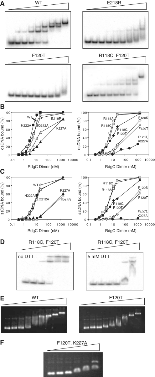
Figure 4. Oligonucleotide binding by RdgC proteins. (A) Representative band-shift assays with dsDNA. Reactions used the RdgC proteins indicated at 0, 0.375, 0.75, 1.5, 3, 6, 12, 24 and 1850 nM (of dimer) and 32P-labelled RGL13/17 DNA at 0.2 nM. Assays with R118A, R118C, Q212A and H222A gave the same band pattern as the WT protein. Assays with F120S and K227A gave the same band pattern as F120T. (B) Quantification of dsDNA binding. RGL13/17 DNA was used at 0.2 nM. Data are means of at least two band-shift experiments. (C) Quantification of ssDNA binding. 32P-labelled RGL13 DNA was used at 0.2 nM. Data are means of at least two band-shift experiments. (D) Effect of DTT on duplex binding by RdgC R118C F120T. Reactions used mutant RdgC protein at 0, 1, 5, 25, 125 and 625 nM (of dimer) and 32P-labelled RGL13/17 DNA at 0.2 nM. (E) Plasmid binding by RdgC proteins. Reactions used the RdgC proteins indicated at 0, 34, 68, 136, 272, 544, 1088, 2176 and 23 400 nM (of dimer) and pGEM-7 Zf(+) circular dsDNA at 2.2 nM plasmid (6480 nM nucleotide pairs). (F) Plasmid binding by the F120T K227A double mutant. Reactions used mutant RdgC at 0, 1000, 2000, 5000, 10 000 and 20 000 nM (of dimer) and pGEM-7 Zf(+) circular dsDNA at 2.2 nM plasmid (6480 nM nucleotide pairs).
Figure 4. Oligonucleotide binding by RdgC proteins. (A) Representative band-shift assays with dsDNA. Reactions used the RdgC proteins indicated at 0, 0.375, 0.75, 1.5, 3, 6, 12, 24 and 1850 nM (of dimer) and 32P-labelled RGL13/17 DNA at 0.2 nM. Assays with R118A, R118C, Q212A and H222A gave the same band pattern as the WT protein. Assays with F120S and K227A gave the same band pattern as F120T. (B) Quantification of dsDNA binding. RGL13/17 DNA was used at 0.2 nM. Data are means of at least two band-shift experiments. (C) Quantification of ssDNA binding. 32P-labelled RGL13 DNA was used at 0.2 nM. Data are means of at least two band-shift experiments. (D) Effect of DTT on duplex binding by RdgC R118C F120T. Reactions used mutant RdgC protein at 0, 1, 5, 25, 125 and 625 nM (of dimer) and 32P-labelled RGL13/17 DNA at 0.2 nM. (E) Plasmid binding by RdgC proteins. Reactions used the RdgC proteins indicated at 0, 34, 68, 136, 272, 544, 1088, 2176 and 23 400 nM (of dimer) and pGEM-7 Zf(+) circular dsDNA at 2.2 nM plasmid (6480 nM nucleotide pairs). (F) Plasmid binding by the F120T K227A double mutant. Reactions used mutant RdgC at 0, 1000, 2000, 5000, 10 000 and 20 000 nM (of dimer) and pGEM-7 Zf(+) circular dsDNA at 2.2 nM plasmid (6480 nM nucleotide pairs).CC BY-NC · PubMed Central
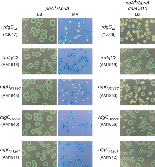
Figure 5. The effect of rdgC mutations on the viability of ΔpriA and ΔpriA dnaC810 strains. Photographs of plates illustrating the results of synthetic lethality assays. All constructs contained a priA+ derivative of the pRC7 plasmid, and white colonies indicate the segregation of plasmid-free cells. Colonies were grown on either LB agar or MA as indicated.
Figure 5. The effect of rdgC mutations on the viability of ΔpriA and ΔpriA dnaC810 strains. Photographs of plates illustrating the results of synthetic lethality assays. All constructs contained a priA+ derivative of the pRC7 plasmid, and white colonies indicate the segregation of plasmid-free cells. Colonies were grown on either LB agar or MA as indicated.CC BY-NC · PubMed Central
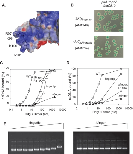
Figure 6. Properties of the finger domain. (A) Electrostatic surface representation of a finger domain, illustrating its electropositive nature and the position of the four conserved basic residues modified in the RdgCfingertip derivative. (B) Synthetic lethality assays showing the effect of finger domain mutations on ΔpriA dnaC810 strain viability. Colonies were grown on LB agar. (C) Quantification of dsDNA binding. Reactions used the RdgC proteins indicated and 32P-labelled RGL13/17 DNA at 0.2 nM. Data are means of at least two band-shift assays. (D) Quantification of ssDNA binding. Reactions used the RdgC proteins indicated and 32P-labelled RGL13 DNA at 0.2 nM. Data are means of at least two band-shift assays. (E) Plasmid binding. Reactions used the RdgC proteins indicated at 0, 34, 68, 136, 272, 544, 1088, 2176 and 23 400 nM (of dimer) and pGEM-7 Zf(+) DNA at 2.2 nM plasmid.
Figure 6. Properties of the finger domain. (A) Electrostatic surface representation of a finger domain, illustrating its electropositive nature and the position of the four conserved basic residues modified in the RdgCfingertip derivative. (B) Synthetic lethality assays showing the effect of finger domain mutations on ΔpriA dnaC810 strain viability. Colonies were grown on LB agar. (C) Quantification of dsDNA binding. Reactions used the RdgC proteins indicated and 32P-labelled RGL13/17 DNA at 0.2 nM. Data are means of at least two band-shift assays. (D) Quantification of ssDNA binding. Reactions used the RdgC proteins indicated and 32P-labelled RGL13 DNA at 0.2 nM. Data are means of at least two band-shift assays. (E) Plasmid binding. Reactions used the RdgC proteins indicated at 0, 34, 68, 136, 272, 544, 1088, 2176 and 23 400 nM (of dimer) and pGEM-7 Zf(+) DNA at 2.2 nM plasmid.CC BY-NC · PubMed Central
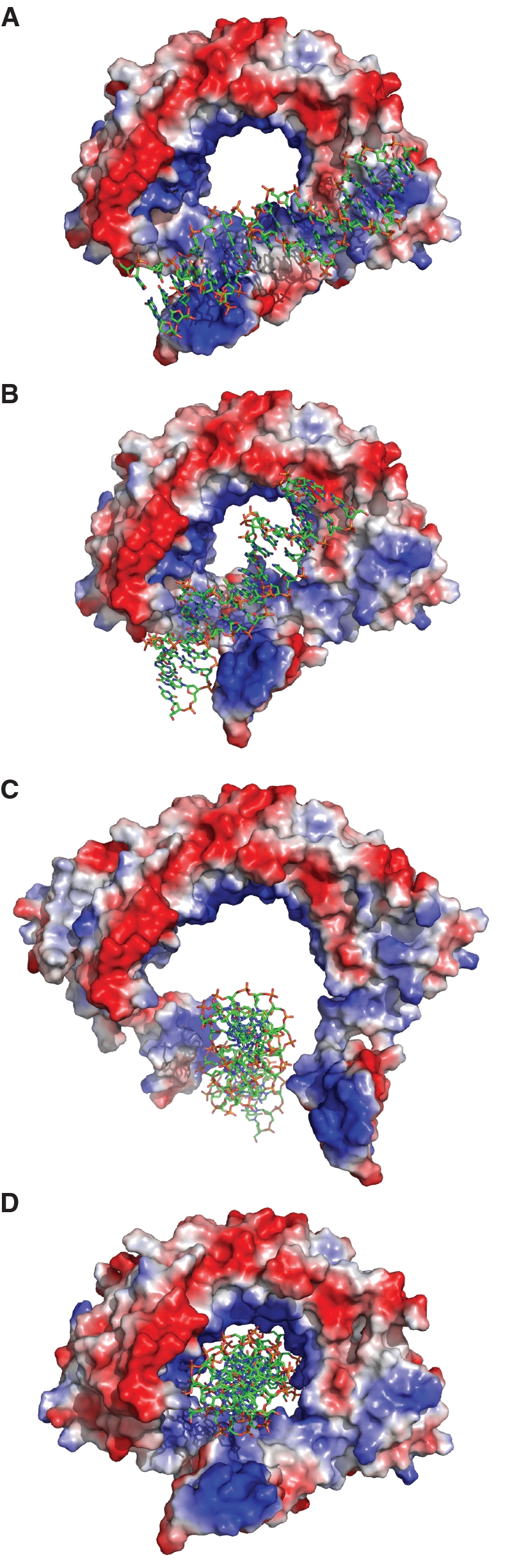
Figure 7. Model illustrating how RdgC may load onto DNA. (A) DNA binds across an exposed positive face, interacting with the finger domain. (B) The finger domain rotates, disrupting the gate dimer interface. (C) The gate opens, and RdgC slides over the DNA. (D) The gate closes, encircling the DNA within the ring.
Figure 7. Model illustrating how RdgC may load onto DNA. (A) DNA binds across an exposed positive face, interacting with the finger domain. (B) The finger domain rotates, disrupting the gate dimer interface. (C) The gate opens, and RdgC slides over the DNA. (D) The gate closes, encircling the DNA within the ring.CC BY-NC · PubMed Central

Verification

Claims the paper makes

The claims and statistics of this paper haven’t been checked yet.

Run the check — free, no account needed. It reads the open-access text and checks the sources it cites.

A few free checks a day · papers already verified open instantly · results are public

Claims other papers make about this one

Each is checked against this paper’s own text: does it say what the citing paper says it does?

Self-check

Percentages, sums and reported differences re-computed from the paper’s own numbers.

The self-check needs the paper’s full text.

Citations

Cites

No references could be resolved yet.

Research works printed but not linked: 45

  1. Murphy, 1999, Journal of Bacteriology, 181, 3842, 10.1128/JB.181.12.3842-3844.1999 · DOI
  2. 1996, 143, 1101, 10.1093/genetics/143.3.1101 · DOI
  3. Moore, 2003, The EMBO Journal, 22, 735, 10.1093/emboj/cdg048 · DOI
  4. Moore, 2004, Journal of Bacteriology, 186, 870, 10.1128/JB.186.3.870-874.2004 · DOI
  5. 2006, Journal of Biological Chemistry, 281, 4708, 10.1074/jbc.M513592200 · DOI
  6. Cox, 2007, Nature reviews. Molecular cell biology, 8, 127, 10.1038/nrm2099 · DOI
  7. Galletto, 2007, Current biology : CB, 17, R395, 10.1016/j.cub.2007.03.009 · DOI
  8. Vos, 2009, Trends in microbiology, 17, 226, 10.1016/j.tim.2009.03.001 · DOI
  9. Hill, 2009, FEMS microbiology reviews, 33, 521, 10.1111/j.1574-6976.2009.00171.x · DOI
  10. 2004, 166, 25, 10.1534/genetics.166.1.25 · DOI
  11. 1991, 9, 154
  12. 2000, 154, 523, 10.1093/genetics/154.2.523 · DOI
  13. Pride, 2002, Journal of molecular biology, 316, 629, 10.1006/jmbi.2001.5311 · DOI
  14. Dworkin, 1997, Molecular microbiology, 26, 433, 10.1046/j.1365-2958.1997.6151958.x · DOI
  15. Sinha, 2000, Journal of Bacteriology, 182, 6532, 10.1128/JB.182.22.6532-6535.2000 · DOI
  16. Hoiseth, 1986, PNAS, 83, 1106, 10.1073/pnas.83.4.1106 · DOI
  17. McCulloch, 1999, Genes & Development, 13, 2875, 10.1101/gad.13.21.2875 · DOI
  18. Cox, 2007, Critical Reviews in Biochemistry and Molecular Biology, 42, 41, 10.1080/10409230701260258 · DOI
  19. West, 2003, Nature reviews. Molecular cell biology, 4, 435, 10.1038/nrm1127 · DOI
  20. Petrova, 2009, Molecular cell, 36, 121, 10.1016/j.molcel.2009.07.026 · DOI
  21. 2007, Journal of Biological Chemistry, 282, 12353, 10.1074/jbc.C700023200 · DOI
  22. 2007, Nucleic Acids Research, 35, 2671, 10.1093/nar/gkm144 · DOI
  23. Hingorani, 2000, Nature reviews. Molecular cell biology, 1, 22, 10.1038/35036044 · DOI
  24. Kong, 1992, Cell, 69, 425, 10.1016/0092-8674(92)90445-I · DOI
  25. Krishna, 1994, Cell, 79, 1233, 10.1016/0092-8674(94)90014-0 · DOI
  26. Bailey, 2007, Science, 318, 459, 10.1126/science.1147353 · DOI
  27. Karow, 1999, Current biology : CB, 9, 597, 10.1016/S0960-9822(99)80264-4 · DOI
  28. O'donnell, 2006, Current opinion in structural biology, 16, 35, 10.1016/j.sbi.2005.12.004 · DOI
  29. Davey, 2002, The EMBO Journal, 21, 3148, 10.1093/emboj/cdf308 · DOI
  30. 2002, Annual review of biochemistry, 71, 333, 10.1146/annurev.biochem.71.110601.135425 · DOI
  31. Ahnert, 2000, The EMBO Journal, 19, 3418, 10.1093/emboj/19.13.3418 · DOI
  32. Bernhardt, 2004, Molecular microbiology, 52, 1255, 10.1111/j.1365-2958.2004.04063.x · DOI
  33. Mahdi, 2006, Genes & Development, 20, 2135, 10.1101/gad.382306 · DOI
  34. 2000, PNAS, 97, 6640, 10.1073/pnas.120163297 · DOI
  35. Al-Deib, 1996, Journal of Bacteriology, 178, 6782, 10.1128/jb.178.23.6782-6789.1996 · DOI
  36. 2005, Journal of Biological Chemistry, 280, 13921, 10.1074/jbc.M412054200 · DOI
  37. 2004, BIOPHOT INT, 11, 36
  38. Szwajkajzer, 2001, Journal of molecular biology, 312, 949, 10.1006/jmbi.2001.4941 · DOI
  39. Vinayagam, 2004, Nucleic Acids Research, 32, D200, 10.1093/nar/gkh026 · DOI
  40. 1996, 143, 5, 10.1093/genetics/143.1.5 · DOI
  41. Sandler, 1999, Molecular microbiology, 34, 91, 10.1046/j.1365-2958.1999.01576.x · DOI
  42. Gregg, 2002, Molecular cell, 9, 241, 10.1016/S1097-2765(02)00455-0 · DOI
  43. Masai, 1994, The EMBO Journal, 13, 5338, 10.1002/j.1460-2075.1994.tb06868.x · DOI
  44. 1996, ESCHERICHIA COLI AND SALMONELLA CELLULAR AND MOLECULAR BIOLOGY, 2, 2460
  45. Nurse, 1991, Journal of Bacteriology, 173, 6686, 10.1128/jb.173.21.6686-6693.1991 · DOI

Cited by

Sorted by citations

  • Ancient DNA analysis

    Orlando, L., Allaby, R., Skoglund, P., Der Sarkissian, C., Stockhammer, P. W., Ávila-Arcos, M. C., Fu, Q., Krause, J., Willerslev, E., Stone, A. C., et al. (2021) · Nature Reviews Methods Primers · doi:10.1038/s43586-020-00011-0 · Cited by 411

See all 13

Metadata

Authors
Briggs, G. S., Yu, J., Mahdi, A. A., & Lloyd, R. G.
Venue
Nucleic Acids Research
Published
2010-06-04
Volume
38
Issue
19
Pages
6433-6446
ISSN
  • 0305-1048
  • 1362-4962
Cited by
13
References
0
Language
en

About these checks

Registry, retraction, Cites and statistics results are computed, not judged, with one exception: a printed reference that no index lists is read once by a model before it counts as a citation error. Claims are extracted by a model and kept only when their quote appears verbatim in the text; each cited source is then checked, with a model’s help, for whether it says what the claim relies on. Those verdicts can be wrong — read the claim’s quote and the cited source before relying on one.

Statistics and arithmetic are recomputed, not judged.

Confidence bands are shown only for fields calibrated on our annotated test set. None is calibrated yet, so no band is shown.

What counts as a claim ›How we measure confidence ›How we check this ›

Are you an author, or do you see a problem with this page? Request a review

How to cite

Cite CoChat for the checks on this page, and the paper for its own findings.

This page
CoChat. (2026). Citation check: The RdgC protein employs a novel mechanism involving a finger domain to bind to circular DNA. https://app.cochat.ai/papers/10.1093/nar/gkq509
The paper
Briggs, G. S., Yu, J., Mahdi, A. A., & Lloyd, R. G. (2010). The RdgC protein employs a novel mechanism involving a finger domain to bind to circular DNA. Nucleic Acids Research. https://doi.org/10.1093/nar/gkq509