

The DOI matches its Crossref record; 2 of the 2 references it cites were found in a registry or an index.
USP14 is a deubiquitinase for Ku70 and critical determinant of non-homologous end joining repair in autophagy and PTEN-deficient cells
Abstract
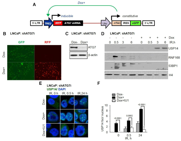
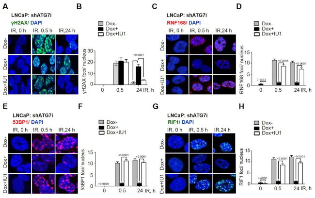
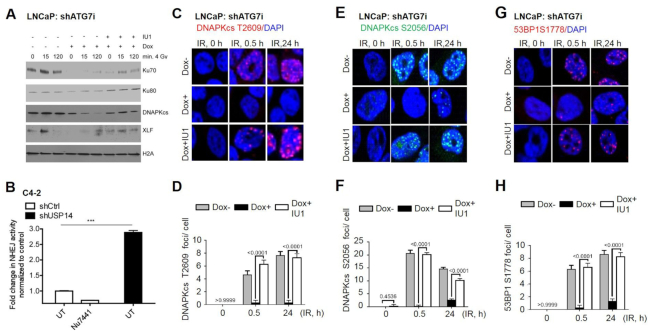
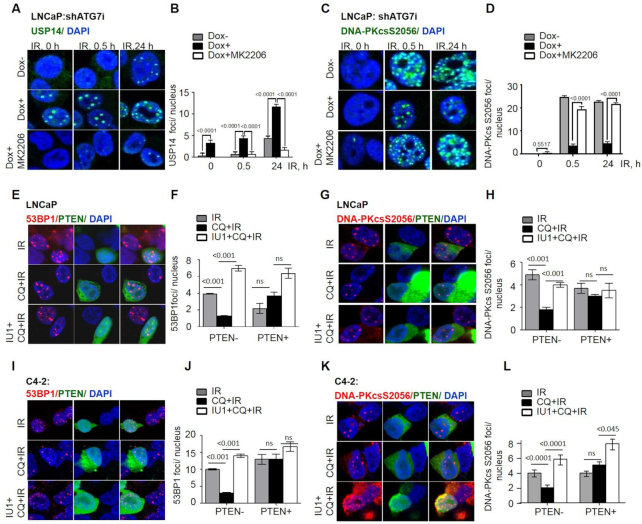
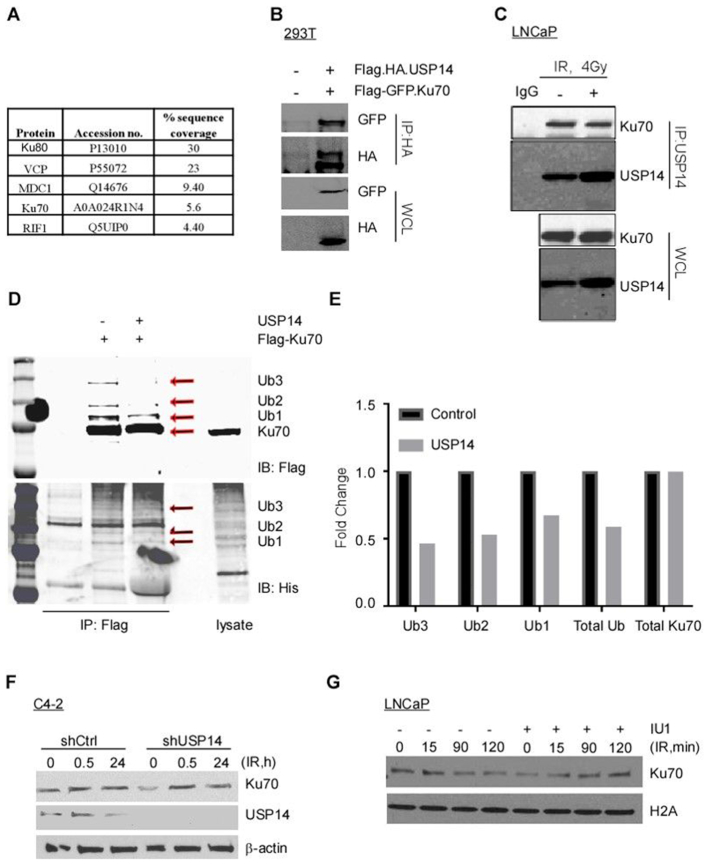
Ionizing radiation (IR)-induced DNA double-strand breaks (DSBs) are predominantly repaired by non-homologous end joining (NHEJ). IR-induced DNA damage activates autophagy, an intracellular degradation process that delivers cytoplasmic components to the lysosome. We identified the deubiquitinase USP14 as a novel autophagy substrate and a regulator of IR-induced DNA damage response (DDR) signaling. Inhibition of autophagy increased levels and DSB recruitment of USP14. USP14 antagonized RNF168-dependent ubiquitin signaling and downstream 53BP1 chromatin recruitment. Here we show that autophagy-deficient cells are defective in NHEJ, as indicated by decreased IR-induced foci (IRIF) formation by pS2056-, pT2609-DNA-PKcs, pS1778-53BP1, RIF1 and a reporter assay activation. Moreover, chromatin recruitment of key NHEJ proteins, including, Ku70, Ku80, DNA-PKcs and XLF was diminished in autophagy-deficient cells. USP14 inhibition rescued the activity of NHEJ-DDR proteins in autophagy-deficient cells. Mass spectrometric analysis identified USP14 interaction with core NHEJ proteins, including Ku70, which was validated by co-immunoprecipitation. An in vitro assay revealed that USP14 targeted Ku70 for deubiquitination. AKT, which mediates Ser432-USP14 phosphorylation, was required for IRIF formation by USP14. Similar to USP14 block, AKT inhibition rescued the activity of NHEJ-DDR proteins in autophagy- and PTEN-deficient cells. These findings reveal a novel negative PTEN/Akt-dependent regulation of NHEJ by USP14.
Overview
The claims and statistics of this paper haven’t been checked yet.
Run the check — free, no account needed. It reads the open-access text and checks the sources it cites.
Integrity signals
Registry
Cites
Statistics
Figures
Verification
Claims the paper makes
The claims and statistics of this paper haven’t been checked yet.
Run the check — free, no account needed. It reads the open-access text and checks the sources it cites.
Claims other papers make about this one
Each is checked against this paper’s own text: does it say what the citing paper says it does?
The check wasn’t confident either way, so it gives no verdict — never disputed.
In the citing paper
“A number of stressors that impair the UPS induce the aggregation/dimerization of SQSTM1, and this can be seen by the detection of a high molecular mass (~150 kDa) protein complex by western blot, which is recognized by SQSTM1 antibodies [561, 605, 606]”
From Guidelines for the use and interpretation of assays for monitoring autophagy (4th edition) 1 (2021) · cites this paper as [605]
Experimental — accuracy not yet measured
Self-check
Percentages, sums and reported differences re-computed from the paper’s own numbers.
The self-check needs the paper’s full text.
Citations
Cites
Guidelines for the use and interpretation of assays for monitoring autophagy (3rd edition).
Klionsky, D. J., Abdelmohsen, K., Abe, A., Abedin, M. J., Abeliovich, H., Acevedo Arozena, A., Adachi, H., Adams, C. M., Adams, P. D., Adeli, K., et al. (2016) · Autophagy · doi:10.1080/15548627.2015.1100356 · Cited by 5,894
Self-citationAutophagy mediates degradation of nuclear lamina
Dou, Z., Xu, C., Donahue, G., Shimi, T., Pan, J.-A., Zhu, J., Ivanov, A., Capell, B. C., Drake, A. M., Shah, P. P., et al. (2015) · Nature · doi:10.1038/nature15548 · Cited by 655
Research works printed but not linked: 51
- Schaue, 2015, Opportunities and challenges of radiotherapy for treating cancer, Nat. Rev.. Clin. Oncol., 12, 527, 10.1038/nrclinonc.2015.120 · DOI · Paper page
- Davis, 2013, DNA double strand break repair via non-homologous end-joining, Transl Cancer Res., 2, 130
- Filippo, 2008, Mechanism of eukaryotic homologous recombination, Annu. Rev. Biochem., 77, 229, 10.1146/annurev.biochem.77.061306.125255 · DOI
- Panier, 2014, Double-strand break repair: 53BP1 comes into focus, Nat. Rev. Mol. Cell Biol., 15, 7, 10.1038/nrm3719 · DOI
- Mizushima, 2008, Autophagy fights disease through cellular self-digestion, Nature, 451, 1069, 10.1038/nature06639 · DOI · Paper page
- Kim, 2009, Combined Bcl-2/mammalian target of rapamycin inhibition leads to enhanced radiosensitization via induction of apoptosis and autophagy in non-small cell lung tumor xenograft model, Clin. Cancer Res., 15, 6096, 10.1158/1078-0432.CCR-09-0589 · DOI
- Cao, 2006, Inhibition of mammalian target of rapamycin or apoptotic pathway induces autophagy and radiosensitizes PTEN null prostate cancer cells, Cancer Res., 66, 10040, 10.1158/0008-5472.CAN-06-0802 · DOI
- Liu, 2015, Loss of autophagy causes a synthetic lethal deficiency in DNA repair, Proc. Nat. Acad. Sci. U.S.A., 112, 773, 10.1073/pnas.1409563112 · DOI
- Robert, 2011, HDACs link the DNA damage response, processing of double-strand breaks and autophagy, Nature, 471, 74, 10.1038/nature09803 · DOI
- Chen, 2015, RAD6 promotes homologous recombination repair by activating the autophagy-mediated degradation of heterochromatin protein HP1, Mol. Cell Biol., 35, 406, 10.1128/MCB.01044-14 · DOI
- Hewitt, 2016, SQSTM1/p62 mediates crosstalk between autophagy and the UPS in DNA repair, Autophagy, 12, 1917, 10.1080/15548627.2016.1210368 · DOI
- Wang, 2016, Autophagy regulates chromatin ubiquitination in DNA damage response through elimination of SQSTM1/p62, Mol. Cell, 63, 34, 10.1016/j.molcel.2016.05.027 · DOI
- Sharma, 2018, USP14 regulates DNA damage repair by targeting RNF168-dependent ubiquitination, Autophagy, 14, 1976, 10.1080/15548627.2018.1496877 · DOI · Paper page
- Lee, 2010, Enhancement of proteasome activity by a small-molecule inhibitor of USP14, Nature, 467, 179, 10.1038/nature09299 · DOI · Paper page
- de Poot, 2017, Meddling with fate: the proteasomal deubiquitinating enzymes, J. Mol. Biol., 429, 3525, 10.1016/j.jmb.2017.09.015 · DOI
- Borodovsky, 2001, A novel active site-directed probe specific for deubiquitylating enzymes reveals proteasome association of USP14, EMBO J., 20, 5187, 10.1093/emboj/20.18.5187 · DOI
- Kim, 2017, The deubiquitinating enzyme Usp14 allosterically inhibits multiple proteasomal activities and ubiquitin-independent proteolysis, J. Biol. Chem., 292, 9830, 10.1074/jbc.M116.763128 · DOI
- Mialki, 2013, Overexpression of USP14 protease reduces I-kappaB protein levels and increases cytokine release in lung epithelial cells, J. Biol. Chem., 288, 15437, 10.1074/jbc.C112.446682 · DOI
- Tian, 2014, A novel small molecule inhibitor of deubiquitylating enzyme USP14 and UCHL5 induces apoptosis in multiple myeloma and overcomes bortezomib resistance, Blood, 123, 706, 10.1182/blood-2013-05-500033 · DOI
- Liao, 2017, Proteasome-associated deubiquitinase ubiquitin-specific protease 14 regulates prostate cancer proliferation by deubiquitinating and stabilizing androgen receptor, Cell Death Dis., 8, e2585, 10.1038/cddis.2016.477 · DOI
- Malik, 2002, Immunohistochemical demonstration of phospho-Akt in high Gleason grade prostate cancer, Clin. Cancer Res., 8, 1168
- Majumder, 2005, Akt-regulated pathways in prostate cancer, Oncogene, 24, 7465, 10.1038/sj.onc.1209096 · DOI
- Xu, 2015, Phosphorylation and activation of ubiquitin-specific protease-14 by Akt regulates the ubiquitin-proteasome system, Elife, 4, e10510, 10.7554/eLife.10510 · DOI
- Shi, 2013, Androgens promote prostate cancer cell growth through induction of autophagy, Mol Endocrin, 27, 280, 10.1210/me.2012-1260 · DOI
- Chatterjee, 2013, Defective chromatin recruitment and retention of NHEJ core components in human tumor cells expressing a Cyclin E fragment, Nucleic Acids Res., 41, 10157, 10.1093/nar/gkt812 · DOI
- Ivanov, 2013, Lysosome-mediated processing of chromatin in senescence, J. Cell Biol., 202, 129, 10.1083/jcb.201212110 · DOI · Paper page
- Chatterjee, 2015, The TMPRSS2-ERG gene fusion blocks XRCC4-mediated non-homologous end-joining repair and radiosensitizes prostate cancer cells to PARP inhibition, Mol. Cancer Ther., 14, 1896, 10.1158/1535-7163.MCT-14-0865 · DOI
- Sulkowski, 2018, PTEN regulates non-homologous end joining by epigenetic induction of NHEJ1/XLF, Mol. Cancer Res., 16, 1241, 10.1158/1541-7786.MCR-17-0581 · DOI
- Karantza-Wadsworth, 2007, Autophagy mitigates metabolic stress and genome damage in mammary tumorigenesis, Genes Dev., 21, 1621, 10.1101/gad.1565707 · DOI
- Mathew, 2007, Autophagy suppresses tumor progression by limiting chromosomal instability, Genes Dev., 21, 1367, 10.1101/gad.1545107 · DOI
- Park, 2009, Autophagic degradation of nuclear components in mammalian cells, Autophagy, 5, 795, 10.4161/auto.8901 · DOI
- Sharma, 2012, DNA Repair Protocols, 613, 10.1007/978-1-61779-998-3_40 · DOI
- Zimmermann, 2013, 53BP1 regulates DSB repair using Rif1 to control 5′ end resection, Science, 339, 700, 10.1126/science.1231573 · DOI
- Chen, 2005, Cell cycle dependence of DNA-dependent protein kinase phosphorylation in response to DNA double strand breaks, J. Biol. Chem., 280, 14709, 10.1074/jbc.M408827200 · DOI
- Cui, 2005, Autophosphorylation of DNA-dependent protein kinase regulates DNA end processing and may also alter double-strand break repair pathway choice, Mol. Cell Biol., 25, 10842, 10.1128/MCB.25.24.10842-10852.2005 · DOI
- Harding, 2012, Discordance between phosphorylation and recruitment of 53BP1 in response to DNA double-strand breaks, Cell Cycle, 11, 1432, 10.4161/cc.19824 · DOI
- Feng, 2012, The E3 ligase RNF8 regulates Ku80 removal and NHEJ repair, Nat. Struct. Mol. Biol., 19, 201, 10.1038/nsmb.2211 · DOI
- Ishida, 2017, Ubiquitylation of Ku80 by RNF126 promotes completion of nonhomologous end joining-mediated DNA repair, Mol. Cell Biol., 37, e00347-16, 10.1128/MCB.00347-16 · DOI
- Ismail, 2015, The RNF138 E3 ligase displaces Ku to promote DNA end resection and regulate DNA repair pathway choice, Nat. Cell Biol., 17, 1446, 10.1038/ncb3259 · DOI
- Postow, 2008, Ku80 removal from DNA through double strand break-induced ubiquitylation, J. Cell Biol., 182, 467, 10.1083/jcb.200802146 · DOI
- Mithal, 2014, PTEN loss in biopsy tissue predicts poor clinical outcomes in prostate cancer, Int. J. Urol., 21, 1209, 10.1111/iju.12571 · DOI
- Testa, 2005, AKT signaling in normal and malignant cells, Oncogene, 24, 7391, 10.1038/sj.onc.1209100 · DOI
- Plo, 2008, AKT1 inhibits homologous recombination by inducing cytoplasmic retention of BRCA1 and RAD51, Cancer Res., 68, 9404, 10.1158/0008-5472.CAN-08-0861 · DOI
- Toulany, 2012, Akt promotes post-irradiation survival of human tumor cells through initiation, progression, and termination of DNA-PKcs-dependent DNA double-strand break repair, Mol. Cancer Res., 10, 945, 10.1158/1541-7786.MCR-11-0592 · DOI
- Xu, 2012, Akt: a double-edged sword in cell proliferation and genome stability, J Oncol, 2012, 951724, 10.1155/2012/951724 · DOI
- Liu, 2015, Akt-mediated phosphorylation of XLF impairs non-homologous end-joining DNA repair, Mol. Cell, 57, 648, 10.1016/j.molcel.2015.01.005 · DOI
- Taylor, 2010, Integrative genomic profiling of human prostate cancer, Cancer Cell, 18, 11, 10.1016/j.ccr.2010.05.026 · DOI
- Vogel, 2016, USP14 is a predictor of recurrence in endometrial cancer and a molecular target for endometrial cancer treatment, Oncotarget, 7, 30962, 10.18632/oncotarget.8821 · DOI
- Zhu, 2016, Function of deubiquitinating enzyme USP14 as Oncogene in different types of cancer, Cell Physiol. Biochem., 38, 993, 10.1159/000443051 · DOI
- Burdelski, 2015, Cytoplasmic accumulation of sequestosome 1 (p62) is a predictor of biochemical recurrence, rapid tumor cell proliferation, and genomic instability in prostate cancer, Clin. Cancer Res., 21, 3471, 10.1158/1078-0432.CCR-14-0620 · DOI
- Singh, 2014, Autophagic flux determines cell death and survival in response to Apo2L/TRAIL (dulanermin), Mol. Cancer, 13, 70, 10.1186/1476-4598-13-70 · DOI
Cited by
Sorted by citations
Guidelines for the use and interpretation of assays for monitoring autophagy (4th edition) 1
Klionsky, D. J., Abdel-Aziz, A. K., Abdelfatah, S., Abdellatif, M., Abdoli, A., Abel, S., Abeliovich, H., Abildgaard, M. H., Abudu, Y. P., Acevedo-Arozena, A., et al. (2021) · Autophagy · doi:10.1080/15548627.2020.1797280 · Cited by 2,897
Self-citation
Metadata
- Authors
- Sharma, A., Alswillah, T., Kapoor, I., Debjani, P., Willard, B., Summers, M. K., Gong, Z., & Almasan, A.
- Affiliations
- Department of Cancer Biology, Cleveland Clinic, 9500 Euclid Avenue, Cleveland, OH 44195, USA
- Department of Radiation Oncology and the Comprehensive Cancer Center, The Ohio State University, Columbus, OH 43017, USA
- Proteomics and Metabolomics Core, Lerner Research Institute, Cleveland Clinic, 9500 Euclid Avenue, Cleveland, OH 44195, USA
- Venue
- Nucleic Acids Research
- Publisher
- Oxford University Press (OUP)
- Published
- 2019-11-19
- ISSN
- 0305-1048
- 1362-4962
- PMC
- PMC7145659
- Cited by
- 15
- References
- 2
- Language
- en
- Funders
- National Institutes of Health (P30CA043703, 1S10OD019972-01, 1S10RR026820-01)
- Mass Spectrometry the Orbitrap Elite instrument (1S10RR031537-01, CA184137, CA222195, GM112895)
About these checks
Registry, retraction, Cites and statistics results are computed, not judged, with one exception: a printed reference that no index lists is read once by a model before it counts as a citation error. Claims are extracted by a model and kept only when their quote appears verbatim in the text; each cited source is then checked, with a model’s help, for whether it says what the claim relies on. Those verdicts can be wrong — read the claim’s quote and the cited source before relying on one.
Statistics and arithmetic are recomputed, not judged.
Confidence bands are shown only for fields calibrated on our annotated test set. None is calibrated yet, so no band is shown.
What counts as a claim ›How we measure confidence ›How we check this ›
Are you an author, or do you see a problem with this page? Request a review
How to cite
Cite CoChat for the checks on this page, and the paper for its own findings.
- This page
- CoChat. (2026). Citation check: USP14 is a deubiquitinase for Ku70 and critical determinant of non-homologous end joining repair in autophagy and PTEN-deficient cells. https://app.cochat.ai/papers/10.1093/nar/gkz1103
- The paper
- Sharma, A., Alswillah, T., Kapoor, I., Debjani, P., Willard, B., Summers, M. K., Gong, Z., & Almasan, A. (2019). USP14 is a deubiquitinase for Ku70 and critical determinant of non-homologous end joining repair in autophagy and PTEN-deficient cells. Nucleic Acids Research. https://doi.org/10.1093/nar/gkz1103
