PreprintCited by 17Biotin and Related StudiesAdvanced Fluorescence Microscopy TechniquesBioinformatics and Genomic Networks

The DOI matches its Crossref record; 1 of the 1 references it cites were found in a registry or an index.

ReLo is a simple and quick colocalization assay to identify and characterize direct protein-protein interactions

Open access
Salgania, Harpreet Kaur, Metz, Jutta, Jeske, Mandy·2022·Nature Communications

Preprint — published as 10.1038/s41467-024-47233-4 · Paper page

Abstract

The characterization of protein-protein interactions (PPIs) is fundamental for understanding biochemical processes. Many methods have been established to identify and study direct PPIs; however, the screening and investigation of PPIs involving large or poorly soluble proteins remain challenging. As a result, we developed ReLo, a simple, rapid, and versatile cell culture-based method for detecting and investigating interactions in a cellular context. Importantly, our data strongly suggest that with ReLo specifically direct binary PPIs are detected. By applying additional bridging experiments ReLo can also be used to determine the binding topology of subunits within multiprotein complexes. Moreover, ReLo has the potential to identify protein domains that mediate complex formation, screen for interfering point mutations, study interactions that depend on conformation or protein arginine methylation, and it is sensitive to drugs that mediate or interfere with an interaction. Taken together, ReLo is a simple and quick alternative for the study of PPIs particularly when established methods fail.

Overview

The claims and statistics of this paper haven’t been checked yet.

Run the check — free, no account needed. It reads the open-access text and checks the sources it cites.

A few free checks a day · papers already verified open instantly · results are public

Integrity signals

Registry

Crossref · 10.1101/2022.03.04.482790

Cites

1 of 1 references verified0 citation errors · 0 not checkable

Statistics

Figures

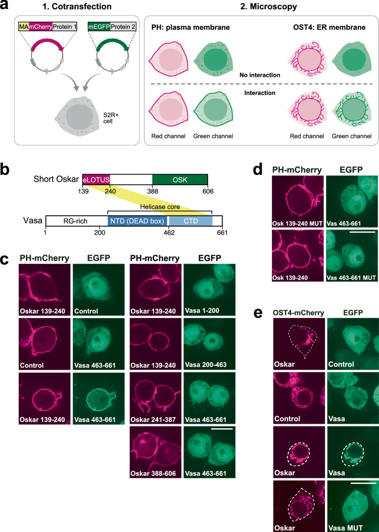
Fig. 1 ReLo assay and its use in PPI mapping and mutational analysis. a Plasmids encoding fluorescently tagged proteins 1 and 2 are cotransfected into S2R+ cells. Protein 1 carries an additional fusion to a membrane-anchoring (MA) domain, i.e., PH or OST4. After 48 h, protein localization is analyzed by confocal fluorescence microscopy. If protein 2 interacts with protein 1, protein 2 is relocalized to the plasma membrane (PH domain) or ER (OST4). The plasmid drawings were created based on BioRender.com icons. b Domain organization of Drosophila Oskar and Vasa proteins. Oskar is expressed in two isoforms, and Short Oskar lacks amino acids 1-138. The eLOTUS domain of Oskar was previously shown to interact with the C-terminal domain (CTD) of Vasa (yellow stripe), and the crystal structure of the complex has been resolved28,30. c Vasa 463-661, but not Vasa 1-200 or 200-463, interacted with the Oskar-eLOTUS domain. Vasa 463-661 did not interact with Oskar 241-387 or Oskar 388-606. n = 5 experiments (left panel) and n = 1 experiment (right panel), of which the data are consistent with the experiment shown in Supplementary Fig. 1e. d Oskar A162E/L228E and Vasa F504E point mutations (MUT) interfered with the Oskar-Vasa interaction. n = 2 experiments. e OST4-mCherry Oskar localized to the ER (top panel), and Vasa relocalized with Oskar to the ER (lower mid panel), while Vasa MUT did not (bottom panel). n = 2 experiments. In the “Control” experiments the respective construct was coexpressed with the vector indicated at the top of the panel lacking an insert. The scale bar is 10 µm.
Fig. 1 ReLo assay and its use in PPI mapping and mutational analysis. a Plasmids encoding fluorescently tagged proteins 1 and 2 are cotransfected into S2R+ cells. Protein 1 carries an additional fusion to a membrane-anchoring (MA) domain, i.e., PH or OST4. After 48 h, protein localization is analyzed by confocal fluorescence microscopy. If protein 2 interacts with protein 1, protein 2 is relocalized to the plasma membrane (PH domain) or ER (OST4). The plasmid drawings were created based on BioRender.com icons. b Domain organization of Drosophila Oskar and Vasa proteins. Oskar is expressed in two isoforms, and Short Oskar lacks amino acids 1-138. The eLOTUS domain of Oskar was previously shown to interact with the C-terminal domain (CTD) of Vasa (yellow stripe), and the crystal structure of the complex has been resolved28,30. c Vasa 463-661, but not Vasa 1-200 or 200-463, interacted with the Oskar-eLOTUS domain. Vasa 463-661 did not interact with Oskar 241-387 or Oskar 388-606. n = 5 experiments (left panel) and n = 1 experiment (right panel), of which the data are consistent with the experiment shown in Supplementary Fig. 1e. d Oskar A162E/L228E and Vasa F504E point mutations (MUT) interfered with the Oskar-Vasa interaction. n = 2 experiments. e OST4-mCherry Oskar localized to the ER (top panel), and Vasa relocalized with Oskar to the ER (lower mid panel), while Vasa MUT did not (bottom panel). n = 2 experiments. In the “Control” experiments the respective construct was coexpressed with the vector indicated at the top of the panel lacking an insert. The scale bar is 10 µm.CC BY · PubMed Central
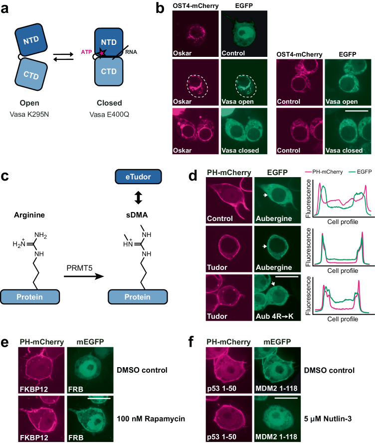
Fig. 2 PPI studies using ReLo with respect to conformation, post-translational modification, and drug sensitivity. a Pictorial representation of Vasa N- and C-terminal RecA-like domains (NTD and CTD, respectively) in the open and closed conformations. Vasa-open and Vasa-closed carry the K295N or E400Q mutations, respectively. b EGFP-Vasa-open, but not EGFP-Vasa-closed, interacted with OST4-mCherry-Oskar. The white dotted lines indicate cell boundaries. n = 2 experiments. c Symmetric dimethyl arginine (sDMA) modifications can be catalyzed by PRMT5 and recognized by eTud domains. d Wild-type Aub interacted with Tudor, whereas Aub carrying nonmethylatable 4R→K point mutations (R11K/R13K/R15K/R17K) did not. The profiles indicate the measured fluorescence distribution across the cell along a line indicated by the white arrows (shown for the green channel only). Red and green fluorescence signals are independent and displayed relatively. n = 3 experiments. e Rapamycin induced an interaction between human FKBP12 and FRB. The control contained 0.0009% dimethyl sulfoxide (DMSO). n = 2 experiments. f The interaction between p53 1–50 and MDM2 1-118 was inhibited by nutlin-3 treatment. The control contained 0.05% DMSO. n = 2 experiments. In the “Control” experiments the respective construct was coexpressed with the vector indicated at the top of the panel lacking an insert. The scale bar is 10 µm.
Fig. 2 PPI studies using ReLo with respect to conformation, post-translational modification, and drug sensitivity. a Pictorial representation of Vasa N- and C-terminal RecA-like domains (NTD and CTD, respectively) in the open and closed conformations. Vasa-open and Vasa-closed carry the K295N or E400Q mutations, respectively. b EGFP-Vasa-open, but not EGFP-Vasa-closed, interacted with OST4-mCherry-Oskar. The white dotted lines indicate cell boundaries. n = 2 experiments. c Symmetric dimethyl arginine (sDMA) modifications can be catalyzed by PRMT5 and recognized by eTud domains. d Wild-type Aub interacted with Tudor, whereas Aub carrying nonmethylatable 4R→K point mutations (R11K/R13K/R15K/R17K) did not. The profiles indicate the measured fluorescence distribution across the cell along a line indicated by the white arrows (shown for the green channel only). Red and green fluorescence signals are independent and displayed relatively. n = 3 experiments. e Rapamycin induced an interaction between human FKBP12 and FRB. The control contained 0.0009% dimethyl sulfoxide (DMSO). n = 2 experiments. f The interaction between p53 1–50 and MDM2 1-118 was inhibited by nutlin-3 treatment. The control contained 0.05% DMSO. n = 2 experiments. In the “Control” experiments the respective construct was coexpressed with the vector indicated at the top of the panel lacking an insert. The scale bar is 10 µm.CC BY · PubMed Central
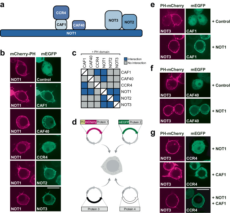
Fig. 3 ReLo identifies direct PPIs. a Subunit organization of the Drosophila CCR4-NOT core complex. b NOT1-mCherry-PH recruited CAF1, CAF40, NOT2, and NOT3 but not CCR4 to the plasma membrane. See Supplementary Fig. 3a for the PPI analysis of the other CCR4-NOT complex subunits. n = 1 experiment, of which the data are fully consistent with the experiments using swapped tags shown in Supplementary Fig. 3a. c Summary of the results from a pairwise screen of CCR4-NOT complex core subunits using ReLo. The blue color indicates an interaction, and the gray color indicates no interaction. d Schematic showing the ReLo assay involving coexpression of four different protein constructs. The plasmid drawings were created based on BioRender.com icons. e NOT3 interacted with CAF1 in the presence of NOT1. n = 2 experiments. f NOT3 interacted with CAF40 in the presence of NOT1. n = 3 experiments. g NOT3 recruited CCR4 to the plasma membrane only in the presence of both NOT1 and CAF1. n = 2 experiments. In the “Control” experiments the respective construct was coexpressed with the vector indicated at the top of the panel lacking an insert. The scale bar is 10 µm.
Fig. 3 ReLo identifies direct PPIs. a Subunit organization of the Drosophila CCR4-NOT core complex. b NOT1-mCherry-PH recruited CAF1, CAF40, NOT2, and NOT3 but not CCR4 to the plasma membrane. See Supplementary Fig. 3a for the PPI analysis of the other CCR4-NOT complex subunits. n = 1 experiment, of which the data are fully consistent with the experiments using swapped tags shown in Supplementary Fig. 3a. c Summary of the results from a pairwise screen of CCR4-NOT complex core subunits using ReLo. The blue color indicates an interaction, and the gray color indicates no interaction. d Schematic showing the ReLo assay involving coexpression of four different protein constructs. The plasmid drawings were created based on BioRender.com icons. e NOT3 interacted with CAF1 in the presence of NOT1. n = 2 experiments. f NOT3 interacted with CAF40 in the presence of NOT1. n = 3 experiments. g NOT3 recruited CCR4 to the plasma membrane only in the presence of both NOT1 and CAF1. n = 2 experiments. In the “Control” experiments the respective construct was coexpressed with the vector indicated at the top of the panel lacking an insert. The scale bar is 10 µm.CC BY · PubMed Central
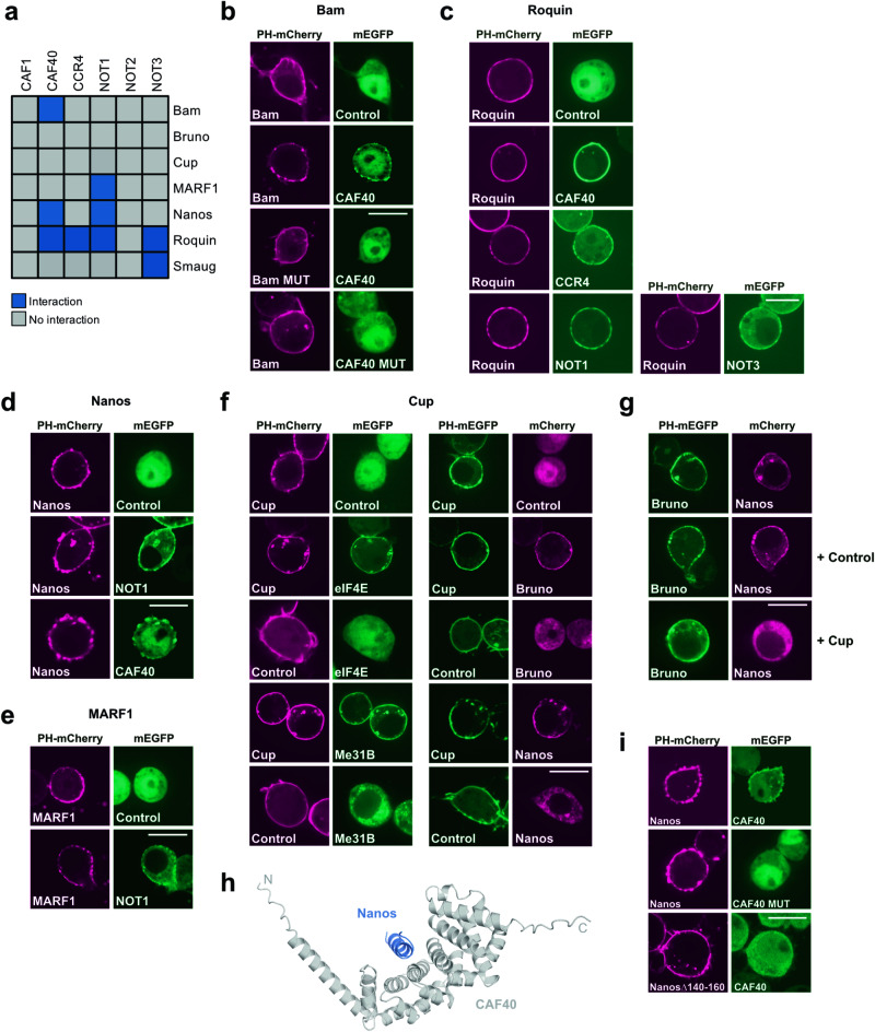
Fig. 4 PPIs between the CCR4-NOT complex and repressor proteins. a Summary of the results from a pairwise screen of CCR4-NOT complex core subunits with RNA-binding proteins as indicated. The blue color indicates an interaction, and the gray color indicates no interaction. Interaction data for Bruno and Cup are shown in Supplementary Fig. 7 and data for Smaug are published in63. b Bam interacted with CAF40. Bam MUT carried the M24E and CAF40 MUT the V186E point mutation. c Roquin interacted with CAF40, CCR4, NOT1, and NOT3. n = 2 experiments. d Nanos interacted with NOT1 and CAF40. e MARF1 interacted with NOT1. f Cup interacted with eIF4E, Me31B, Bruno, and Nanos. g Bruno interacted with Nanos in the absence but not in the presence of Cup. h Structural model of the CAF40-Nanos 140–160 complex obtained using AlphaFold-Multimer version 3. The predicted aligned error (PAE) plot is shown in Supplementary Fig. 9. i CAF40 MUT (V186E) did not interact with Nanos and Nanos lacking residues 140-160 did not interact with CAF40. In the “Control” experiments the respective construct was coexpressed with the vector indicated at the top of the panel lacking an insert. For all, n = 2 experiments. The scale bar is 10 µm.
Fig. 4 PPIs between the CCR4-NOT complex and repressor proteins. a Summary of the results from a pairwise screen of CCR4-NOT complex core subunits with RNA-binding proteins as indicated. The blue color indicates an interaction, and the gray color indicates no interaction. Interaction data for Bruno and Cup are shown in Supplementary Fig. 7 and data for Smaug are published in63. b Bam interacted with CAF40. Bam MUT carried the M24E and CAF40 MUT the V186E point mutation. c Roquin interacted with CAF40, CCR4, NOT1, and NOT3. n = 2 experiments. d Nanos interacted with NOT1 and CAF40. e MARF1 interacted with NOT1. f Cup interacted with eIF4E, Me31B, Bruno, and Nanos. g Bruno interacted with Nanos in the absence but not in the presence of Cup. h Structural model of the CAF40-Nanos 140–160 complex obtained using AlphaFold-Multimer version 3. The predicted aligned error (PAE) plot is shown in Supplementary Fig. 9. i CAF40 MUT (V186E) did not interact with Nanos and Nanos lacking residues 140-160 did not interact with CAF40. In the “Control” experiments the respective construct was coexpressed with the vector indicated at the top of the panel lacking an insert. For all, n = 2 experiments. The scale bar is 10 µm.CC BY · PubMed Central

Verification

Claims the paper makes

The claims and statistics of this paper haven’t been checked yet.

Run the check — free, no account needed. It reads the open-access text and checks the sources it cites.

A few free checks a day · papers already verified open instantly · results are public

Self-check

Percentages, sums and reported differences re-computed from the paper’s own numbers.

The self-check needs the paper’s full text.

Citations

Cites

Research works printed but not linked: 81

  1. 2004, Co-immunoprecipitation from transfected cells, Methods Mol Biol, 261, 337
  2. 1995, Detection of protein-protein interactions by coimmunoprecipitation and dimerization, Methods Enzymol, 254, 491, 10.1016/0076-6879(95)54034-2 · DOI
  3. 10.1038/13732 · DOI · Paper page
  4. 10.1016/j.cbpa.2018.10.017 · DOI
  5. 10.1002/wdev.399 · DOI
  6. 10.1007/s00018-013-1287-3 · DOI
  7. 10.1111/j.1365-313X.2007.03332.x · DOI
  8. 2021, The characterization of protein interactions – what, how and how much?, Chem Soc Rev, 50, 12292, 10.1039/D1CS00548K · DOI
  9. 10.2144/000112396 · DOI
  10. 10.1007/s11427-018-9500-7 · DOI
  11. 10.1042/BST20201058 · DOI
  12. 2020, Detecting Protein-Protein Interaction Based on Protein Fragment Complementation Assay, Curr Protein Pept Sci, 21, 598, 10.2174/1389203721666200213102829 · DOI
  13. 2017, CAPPI: A Cytoskeleton-Based Localization Assay Reports Protein-Protein Interaction in Living Cells by Fluorescence Microscopy, Mol Plant, 10, 1473, 10.1016/j.molp.2017.09.006 · DOI
  14. 10.1093/mp/ssm015 · DOI
  15. 10.1093/plcell/koab004 · DOI
  16. 10.2144/000113452 · DOI
  17. 10.1073/pnas.0911262107 · DOI
  18. 10.1371/journal.pone.0054583 · DOI · Paper page
  19. 10.1038/ncomms5925 · DOI
  20. 10.1016/j.chembiol.2007.07.017 · DOI
  21. 10.1038/srep41926 · DOI
  22. 10.1242/dev.27.2.353 · DOI
  23. 1999, Identification and characterization of a novel line of Drosophila Schneider s2 cells that respond to wingless signaling, Journal of Biological Chemistry, 273, 32353
  24. 10.1021/bi00049a039 · DOI
  25. 10.1073/pnas.92.23.10472 · DOI · Paper page
  26. 10.1073/pnas.1332735100 · DOI
  27. 10.2144/000112455 · DOI
  28. 10.1101/gad.297051.117 · DOI
  29. 10.1101/gad.10.17.2179 · DOI
  30. 10.1016/j.celrep.2015.06.055 · DOI
  31. 10.1073/pnas.1515568112 · DOI
  32. 10.1038/nrm3154 · DOI
  33. 10.1002/j.1460-2075.1982.tb01276.x · DOI · Paper page
  34. 1988, A conserved NTP-motif in putative helicases, Nature, 333, 22, 10.1038/333022a0 · DOI
  35. 10.1016/j.cell.2014.05.018 · DOI
  36. 10.1007/s00018-015-1847-9 · DOI
  37. 10.1016/j.cub.2006.04.037 · DOI
  38. 10.1101/gad.1956010 · DOI
  39. 10.1261/rna.1869710 · DOI
  40. 10.1038/emboj.2009.365 · DOI · Paper page
  41. 10.1038/ncb1872 · DOI
  42. 10.1126/science.274.5285.239 · DOI
  43. 10.1126/science.1092472 · DOI · Paper page
  44. 10.1126/science.274.5289.948 · DOI · Paper page
  45. 10.1007/978-3-319-46503-6_13 · DOI
  46. 10.3389/fgene.2014.00143 · DOI
  47. 10.1038/nature12962 · DOI
  48. 10.1016/j.tcb.2016.08.001 · DOI
  49. Hu, Y. , Comjean, A. , Perrimon, N. & Mohr, S. E . The Drosophila Gene Expression Tool (DGET) for expression analyses. BMC Bioinformatics 18, 98 (2017). · DOI
  50. 10.1126/science.1066333 · DOI · Paper page
  51. 10.1006/bbrc.2000.4151 · DOI
  52. 10.1073/pnas.95.9.5187 · DOI
  53. 10.1007/978-1-61779-412-4_20 · DOI
  54. 10.1261/rna.064584.117 · DOI
  55. 10.1038/ncomms14307 · DOI
  56. 10.15252/embj.201593634 · DOI
  57. 10.1038/s41467-018-06404-w · DOI · Paper page
  58. 10.1016/j.cub.2005.01.048 · DOI
  59. 10.1242/dev.02649 · DOI
  60. 10.1101/gad.10.20.2600 · DOI
  61. 10.1101/gad.10.20.2610 · DOI
  62. 10.1016/S1097-2765(00)80368-8 · DOI
  63. 2023, RNA binding proteins Smaug and Cup induce CCR4–NOT- dependent deadenylation of the nanos mRNA in a reconstituted system, Nucleic Acids Res, 51, 3950, 10.1093/nar/gkad159 · DOI
  64. 10.1038/sj.emboj.7600026 · DOI
  65. 10.1016/S1534-5807(03)00400-3 · DOI
  66. 10.1083/jcb.200309088 · DOI
  67. 10.1073/pnas.0406451101 · DOI
  68. 10.1093/nar/gkw565 · DOI
  69. 10.1016/j.celrep.2015.09.033 · DOI
  70. 10.1261/rna.033639.112 · DOI
  71. 10.1101/gad.17136311 · DOI
  72. 2000, Nanos interacts with Cup in the female germline of Drosophila, Development, 127, 5225, 10.1242/dev.127.23.5225 · DOI
  73. 10.1371/journal.pgen.1005199 · DOI
  74. 2020, An Interaction Network of RNA-Binding Proteins Involved in Drosophila Oogenesis, Molecular and Cellular Proteomics, 19, 1485, 10.1074/mcp.RA119.001912 · DOI
  75. Evans, R. , et al. Protein complex prediction with AlphaFold-Multimer. bioRxiv 2021.10.04.463034 (2021) doi:https://doi.org/10.1101/2021.10.04.463034. · DOI · Paper page
  76. 2022, ColabFold: making protein folding accessible to all, Nature Methods, 19, 679, 10.1038/s41592-022-01488-1 · DOI · Paper page
  77. 10.1016/j.tips.2013.04.007 · DOI · Paper page
  78. 10.1002/iub.383 · DOI · Paper page
  79. 10.1101/gad.1424106 · DOI
  80. 10.1016/j.celrep.2015.07.054 · DOI
  81. 10.1038/nmeth.2019 · DOI · Paper page

Metadata

Authors
Salgania, H. K., Metz, J., & Jeske, M.
Affiliations
Heidelberg University (DE)
Corresponding author
Jeske, Mandy
Venue
Nature Communications
Publisher
openRxiv
Published
2022-03-04
Volume
15
Issue
1
Pages
2875-2875
ISSN
2041-1723 (ISSN-L)
OpenAlex
W4393872440
Cited by
17
References
1
Citations by year
2026: 5 · 2025: 6 · 2024: 4 · 2023: 2
FWCI
3.37
Type
journal article
Language
en
Open access
Licence
cc-by
Locations
  • Nature Communications (publishedVersion, cc-by)
  • bioRxiv (Cold Spring Harbor Laboratory) (acceptedVersion, cc-by-nc)
  • PubMed Central (publishedVersion, cc-by)
  • PubMed (publishedVersion)
  • RePEc: Research Papers in Economics (submittedVersion)
  • DOAJ (DOAJ: Directory of Open Access Journals) (submittedVersion, cc-by-sa)
Funders
  • Deutsche Forschungsgemeinschaft (INST 35/1314-1 FUGG and INST 35/1503-1 FUGG, INST 35/1314-1 FUGG., INST35/1503‐1FUGG, INST 35/1314‐1, JE827/1-1, 35/1503-1, 35/1503-1 FUGG, 35/1314-1 FUGG, INST 35, INST 35/1503‐1, 35/1314-1)
  • Universität Heidelberg
Topics
  • Life Sciences › Biochemistry, Genetics and Molecular Biology › Cell Biology › Biotin and Related Studies
  • Life Sciences › Biochemistry, Genetics and Molecular Biology › Biophysics › Advanced Fluorescence Microscopy Techniques
  • Life Sciences › Biochemistry, Genetics and Molecular Biology › Molecular Biology › Bioinformatics and Genomic Networks
Keywords
protein-protein interaction, protein complexes, protein domains, drug screening
MeSH terms
Proteins, Protein Interaction Mapping

About these checks

Registry, retraction, Cites and statistics results are computed, not judged, with one exception: a printed reference that no index lists is read once by a model before it counts as a citation error. Claims are extracted by a model and kept only when their quote appears verbatim in the text; each cited source is then checked, with a model’s help, for whether it says what the claim relies on. Those verdicts can be wrong — read the claim’s quote and the cited source before relying on one.

Statistics and arithmetic are recomputed, not judged.

Confidence bands are shown only for fields calibrated on our annotated test set. None is calibrated yet, so no band is shown.

What counts as a claim ›How we measure confidence ›How we check this ›

Are you an author, or do you see a problem with this page? Request a review