

The DOI matches its Crossref record; 1 of the 1 references it cites were found in a registry or an index.
Evaluating the breadth of neutralizing antibody responses elicited by infectious bursal disease virus (IBDV) genogroup A1 strains using a novel chicken B-cell rescue system and neutralization assay
Open accessPreprint — published as 10.1128/jvi.01255-22 · Paper page
Abstract
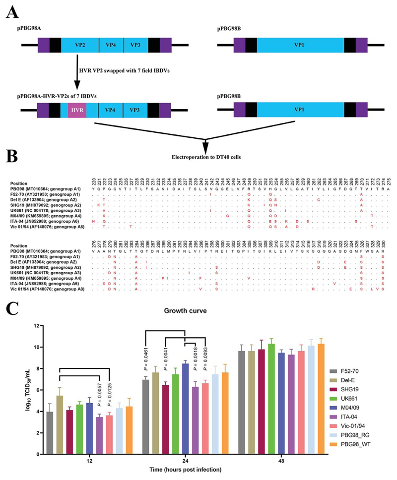
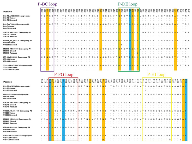
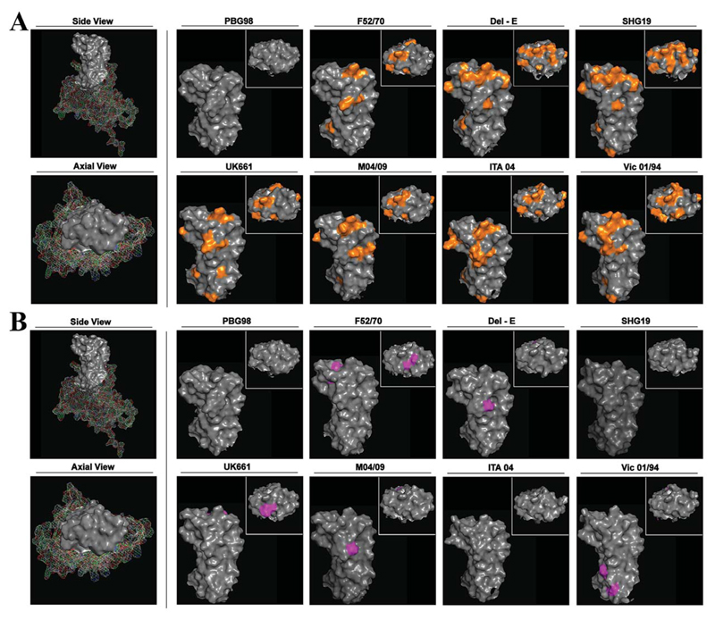
Eight infectious bursal disease virus (IBDV) genogroups have been identified based on the sequence of the capsid hypervariable region (HVR) (A1-8), yet many vaccines are based on A1 strains. Given reported vaccine failures, there is a need to evaluate the ability of vaccines to neutralize the different genogroups. To address this, we used a reverse genetics system and the chicken B-cell line DT40 to rescue a panel of chimeric IBDVs and perform neutralization assays. Chimeric viruses had the backbone of a lab-adapted strain (PBG98) and the HVRs from diverse field strains: classical F52-70 (A1), US-variant Del-E (A2), Chinese-variant SHG19 (A2), very-virulent UK661 (A3), M04/09 distinct (A4), Italian ITA-04 (A6), and Australian-variant Vic-01/94 (A8). Rescued viruses showed no substitutions at amino-acid positions 253, 284, or 330, previously associated with cell-culture adaptation. Sera from chickens inoculated with wt (F52-70) or vaccine (228E) A1 strains had the highest mean virus neutralization (VN) titers against the A1 virus (log 2 15.41 and 12.66), and the lowest against A2 viruses (log 2 7.41-7.91, p=0.0001-0.0274), consistent with A1 viruses being most antigenically distant from A2 strains, which correlated with the extent of differences in the predicted HVR structure. VN titers against the other genogroups ranged from log 2 9.32-13.32, and A1 strains were likely more closely antigenically related to genogroups A3 and A4 than A6 and A8. Our data are consistent with field observations, validating our method, which can used to screen future vaccine candidates for breadth of neutralizing antibodies, and evaluate the antigenic relatedness of different genogroups. Importance: There is a need to evaluate the ability of vaccines to neutralize diverse IBDV genogroups, and to better understand the relationship between HVR sequence, structure, and antigenicity. Here, we used a chicken B cell-line to rescue a panel of chimeric IBDVs with the HVR from seven diverse IBDV field strains, and conduct neutralization assays and protein modelling. We evaluated the ability of serum from vaccinated or infected birds to neutralize the different genogroups. Our novel chicken B-cell rescue system and neutralization assay can be used to screen IBDV vaccine candidates, platforms, and regimens for the breadth of neutralizing antibody responses elicited, evaluate the antigenic relatedness of diverse IBDV strains, and when coupled with structural modelling, elucidate immunodominant and conserved epitopes to strategically design novel IBDV vaccines in the future.
Overview
The claims and statistics of this paper haven’t been checked yet.
Run the check — free, no account needed. It reads the open-access text and checks the sources it cites.
Integrity signals
Registry
Cites
Statistics
Figures
Verification
Claims the paper makes
The claims and statistics of this paper haven’t been checked yet.
Run the check — free, no account needed. It reads the open-access text and checks the sources it cites.
Self-check
Percentages, sums and reported differences re-computed from the paper’s own numbers.
The self-check needs the paper’s full text.
Citations
Cites
Highly accurate protein structure prediction with AlphaFold
Jumper, J., Evans, R., Pritzel, A., Green, T., Figurnov, M., Ronneberger, O., Tunyasuvunakool, K., Bates, R., Žídek, A., Potapenko, A., et al. (2021) · Nature · doi:10.1038/s41586-021-03819-2 · Cited by 43,764
Research works printed but not linked: 52
- 10.1637/8909-043009-Review.1 · DOI
- 10.1371/journal.pone.0170080 · DOI
- 10.1099/0022-1317-81-4-983 · DOI
- 10.1016/j.virol.2008.12.036 · DOI
- 10.1016/j.jmb.2008.11.029 · DOI
- 10.1073/pnas.0704447104 · DOI
- 10.1128/JVI.02360-12 · DOI
- 2021, A unified genotypic classification of infectious bursal disease virus based on both genome segments, Avian Pathol, 50, 190, 10.1080/03079457.2021.1873245 · DOI
- 10.1007/s00705-017-3500-4 · DOI
- 10.1128/JVI.01501-07 · DOI
- 10.1099/0022-1317-83-1-121 · DOI
- 10.1128/JVI.73.4.2854-2862.1999 · DOI
- 10.1099/0022-1317-80-8-2067 · DOI
- 10.1016/j.vetmic.2019.01.023 · DOI
- 2021, Genetic Diversity of Recent Infectious Bursal Disease Viruses Isolated From Vaccinated Poultry Flocks in Malaysia, Front Vet Sci, 8, 643976, 10.3389/fvets.2021.643976 · DOI
- 2016, Isolation of novel variants of infectious bursal disease virus from different outbreaks in Northeast India, Microb Pathog, 93, 131, 10.1016/j.micpath.2016.02.004 · DOI
- 2020, The Stronger Downregulation of in vitro and in vivo Innate Antiviral Responses by a Very Virulent Strain of Infectious Bursal Disease Virus (IBDV), Compared to a Classical Strain, Is Mediated, in Part, by the VP4 Protein, Front Cell Infect Microbiol, 10, 315, 10.3389/fcimb.2020.00315 · DOI
- 2018, Propagation and titration of infectious bursal disease virus, including non-cell-culture-adapted strains, using ex vivo-stimulated chicken bursal cells, Avian Pathol, 47, 179, 10.1080/03079457.2017.1393044 · DOI
- Eterradossi N , Saif M. 2013. Infectious Bursal Disease, p 219–246. In Glisson J , McDougald L , Nolan L , Suarez D , Nair V , Swayne D (ed), Diseases of Poultry 13 ed. Ames, Iowa: Blackwell Pub Professional. · DOI
- 2017, Differential gene expression in chicken primary B cells infected ex vivo with attenuated and very virulent strains of infectious bursal disease virus (IBDV), J Gen Virol, 98, 2918, 10.1099/jgv.0.000979 · DOI
- 10.3791/58489 · DOI
- 2019, Macrophage Migration Inhibitory Factor Triggers Inflammatory Responses During Very Virulent Infectious Bursal Disease Virus Infection, Front Microbiol, 10, 2225, 10.3389/fmicb.2019.02225 · DOI
- 10.1292/jvms.70.407 · DOI
- 10.1099/vir.0.008870-0 · DOI
- 2020, Ex vivo rescue of recombinant very virulent IBDV using a RNA polymerase II driven system and primary chicken bursal cells, Sci Rep, 10, 13298, 10.1038/s41598-020-70095-x · DOI
- Campbell EA , Reddy V , Gray AG , Wells J , Simpson J , Skinner MA , Hawes PC , Broadbent AJ. 2020. Discrete Virus Factories Form in the Cytoplasm of Cells Coinfected with Two Replication-Competent Tagged Reporter Birnaviruses That Subsequently Coalesce over Time. J Virol 94. · DOI
- 10.1016/0042-6822(85)90312-5 · DOI
- 1990, A comparison of the sequences of segment A of four infectious bursal disease virus strains and identification of a variable region in VP2, J Gen Virol, 71, 1303
- 10.1016/0168-1702(95)01253-2 · DOI
- 10.1093/oxfordjournals.aje.a118408 · DOI · Paper page
- 2014, Further evidence for the association of distinct amino acid residues with in vitro and in vivo growth of infectious bursal disease virus, Arch Virol, 159, 701, 10.1007/s00705-013-1885-2 · DOI
- 10.1099/0022-1317-70-6-1473 · DOI
- 10.1080/03079457.2012.661403 · DOI
- 2021, Efficacy of a turkey herpesvirus double construct vaccine (HVT-ND-IBD) against challenge with different strains of Newcastle disease, infectious bursal disease and Marek’s disease viruses, Avian Pathol, 50, 18, 10.1080/03079457.2020.1828567 · DOI
- 2018, Field and laboratory findings following the large-scale use of intermediate type infectious bursal disease vaccines in Denmark, Avian Pathol, 47, 595, 10.1080/03079457.2018.1520388 · DOI
- 10.1637/8687-31009RESNOTE.1 · DOI
- 10.1637/8902-042809-Reg.1 · DOI
- 2013, An in vivo experimental model to determine antigenic variations among infectious bursal disease viruses, Avian Pathol, 42, 309, 10.1080/03079457.2013.793783 · DOI
- 10.2307/1588880 · DOI
- 10.1007/s00705-003-0230-6 · DOI
- 10.2307/1590924 · DOI
- 10.2307/1591345 · DOI
- 2020, Novel variants of infectious bursal disease virus can severely damage the bursa of fabricius of immunized chickens, Vet Microbiol, 240, 108507, 10.1016/j.vetmic.2019.108507 · DOI
- Tomas G , Hernandez M , Marandino A , Hernandez D , Techera C , Grecco S , Panzera Y , Perez R. 2015. Genome Sequence of a Distinct Infectious Bursal Disease Virus. Genome Announc 3. · DOI
- 10.1080/03079457.2016.1165792 · DOI
- 2002, Confirmation of the existence of two distinct genetic groups of infectious bursal disease virus in Australia, Aust Vet J, 80, 689, 10.1111/j.1751-0813.2002.tb11299.x · DOI
- 10.1007/s007050050670 · DOI
- 2017, Transcriptional profiles in bursal B-lymphoid DT40 cells infected with very virulent infectious bursal disease virus, Virol J, 14, 7, 10.1186/s12985-016-0668-2 · DOI
- 2003, Identification of crucial residues of conformational epitopes on VP2 protein of infectious bursal disease virus by phage display, J Virol Methods, 109, 75, 10.1016/S0166-0934(03)00049-1 · DOI
- 10.1080/03079450801910206 · DOI
- 10.1006/viro.1996.0470 · DOI
- 1993, Infectious bursal disease virus structural proteins expressed in a baculovirus recombinant confer protection in chickens, J Gen Virol, 74, 1201
Metadata
- Authors
- Reddy, V. R. A. P., Nazki, S., Brodrick, A. J., Asfor, A., Urbaniec, J., Morris, Y., & Broadbent, A. J.
- Affiliations
- The Pirbright Institute (GB)
- University of Maryland, College Park (US)
- Corresponding author
- Reddy, Vishwanatha R. A. P.
- Broadbent, Andrew J.
- ORCID
- Venue
- Journal of Virology
- Publisher
- openRxiv
- Published
- 2022-06-04
- Volume
- 96
- Issue
- 18
- Pages
- e0125522-e0125522
- ISSN
- 0022-538X (ISSN-L)
- PMC
- PMC9517715
- OpenAlex
- W4294921245
- Cited by
- 12
- References
- 1
- Citations by year
- 2026: 3 · 2025: 1 · 2024: 2 · 2023: 3 · 2022: 3
- FWCI
- 1.40
- Type
- journal article
- Language
- en
- Open access
- hybrid
- doi.org
- Licence
- cc-by
- Locations
- Journal of Virology (publishedVersion, cc-by)
- bioRxiv (Cold Spring Harbor Laboratory) (acceptedVersion, cc-by)
- PubMed Central (publishedVersion)
- PubMed (publishedVersion)
- Funders
- UK Research and Innovation (BBS/E/I/00001845, BBS/E/I/00007039, BB/S014594/1, BBS/E/I/00007034)
- Directorate for Biological Sciences
- Biotechnology and Biological Sciences Research Council (BBS/E/I/00007034, BBS/E/I/00007039, BB/S014594/1, BBS/E/I/00001845)
- Topics
- Life Sciences › Agricultural and Biological Sciences › Animal Science and Zoology › Animal Virus Infections Studies
- Health Sciences › Medicine › Epidemiology › Virology and Viral Diseases
- Life Sciences › Biochemistry, Genetics and Molecular Biology › Molecular Biology › vaccines and immunoinformatics approaches
- Keywords
- infectious bursal disease virus, neutralizing antibodies, antigenic relatedness, vaccine design, immunodominant epitopes, conserved epitopes, protein structure modeling, vaccine screening
- MeSH terms
- Animals, Epitopes, Australia, Chickens, Genotype, Infectious bursal disease virus, Poultry Diseases, Birnaviridae Infections, Antibodies, Neutralizing
About these checks
Registry, retraction, Cites and statistics results are computed, not judged, with one exception: a printed reference that no index lists is read once by a model before it counts as a citation error. Claims are extracted by a model and kept only when their quote appears verbatim in the text; each cited source is then checked, with a model’s help, for whether it says what the claim relies on. Those verdicts can be wrong — read the claim’s quote and the cited source before relying on one.
Statistics and arithmetic are recomputed, not judged.
Confidence bands are shown only for fields calibrated on our annotated test set. None is calibrated yet, so no band is shown.
What counts as a claim ›How we measure confidence ›How we check this ›
Are you an author, or do you see a problem with this page? Request a review
Data sources
Bibliographic data: Semantic Scholar, OpenAlex, Crossref.
Contains information from the Semantic Scholar Academic Graph Datasets, made available under the ODC Attribution License (ODC-BY 1.0).
Semantic Scholar’s data is described in: Kinney, R., et al. (2023). The Semantic Scholar Open Data Platform. arXiv:2301.10140.
