Student or educator? Sign up with your school email and get 6 months of Pro free.
PreprintCited by 6Escherichia coli research studiesBiochemical and Structural CharacterizationBacterial Genetics and Biotechnology

The DOI matches its Crossref record; 1 of the 1 references it cites were found in a registry or an index.

Cysteine dependent conformation heterogeneity of Shigella flexneri autotransporter IcsA and implications in its function

Open access

Preprint — published as 10.1128/spectrum.03410-22 · Paper page

Abstract

Shigella IcsA is a versatile surface virulence factor required for both early and late pathogenesis stages, extracellularly to intracellularly. Despite IcsA serving as a model Type V secretion system (T5SS) autotransporter to study host pathogen interactions, its detailed molecular architecture is poorly understood. Recently, IcsA was found to switch to a different conformation for its adhesin activity upon sensing of the host stimuli by Shigella Type III secretion system (T3SS). Here, we report that the single cysteine residue (C130) near the N-terminus of IcsA passenger has a role in IcsA adhesin activity. We also show that the IcsA passenger (IcsAp) exists in multiple conformations, and the conformation populations are influenced by a central pair of cysteine residues (C375 and C379), which is not previously reported for any Type V autotransporter passengers. Disruption of either or both central cysteine residues alters the exposure of IcsA epitopes to polyclonal anti-IcsA antibodies previously shown to block Shigella adherence, yet without loss of IcsA intracellular functions in actin-based motility (ABM). Anti-IcsA antibody reactivity was restored when the IcsA paired cysteine substitution mutants were expressed in a ∆ipaD background with a constitutively active T3SS, highlighting an interplay between T3SS and T5SS. The work here uncovers a novel molecular switch empowered by a centrally localised, short-spaced cysteine pair in the Type V autotransporter IcsA that ensures conformational heterogeneity to aid IcsA evasion of host immunity. Importance: Shigella species are the leading cause of diarrheal related death globally by causing bacillary dysentery. The surface virulence factor IcsA which is essential for Shigella pathogenesis is a unique multi-functional autotransporter that is responsible for cell adhesion, and actin-based motility, yet detailed mechanistic understanding is lacking. Here, we show that the three cysteine residues in IcsA contribute to the protein’s distinct functions. The N terminus cysteine residue within the IcsA passenger domain plays a role in adhesin function, while a centrally localised cysteine pair provides conformational heterogeneity resulting in IcsA molecules with different reactivity to adhesion-blocking anti-IcsA antibodies. In synergy with the Type III secretion system, this molecular switch preserves biological function in distinct IcsA conformations for cell adhesion, actin-based motility and autophagy escape, providing a potential strategy by which Shigella evade host immunity targeting of this essential virulence factor.

Overview

The claims and statistics of this paper haven’t been checked yet.

Run the check — free, no account needed. It reads the open-access text and checks the sources it cites.

A few free checks a day · papers already verified open instantly · results are public

Integrity signals

Registry

Crossref · 10.1101/2022.07.21.501072

Cites

1 of 1 references verified0 citation errors · 0 not checkable

Statistics

Figures

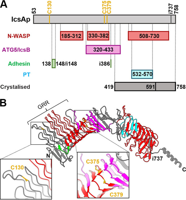
FIG 1 The autotransporter protein IcsA passenger domain. (A) Schematic representation of defined subregions on IcsA passenger affecting its known biological functions with all three cysteine positions shown. PT, polar targeting. (B) IcsA passenger tertiary structure predicted by AlphaFold (AF-Q7BCK4-F1) with defined functional subregions marked in color as in (A) and cysteine residue locations is shown in a close-up view. GRR, glycine-rich repeats (aa 117 to 307).
FIG 1 The autotransporter protein IcsA passenger domain. (A) Schematic representation of defined subregions on IcsA passenger affecting its known biological functions with all three cysteine positions shown. PT, polar targeting. (B) IcsA passenger tertiary structure predicted by AlphaFold (AF-Q7BCK4-F1) with defined functional subregions marked in color as in (A) and cysteine residue locations is shown in a close-up view. GRR, glycine-rich repeats (aa 117 to 307).CC BY · PubMed Central
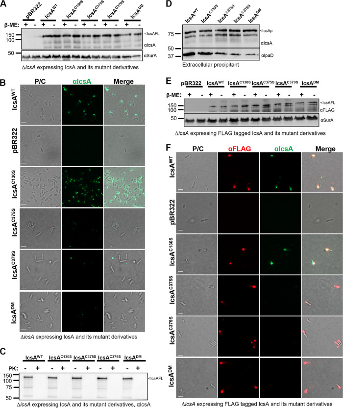
FIG 2 Cysteine substitutions in IcsAp did not affect surface display but influenced protein conformation. (A) Western immunoblotting of bacterial cell lysates expressing IcsAWT and cysteine mutant detected with anti-IcsA and anti-SurA antibodies. IcsAFL, IcsA full-length. (B) Indirect bacterial surface immunofluorescent labeling of S. flexneri expressing IcsAWT and cysteine substitution mutants with anti-IcsA antibodies. (C) Western immunoblotting of IcsA with lysates of bacterial cells with and without proteinase K surface shaving. (D) Western immunoblotting of precipitated extracellular cleaved IcsAp with anti-IcsA and anti-IpaD antibodies. (E) Western immunoblotting of lysates of bacterial cells expressed FLAG-tagged IcsAWT and cysteine substitution mutants with anti-FLAG and anti-SurA antibodies. (F) Indirect bacterial surface immunofluorescent labeling of FLAG-tagged IcsA passenger with anti-IcsA antibodies and anti-FLAG antibody. Scale bars are shown as 2 μm in (B and F).
FIG 2 Cysteine substitutions in IcsAp did not affect surface display but influenced protein conformation. (A) Western immunoblotting of bacterial cell lysates expressing IcsAWT and cysteine mutant detected with anti-IcsA and anti-SurA antibodies. IcsAFL, IcsA full-length. (B) Indirect bacterial surface immunofluorescent labeling of S. flexneri expressing IcsAWT and cysteine substitution mutants with anti-IcsA antibodies. (C) Western immunoblotting of IcsA with lysates of bacterial cells with and without proteinase K surface shaving. (D) Western immunoblotting of precipitated extracellular cleaved IcsAp with anti-IcsA and anti-IpaD antibodies. (E) Western immunoblotting of lysates of bacterial cells expressed FLAG-tagged IcsAWT and cysteine substitution mutants with anti-FLAG and anti-SurA antibodies. (F) Indirect bacterial surface immunofluorescent labeling of FLAG-tagged IcsA passenger with anti-IcsA antibodies and anti-FLAG antibody. Scale bars are shown as 2 μm in (B and F).CC BY · PubMed Central
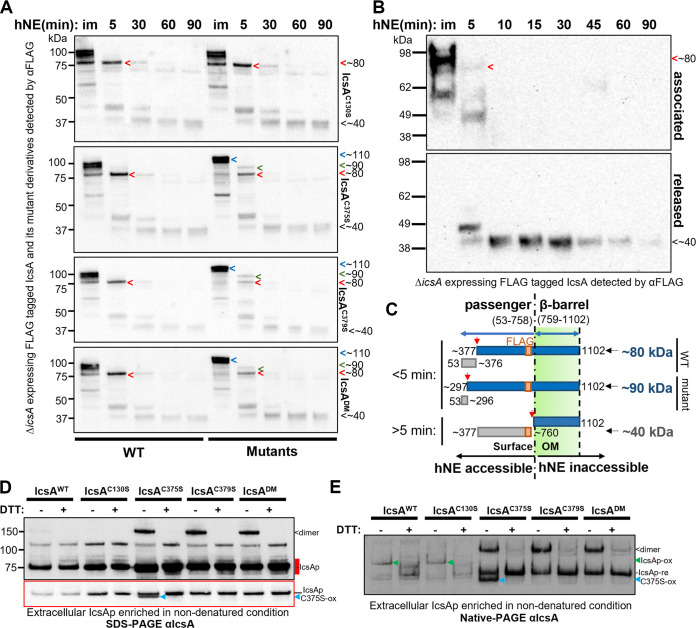
FIG 3 The IcsA N terminus had the altered conformation and was maintained in the cleaved IcsAp. (A) Western immunoblotting with bacterial cells expressing IcsA and its cysteine substitution mutants treated with hNE. Samples were taken at different time points as indicated above the blot. im, immediate recovery of the digestion sample upon the addition of hNE. Samples from bacteria expressing IcsAWT were electrophoresed in parallel (left) with mutants (right) and blotted together to ensure equal exposure for direct comparison. (B) Western immunoblotting of fractionated hNE digestion samples from the bacteria expressing IcsAWT. (C) Model of the digestion differences due to altered conformation in IcsA mutant with estimated cutting sites shown. (D) Western immunoblotting of IcsAp separated in semidenatured conditions. Extracellular IcsA was precipitated in ammonium sulfate and was solubilized in SDS-PAGE buffer in the presence or absence of DTT and was subjected to SDS-PAGE and immunoblotting without heating. The underexposure image in the red border correlates to the region in the long exposure image marked red. re, reduced; ox, oxidized. <, IcsAp dimer; blue arrow, oxidized IcsAC375S, C375S-ox. (E) Culture supernatant IcsAp protein recovered as above was analyzed by Native-PAGE and detected with anti-IcsA antibodies. Green arrow, oxidized IcsAp, IcsAp-ox.
FIG 3 The IcsA N terminus had the altered conformation and was maintained in the cleaved IcsAp. (A) Western immunoblotting with bacterial cells expressing IcsA and its cysteine substitution mutants treated with hNE. Samples were taken at different time points as indicated above the blot. im, immediate recovery of the digestion sample upon the addition of hNE. Samples from bacteria expressing IcsAWT were electrophoresed in parallel (left) with mutants (right) and blotted together to ensure equal exposure for direct comparison. (B) Western immunoblotting of fractionated hNE digestion samples from the bacteria expressing IcsAWT. (C) Model of the digestion differences due to altered conformation in IcsA mutant with estimated cutting sites shown. (D) Western immunoblotting of IcsAp separated in semidenatured conditions. Extracellular IcsA was precipitated in ammonium sulfate and was solubilized in SDS-PAGE buffer in the presence or absence of DTT and was subjected to SDS-PAGE and immunoblotting without heating. The underexposure image in the red border correlates to the region in the long exposure image marked red. re, reduced; ox, oxidized. <, IcsAp dimer; blue arrow, oxidized IcsAC375S, C375S-ox. (E) Culture supernatant IcsAp protein recovered as above was analyzed by Native-PAGE and detected with anti-IcsA antibodies. Green arrow, oxidized IcsAp, IcsAp-ox.CC BY · PubMed Central
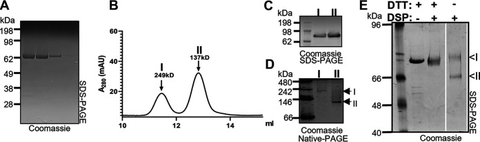
FIG 4 Purified IcsA existed in two different conformations. (A) Affinity purified IcsAp resolved by SDS-PAGE and stained with Coomassie blue. (B) SEC separation of IcsAp into populations I and II. (C) SDS-PAGE of IcsAp populations I and II isolated by SEC. (D) Native-PAGE of IcsAp populations I and II isolated by SEC. (E) SDS-PAGE of IcsAp cross-linked with 0.1 mM DSP in the presence or absence of 10 mM DTT. The image was from the same gel with the relevant area shown.
FIG 4 Purified IcsA existed in two different conformations. (A) Affinity purified IcsAp resolved by SDS-PAGE and stained with Coomassie blue. (B) SEC separation of IcsAp into populations I and II. (C) SDS-PAGE of IcsAp populations I and II isolated by SEC. (D) Native-PAGE of IcsAp populations I and II isolated by SEC. (E) SDS-PAGE of IcsAp cross-linked with 0.1 mM DSP in the presence or absence of 10 mM DTT. The image was from the same gel with the relevant area shown.CC BY · PubMed Central
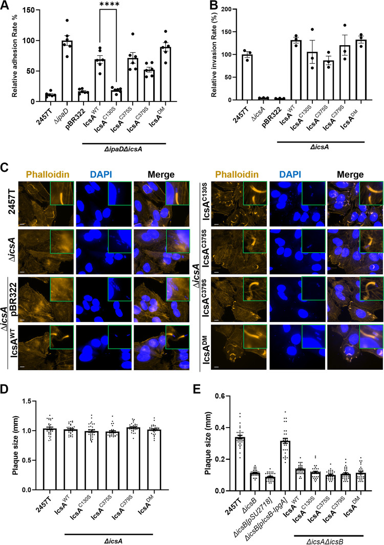
FIG 5 Effect of cysteine substitution mutation on IcsA function. (A) Cell adhesion assay of indicated S. flexneri strains with HeLa cells. Mean values from 6 independent assays with standard error mean (SEM) are shown. Statistical significance was calculated using one-way analysis of variance (ANOVA) followed by Dunnett’s multiple-comparison test against ΔipaDΔicsA pIcsAWT. ****, P < 0.0001. (B) HeLa cell invasion assay of indicated S. flexneri strains treated with 2.5 mM DOC. Data represent 3 independent assays. (C) Fluorescent labeling of host cell F-actin by phalloidin and host and bacterial nuclei by DAPI of HeLa cells infected with indicated S. flexneri strains. Scale bar is 10 μm. (D and E) Plaque sizes formed by indicated S. flexneri strains in MDCK-2 monolayers. Data were from two independent assays with ~30 plaque sizes plotted for each strain.
FIG 5 Effect of cysteine substitution mutation on IcsA function. (A) Cell adhesion assay of indicated S. flexneri strains with HeLa cells. Mean values from 6 independent assays with standard error mean (SEM) are shown. Statistical significance was calculated using one-way analysis of variance (ANOVA) followed by Dunnett’s multiple-comparison test against ΔipaDΔicsA pIcsAWT. ****, P < 0.0001. (B) HeLa cell invasion assay of indicated S. flexneri strains treated with 2.5 mM DOC. Data represent 3 independent assays. (C) Fluorescent labeling of host cell F-actin by phalloidin and host and bacterial nuclei by DAPI of HeLa cells infected with indicated S. flexneri strains. Scale bar is 10 μm. (D and E) Plaque sizes formed by indicated S. flexneri strains in MDCK-2 monolayers. Data were from two independent assays with ~30 plaque sizes plotted for each strain.CC BY · PubMed Central
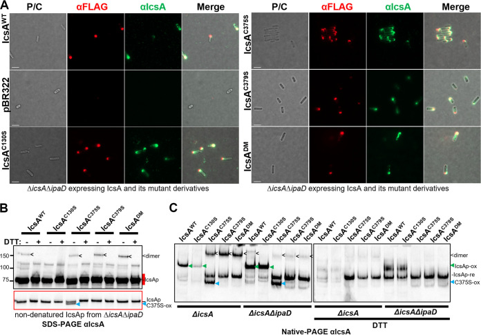
FIG 6 Effect of ΔipaD on IcsA conformation. (A) Indirect bacterial surface immunofluorescent labeling of FLAG-tagged IcsA passenger with anti-IcsA antibodies and anti-FLAG antibody. (B) Western immunoblotting of IcsAp recovered from the culture supernatant of indicated S. flexneri strains expressing IcsA and IcsA mutants in nondenatured conditions. Samples were solubilized in SDS-PAGE buffer with or without DTT and subjected to SDS-PAGE and Western immunoblotting without heating. The underexposure image in the red border correlates to the region in the long exposure image marked red. re: reduced, ox: oxidized, <: IcsAp dimer; blue arrow: oxidized IcsAC375S, C375S-ox. (C) IcsAp protein recovered as above and analyzed by Native-PAGE and detected with anti-IcsA antibodies. Green arrow: oxidized IcsAp, IcsAp-ox.
FIG 6 Effect of ΔipaD on IcsA conformation. (A) Indirect bacterial surface immunofluorescent labeling of FLAG-tagged IcsA passenger with anti-IcsA antibodies and anti-FLAG antibody. (B) Western immunoblotting of IcsAp recovered from the culture supernatant of indicated S. flexneri strains expressing IcsA and IcsA mutants in nondenatured conditions. Samples were solubilized in SDS-PAGE buffer with or without DTT and subjected to SDS-PAGE and Western immunoblotting without heating. The underexposure image in the red border correlates to the region in the long exposure image marked red. re: reduced, ox: oxidized, <: IcsAp dimer; blue arrow: oxidized IcsAC375S, C375S-ox. (C) IcsAp protein recovered as above and analyzed by Native-PAGE and detected with anti-IcsA antibodies. Green arrow: oxidized IcsAp, IcsAp-ox.CC BY · PubMed Central

Verification

Claims the paper makes

The claims and statistics of this paper haven’t been checked yet.

Run the check — free, no account needed. It reads the open-access text and checks the sources it cites.

A few free checks a day · papers already verified open instantly · results are public

Self-check

Percentages, sums and reported differences re-computed from the paper’s own numbers.

The self-check needs the paper’s full text.

Citations

Cites

Research works printed but not linked: 45

  1. 2018, Shigellosis. Lancet, 391, 801, 10.1016/S0140-6736(17)33296-8 · DOI · Paper page
  2. 10.1016/0092-8674(86)90880-9 · DOI
  3. 1993, Unipolar localization and ATPase activity of IcsA, a Shigella flexneri protein involved in intracellular movement, Infect Agents Dis, 2, 210
  4. 10.1128/IAI.63.9.3642-3648.1995 · DOI
  5. 2017, Deoxycholate-Enhanced Shigella Virulence Is Regulated by a Rare pi-Helix in the Type Three Secretion System Tip Protein IpaD, Biochemistry, 56, 6503, 10.1021/acs.biochem.7b00836 · DOI
  6. 10.1016/j.chom.2014.03.001 · DOI
  7. 10.1093/emboj/17.10.2767 · DOI
  8. 10.1126/science.1106036 · DOI · Paper page
  9. 10.1128/MMBR.68.4.692-744.2004 · DOI
  10. 10.1046/j.1365-2958.1997.4681827.x · DOI
  11. 2020, The virulence domain of Shigella IcsA contains a subregion with specific host cell adhesion function, PLoS One, 15, e0227425, 10.1371/journal.pone.0227425 · DOI
  12. 10.1128/JB.00093-08 · DOI
  13. 2013, Identification of Shigella flexneri IcsA residues affecting interaction with N-WASP, and evidence for IcsA-IcsA co-operative interaction, PLoS One, 8, e55152, 10.1371/journal.pone.0055152 · DOI
  14. 10.1099/mic.0.000165 · DOI
  15. 10.1128/JB.00790-10 · DOI
  16. 10.1016/j.jsb.2017.03.003 · DOI
  17. 10.1038/nrmicro2733 · DOI
  18. 10.1074/jbc.M111.306118 · DOI
  19. 10.1016/0378-1119(96)00343-5 · DOI
  20. 10.1074/jbc.270.52.30874 · DOI
  21. 10.1111/j.1365-2958.2007.05605.x · DOI
  22. 10.1111/j.1365-2958.1995.mmi_18020378.x · DOI
  23. 10.1046/j.1365-2958.1999.01571.x · DOI
  24. 10.1074/jbc.M112.388199 · DOI
  25. 10.1099/mic.0.28548-0 · DOI
  26. 10.1128/jb.171.12.6566-6572.1989 · DOI
  27. 10.1128/JB.183.3.951-958.2001 · DOI
  28. 2018, Extended cleavage specificity of human neutrophil elastase, human proteinase 3, and their distant ortholog clawed frog PR3–three elastases with similar primary but different extended specificities and stability, Front Immunol, 9, 2387, 10.3389/fimmu.2018.02387 · DOI
  29. 10.1099/mic.0.26141-0 · DOI
  30. 10.1074/jbc.M116.758003 · DOI
  31. 10.1046/j.1365-2958.2003.03489.x · DOI
  32. 10.1186/1471-2180-11-85 · DOI
  33. 10.1099/mic.0.056465-0 · DOI
  34. 10.1093/protein/14.11.939 · DOI
  35. 10.1021/ja962005v · DOI
  36. Shimanovich AA , Buskirk AD , Heine SJ , Blackwelder WC , Wahid R , Kotloff KL , Pasetti MF . 2017. Functional and Antigen-Specific Serum Antibody Levels as Correlates of Protection against Shigellosis in a Controlled Human Challenge Study. Clin Vaccine Immunol 24. · DOI
  37. 2021, Repertoire of Naturally Acquired Maternal Antibodies Transferred to Infants for Protection Against Shigellosis, Front Immunol, 12, 725129, 10.3389/fimmu.2021.725129 · DOI
  38. 1996, Lack of cleavage of IcsA in Shigella flexneri causes aberrant movement and allows demonstration of a cross-reactive eukaryotic protein, Infect Immun, 64, 511, 10.1128/iai.64.2.511-517.1996 · DOI
  39. 10.1016/0042-6822(55)90016-7 · DOI
  40. 10.1073/pnas.120163297 · DOI · Paper page
  41. 10.1016/S0378-1097(03)00217-9 · DOI
  42. 10.1016/j.jbbm.2005.12.008 · DOI · Paper page
  43. 10.1016/0014-5793(75)80272-9 · DOI · Paper page
  44. 10.1046/j.1365-2958.1997.2541625.x · DOI
  45. 10.1002/PRO.3943 · DOI · Paper page

Metadata

Authors
Qin, J., Hong, Y., Morona, R., & Totsika, M.
Affiliations
  • Queensland University of Technology (AU)
  • The University of Adelaide (AU)
Corresponding author
  • Qin, Jilong
  • Totsika, Makrina
Venue
Microbiology Spectrum
Publisher
openRxiv
Published
2022-07-22
Volume
10
Issue
6
Pages
e0341022-e0341022
ISSN
2165-0497 (ISSN-L)
OpenAlex
W4308957989
Cited by
6
References
1
Citations by year
2025: 1 · 2024: 2 · 2023: 2 · 2022: 1
FWCI
0.94
Type
journal article
Language
en
Open access
Licence
cc-by
Locations
  • Microbiology Spectrum (publishedVersion, cc-by)
  • bioRxiv (Cold Spring Harbor Laboratory) (acceptedVersion, cc-by-nc-nd)
  • PubMed Central (publishedVersion, cc-by)
  • PubMed (publishedVersion)
  • ResearchOnline - JCU (James Cook University) (submittedVersion)
  • Adelaide Research & Scholarship (AR&S) (University of Adelaide) (submittedVersion, other-oa)
  • DOAJ (DOAJ: Directory of Open Access Journals) (submittedVersion, cc-by-sa)
  • QUT ePrints (Queensland University of Technology) (submittedVersion)
Funders
  • Ramaciotti Foundations (2017HIG0119)
  • University of Adelaide
  • Department of Health and Aged Care, Australian Government (GNT1144046)
  • Medical Research Council
  • National Health and Medical Research Council (1144046)
Topics
  • Life Sciences › Immunology and Microbiology › Microbiology › Escherichia coli research studies
  • Life Sciences › Biochemistry, Genetics and Molecular Biology › Molecular Biology › Biochemical and Structural Characterization
  • Life Sciences › Biochemistry, Genetics and Molecular Biology › Genetics › Bacterial Genetics and Biotechnology
Keywords
Shigella flexneri, IcsA, autotransporters, shigellosis, cell adhesion, actin-based motility, protein conformation
MeSH terms
Type III Secretion Systems, Type V Secretion Systems, Actins, Bacterial Proteins, Cysteine, DNA-Binding Proteins, Shigella, Shigella flexneri, Transcription Factors, Adhesins, Bacterial, Virulence Factors

About these checks

Registry, retraction, Cites and statistics results are computed, not judged, with one exception: a printed reference that no index lists is read once by a model before it counts as a citation error. Claims are extracted by a model and kept only when their quote appears verbatim in the text; each cited source is then checked, with a model’s help, for whether it says what the claim relies on. Those verdicts can be wrong — read the claim’s quote and the cited source before relying on one.

Statistics and arithmetic are recomputed, not judged.

Confidence bands are shown only for fields calibrated on our annotated test set. None is calibrated yet, so no band is shown.

What counts as a claim ›How we measure confidence ›How we check this ›

Are you an author, or do you see a problem with this page? Request a review

Data sources

Bibliographic data: Semantic Scholar, OpenAlex, Crossref.

Contains information from the Semantic Scholar Academic Graph Datasets, made available under the ODC Attribution License (ODC-BY 1.0).

Semantic Scholar’s data is described in: Kinney, R., et al. (2023). The Semantic Scholar Open Data Platform. arXiv:2301.10140.