PreprintCited by 24Plant Virus Research StudiesEnzyme Structure and FunctionAdvanced Electron Microscopy Techniques and Applications

The DOI matches its Crossref record; 1 of the 1 references it cites were found in a registry or an index.

Nodavirus RNA Replication Crown Architecture Reveals Proto-Crown Precursor and Viral Protein A Conformational Switching

Open access

Preprint — published as 10.1073/pnas.2217412120

Abstract

Positive-strand RNA viruses replicate their genomes in virus-induced membrane vesicles, and the resulting RNA replication complexes are a major target for virus control. Nodavirus studies first revealed viral RNA replication proteins forming a 12-fold symmetric “crown” at the vesicle opening to the cytosol, an arrangement recently confirmed to extend to distantly related alphaviruses. Using cryo-electron microscopy (cryo-EM), we show that mature nodavirus crowns comprise two stacked 12-mer rings of multi-domain viral RNA replication protein A. Each ring contains an ~ 19 nm circle of C-proximal polymerase domains, differentiated by strikingly diverged positions of N-proximal RNA capping/membrane binding domains. The lower ring is a “proto-crown” precursor that assembles prior to RNA template recruitment, RNA synthesis and replication vesicle formation. In this proto-crown, the N-proximal segments interact to form a toroidal central floor, whose 3.1 Å resolution structure reveals many mechanistic details of the RNA capping/membrane binding domains. In the upper ring, cryo-EM fitting indicates that the N-proximal domains extend radially outside the polymerases, forming separated, membrane-binding “legs.” The polymerase and N-proximal domains are connected by a long linker accommodating the conformational switch between the two rings and possibly also polymerase movements associated with RNA synthesis and non-symmetric electron density in the lower center of mature crowns. The results reveal remarkable viral protein multifunctionality, conformational flexibility and evolutionary plasticity and new insights into (+)RNA virus replication and control. Significance: Positive-strand RNA viruses - including coronaviruses, alphaviruses, flaviviruses and many other medically and economically important pathogens - replicate their RNA genomes by virus-encoded machinery that has been poorly characterized. Using an advanced nodavirus model, we identify a major precursor in RNA replication complex assembly and show it to be a 12-mer ring of viral RNA replication protein A, whose single particle cryo-EM structure reveals functional features of its membrane interaction, assembly, polymerase and RNA capping domains. We further show that fully functional RNA replication complexes acquire a second 12-mer ring of protein A in alternate conformation atop the first, and a central density likely to represent another polymerase conformation. These findings provide strong foundations for understanding, controlling and beneficially using such viruses.

Overview

The claims and statistics of this paper haven’t been checked yet.

Run the check — free, no account needed. It reads the open-access text and checks the sources it cites.

A few free checks a day · papers already verified open instantly · results are public

Integrity signals

Registry

Crossref · 10.1101/2022.12.16.520638

Cites

1 of 1 references verified0 citation errors · 0 not checkable

Statistics

Figures

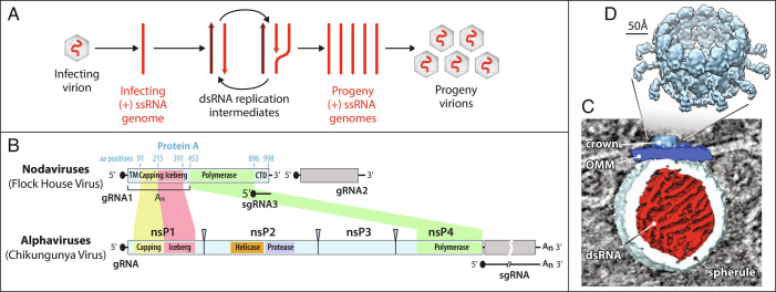
Fig. 1. Nodavirus genome replication overview. (A) General mechanism of RNA genome replication by (+) strand RNA viruses. Incoming (+) RNA is used as a template for (−) strand synthesis, yielding a dsRNA replicative intermediate that templates synthesis of many copies of (+) strand genomic RNA for packaging in progeny virions. (B) The bipartite FHV RNA genome encodes RNA replication protein A on RNA1 (3.1 kb) and the capsid protein precursor on RNA2 (1.4 kb). Subgenomic RNA3, transcribed from RNA1, encodes RNAi suppressor B2 and an overlapping ORF B1, comprising the C-terminal 102 aa of protein A. Aligning FHV protein A and the alphavirus RNA replication polyprotein highlights conserved RNA capping and polymerase domains and FHV’s lack of helicase and protease domains. The N-terminal half of protein A, denoted segment AN, and alphavirus nsP1 show particular parallels. (C) Cryo-ET imaging of a nodavirus RC spherule in infected Drosophila S2 cells with the mitochondrial outer membrane in dark blue, invaginated spherule membrane in white, interior spherule fibrils corresponding to viral dsRNA in red, and the protein A-containing crown complex in light blue. (D) Reconstruction of the 12-fold symmetric nodavirus RC crown by subtomogram averaging (2).
Fig. 1. Nodavirus genome replication overview. (A) General mechanism of RNA genome replication by (+) strand RNA viruses. Incoming (+) RNA is used as a template for (−) strand synthesis, yielding a dsRNA replicative intermediate that templates synthesis of many copies of (+) strand genomic RNA for packaging in progeny virions. (B) The bipartite FHV RNA genome encodes RNA replication protein A on RNA1 (3.1 kb) and the capsid protein precursor on RNA2 (1.4 kb). Subgenomic RNA3, transcribed from RNA1, encodes RNAi suppressor B2 and an overlapping ORF B1, comprising the C-terminal 102 aa of protein A. Aligning FHV protein A and the alphavirus RNA replication polyprotein highlights conserved RNA capping and polymerase domains and FHV’s lack of helicase and protease domains. The N-terminal half of protein A, denoted segment AN, and alphavirus nsP1 show particular parallels. (C) Cryo-ET imaging of a nodavirus RC spherule in infected Drosophila S2 cells with the mitochondrial outer membrane in dark blue, invaginated spherule membrane in white, interior spherule fibrils corresponding to viral dsRNA in red, and the protein A-containing crown complex in light blue. (D) Reconstruction of the 12-fold symmetric nodavirus RC crown by subtomogram averaging (2).CC BY-NC-ND · PubMed Central
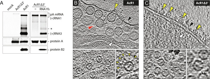
Fig. 2. Baculovirus-launched nodavirus RNA1 replication. (A) Northern and western blots of Nodavirus RNAs and proteins in Sf9 cells at 48 h post-infection with baculoviruses AcR1 or AcR1∆3′ at an m.o.i. of 20. AcR1 launches wt RNA1 that self-replicates through cis-recognition by its product replicase protein A and produces subgenomic RNA3 and its translation product protein B2. AcR1∆3′, lacking the RNA1 3′ noncoding region, fails to launch self-replicating RNA1, but its expressed protein A supports replication of a trans-supplied RNA1 derivative (RNA1fs) unable to produce protein A due to an N-proximal frameshift. The FHV RNA probe also detects the primary mRNA transcript expressed from the baculovirus DNA genome and a regularly-observed virus-specific RNA species roughly twice the size of sgRNA3 (asterisk). (B) Cryo-ET imaging shows that AcR1-launched wt FHV RNA1 induces active RNA RCs bearing spherule membrane vesicles and mature crowns, while (C) AcR1∆3′-expressed protein A, lacking a functional template RNA, forms crown-like structures on the OMM but no replication vesicles. Arrowheads: black = OMM, white = inner mitochondrial membrane, red = RC vesicle, yellow = crown-like structures. Higher magnification top views of the OMM show AcR1-induced crowns with leg-like extensions (yellow arrowheads), and “leg-less” and more tightly packed (white arrowheads) AcR1∆3′ crowns.
Fig. 2. Baculovirus-launched nodavirus RNA1 replication. (A) Northern and western blots of Nodavirus RNAs and proteins in Sf9 cells at 48 h post-infection with baculoviruses AcR1 or AcR1∆3′ at an m.o.i. of 20. AcR1 launches wt RNA1 that self-replicates through cis-recognition by its product replicase protein A and produces subgenomic RNA3 and its translation product protein B2. AcR1∆3′, lacking the RNA1 3′ noncoding region, fails to launch self-replicating RNA1, but its expressed protein A supports replication of a trans-supplied RNA1 derivative (RNA1fs) unable to produce protein A due to an N-proximal frameshift. The FHV RNA probe also detects the primary mRNA transcript expressed from the baculovirus DNA genome and a regularly-observed virus-specific RNA species roughly twice the size of sgRNA3 (asterisk). (B) Cryo-ET imaging shows that AcR1-launched wt FHV RNA1 induces active RNA RCs bearing spherule membrane vesicles and mature crowns, while (C) AcR1∆3′-expressed protein A, lacking a functional template RNA, forms crown-like structures on the OMM but no replication vesicles. Arrowheads: black = OMM, white = inner mitochondrial membrane, red = RC vesicle, yellow = crown-like structures. Higher magnification top views of the OMM show AcR1-induced crowns with leg-like extensions (yellow arrowheads), and “leg-less” and more tightly packed (white arrowheads) AcR1∆3′ crowns.CC BY-NC-ND · PubMed Central
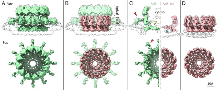
Fig. 3. Subtomogram averages of (A) the baculovirus AcR1-induced, RNA replication-active mature crown (green), and (D) the AcR1∆3′-induced proto-crown (pink). Upper images show side views superimposed on low-threshold density contours of the OMM, with top views below. The superposition in (B) and cross-sectional views in (C) show how the proto-crown corresponds directly to the floor and basal lobe sections of the mature crown.
Fig. 3. Subtomogram averages of (A) the baculovirus AcR1-induced, RNA replication-active mature crown (green), and (D) the AcR1∆3′-induced proto-crown (pink). Upper images show side views superimposed on low-threshold density contours of the OMM, with top views below. The superposition in (B) and cross-sectional views in (C) show how the proto-crown corresponds directly to the floor and basal lobe sections of the mature crown.CC BY-NC-ND · PubMed Central
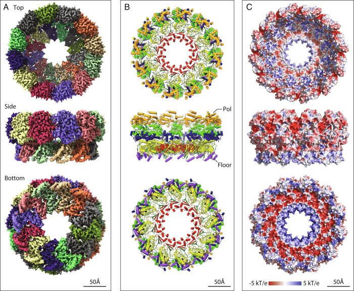
Fig. 4. Single particle cryo-EM proto-crown structure. Top, side, and bottom views of the C12 symmetric floor section in crowns from baculovirus AcR1-infected cells are shown merged with 12 copies of the Protein A Pol domain locally refined from the C12 proto-crown. (A) EM density maps with individually colored floor and basal lobe segments. (B) Ribbon diagrams of the protein A structure. The lower floor segments include domains for membrane-association (purple), capping (yellow), and central ring (red) and a functionally unassigned region (dark green). The upper Pol domain includes thumb (gold), palm (light green), and fingers (blue) domains. (C) Surface electrostatics calculated using APBS plugin in PyMol.
Fig. 4. Single particle cryo-EM proto-crown structure. Top, side, and bottom views of the C12 symmetric floor section in crowns from baculovirus AcR1-infected cells are shown merged with 12 copies of the Protein A Pol domain locally refined from the C12 proto-crown. (A) EM density maps with individually colored floor and basal lobe segments. (B) Ribbon diagrams of the protein A structure. The lower floor segments include domains for membrane-association (purple), capping (yellow), and central ring (red) and a functionally unassigned region (dark green). The upper Pol domain includes thumb (gold), palm (light green), and fingers (blue) domains. (C) Surface electrostatics calculated using APBS plugin in PyMol.CC BY-NC-ND · PubMed Central
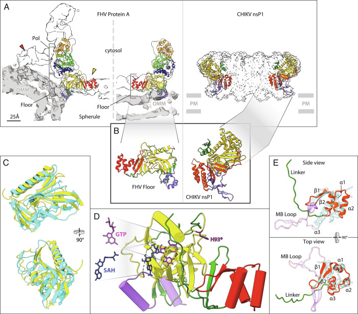
Fig. 5. Structure of the nodavirus protein A proto-crown and selected comparisons to alphavirus nsP1. These and other zoomed-in views in figures below were taken from the highest resolution protein A structure obtained, which was the baculovirus-expressed C11 ring, but as noted in the Results no significant variations were detected between C11 and C12 subunit structures. (A) Fit of the atomic build of the FHV proto-crown in central cross-sections of the cryo-ET densities of the mature replication crown and the proto-crown (Left side) and atomic build of the CHIKV nsP1 crown (Right side) (13). Overlaying light gray segmentations define the OMM for the FHV crowns; positioning of the CHIKV crown on the plasma membrane (PM) is inferred from the detergent belt in the published structure. Color coding is as defined in the legend to Fig. 4B. (B) Twelve copies of the N-terminal half of FHV protein A (segment AN, aa 1 to 452) form a flat floor in the mature crown and the proto-crown, while the equivalent CHIKV nsP1 forms a cone-shaped crown. (C) Superposition of the capping domains of FHV protein A (yellow) and CHIKV nsP1 (cyan). The close superposition of FHV and CHIKV capping and central channel domains and their distinct orientations in their respective crowns are further illustrated in Movie S3. (D) Positions of SAH and GTP in the capping domain (yellow) of the FHV proto-crown floor were estimated based on superposition of the FHV protein A structural elements on the published CHIKV nsP1-ligand interacting structures and fungal pathogen E. cuniculi. The SAM/SAH binding site is composed of R100, R104, N122, and V162, while D161, Y165, P186, W215, and R271 bind GTP. Note the 16 Å distance between the GTP α phosphate and GMP acceptor site H93 (dark purple). (E) Similarity of central ring domains in FHV protein A and CHIKV nsP1. Side- and top-views of the FHV protein A central ring domain (red) including an 18 aa linker loop (green), super-imposed on the CHIKV nsP1 RAMBO domain (cyan). Three central helical bundles (α1, α2, and α3) are sandwiched between two conserved upstream beta strands (β1 and β2) and the downstream polymerase linker in FHV protein A, whereas in CHIKV nsP1, the central helical bundles (α1, α2, and α3) are connected with two upstream beta strands (β1 and β2) followed by a membrane-binding loop (MB Loop, purple).
Fig. 5. Structure of the nodavirus protein A proto-crown and selected comparisons to alphavirus nsP1. These and other zoomed-in views in figures below were taken from the highest resolution protein A structure obtained, which was the baculovirus-expressed C11 ring, but as noted in the Results no significant variations were detected between C11 and C12 subunit structures. (A) Fit of the atomic build of the FHV proto-crown in central cross-sections of the cryo-ET densities of the mature replication crown and the proto-crown (Left side) and atomic build of the CHIKV nsP1 crown (Right side) (13). Overlaying light gray segmentations define the OMM for the FHV crowns; positioning of the CHIKV crown on the plasma membrane (PM) is inferred from the detergent belt in the published structure. Color coding is as defined in the legend to Fig. 4B. (B) Twelve copies of the N-terminal half of FHV protein A (segment AN, aa 1 to 452) form a flat floor in the mature crown and the proto-crown, while the equivalent CHIKV nsP1 forms a cone-shaped crown. (C) Superposition of the capping domains of FHV protein A (yellow) and CHIKV nsP1 (cyan). The close superposition of FHV and CHIKV capping and central channel domains and their distinct orientations in their respective crowns are further illustrated in Movie S3. (D) Positions of SAH and GTP in the capping domain (yellow) of the FHV proto-crown floor were estimated based on superposition of the FHV protein A structural elements on the published CHIKV nsP1-ligand interacting structures and fungal pathogen E. cuniculi. The SAM/SAH binding site is composed of R100, R104, N122, and V162, while D161, Y165, P186, W215, and R271 bind GTP. Note the 16 Å distance between the GTP α phosphate and GMP acceptor site H93 (dark purple). (E) Similarity of central ring domains in FHV protein A and CHIKV nsP1. Side- and top-views of the FHV protein A central ring domain (red) including an 18 aa linker loop (green), super-imposed on the CHIKV nsP1 RAMBO domain (cyan). Three central helical bundles (α1, α2, and α3) are sandwiched between two conserved upstream beta strands (β1 and β2) and the downstream polymerase linker in FHV protein A, whereas in CHIKV nsP1, the central helical bundles (α1, α2, and α3) are connected with two upstream beta strands (β1 and β2) followed by a membrane-binding loop (MB Loop, purple).CC BY-NC-ND · PubMed Central
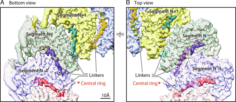
Fig. 6. N-end of the polymerase linker is interposed between adjacent floor segments. (A) Bottom and (B) top views of the crown floor density, showing the first 18 aa (dark shaded region of each colored segment) of the 74 aa linker (protein A aa 379 to 452) between each floor segment and its corresponding Pol domain. See main text for further details of the linker path under and between adjacent floor segments, rising to closely approach the Pol domain. Following these 18 aa, the remainder of the linker is too flexible to visualize by cryo-EM.
Fig. 6. N-end of the polymerase linker is interposed between adjacent floor segments. (A) Bottom and (B) top views of the crown floor density, showing the first 18 aa (dark shaded region of each colored segment) of the 74 aa linker (protein A aa 379 to 452) between each floor segment and its corresponding Pol domain. See main text for further details of the linker path under and between adjacent floor segments, rising to closely approach the Pol domain. Following these 18 aa, the remainder of the linker is too flexible to visualize by cryo-EM.CC BY-NC-ND · PubMed Central
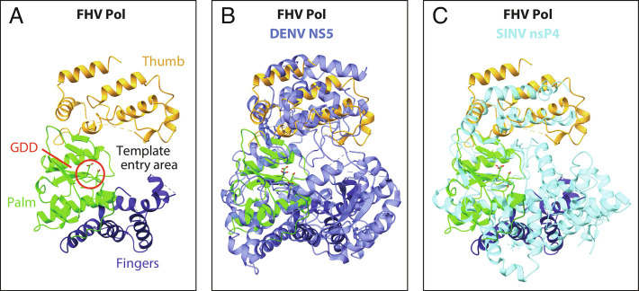
Fig. 7. Structural comparison of Pol domains of FHV protein A, Dengue virus NS5 (PDB 4C11), and Sindbis virus nsP4 (PDB 7VW5). (A) Annotated ribbon diagram of the FHV protein A polymerase, in a radial view from the crown’s central axis, showing the finger (dark blue), palm (light green), and thumb regions (gold), the catalytic site (GDD, circled in red) and the RNA template entry area. (B) Superposition of FHV protein A Pol and Dengue virus NS5 (blue). (C) Superposition of FHV protein A Pol and Sindbis virus nsP4 (cyan).
Fig. 7. Structural comparison of Pol domains of FHV protein A, Dengue virus NS5 (PDB 4C11), and Sindbis virus nsP4 (PDB 7VW5). (A) Annotated ribbon diagram of the FHV protein A polymerase, in a radial view from the crown’s central axis, showing the finger (dark blue), palm (light green), and thumb regions (gold), the catalytic site (GDD, circled in red) and the RNA template entry area. (B) Superposition of FHV protein A Pol and Dengue virus NS5 (blue). (C) Superposition of FHV protein A Pol and Sindbis virus nsP4 (cyan).CC BY-NC-ND · PubMed Central
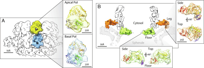
Fig. 8. Domain assignment in the upper mature crown. (A) Superposition of the Pol ribbon diagram with both lobes of the central turret of the mature crown cryo-ET density, consistent with independent identification of these apical (2) and basal (Figs. 3–5) lobes as stacked Pol domains. (B) As shown in Figs. 3–5 and matching the AN ribbon diagram superposition shown, the 12 floor segments (green) are the N-proximal AN portions of protein A (aa 1 to 452, Fig. 1B) that are contiguous with each of the 12 basal Pol domains that constitute protein A’s C-proximal portion. Similarly, as detailed further in the main text, the legs (orange) are singled out as the AN segments linked to the 12 basal Pol domains by numerous criteria, including their 12-fold repetition, position, volume, appropriately oriented membrane binding, the correspondingly good superposition of the AN ribbon diagram illustrated, and the absence of any other unassigned density.
Fig. 8. Domain assignment in the upper mature crown. (A) Superposition of the Pol ribbon diagram with both lobes of the central turret of the mature crown cryo-ET density, consistent with independent identification of these apical (2) and basal (Figs. 3–5) lobes as stacked Pol domains. (B) As shown in Figs. 3–5 and matching the AN ribbon diagram superposition shown, the 12 floor segments (green) are the N-proximal AN portions of protein A (aa 1 to 452, Fig. 1B) that are contiguous with each of the 12 basal Pol domains that constitute protein A’s C-proximal portion. Similarly, as detailed further in the main text, the legs (orange) are singled out as the AN segments linked to the 12 basal Pol domains by numerous criteria, including their 12-fold repetition, position, volume, appropriately oriented membrane binding, the correspondingly good superposition of the AN ribbon diagram illustrated, and the absence of any other unassigned density.CC BY-NC-ND · PubMed Central
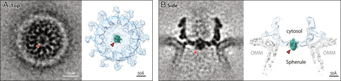
Fig. 9. Asymmetric density in the crown floor central channel. (A) Top and (B) side views of the cryo-ET density of the RNA replication-active mature crown from FHV-infected cells refined by subtomogram averaging without imposed symmetry. In both graded density (Left) and density-thresholded views (Right), red arrowheads point to an asymmetric density within the 7 nm diameter crown floor central channel, distinct from the floor segment densities of Figs. 3–6. This density (colored teal in the right side views) occupies a volume closely similar to that of the apical and basal polymerase lobes.
Fig. 9. Asymmetric density in the crown floor central channel. (A) Top and (B) side views of the cryo-ET density of the RNA replication-active mature crown from FHV-infected cells refined by subtomogram averaging without imposed symmetry. In both graded density (Left) and density-thresholded views (Right), red arrowheads point to an asymmetric density within the 7 nm diameter crown floor central channel, distinct from the floor segment densities of Figs. 3–6. This density (colored teal in the right side views) occupies a volume closely similar to that of the apical and basal polymerase lobes.CC BY-NC-ND · PubMed Central

Verification

Claims the paper makes

The claims and statistics of this paper haven’t been checked yet.

Run the check — free, no account needed. It reads the open-access text and checks the sources it cites.

A few free checks a day · papers already verified open instantly · results are public

Self-check

Percentages, sums and reported differences re-computed from the paper’s own numbers.

The self-check needs the paper’s full text.

Citations

Cites

Research works printed but not linked: 46

  1. M. Nishikiori , J. A. den Boon , N. Unchwaniwala , P. Ahlquist , Crowning Touches in Positive-Strand RNA Virus Genome Replication Complex Structure and Function. Annu. Rev. Virol. (2022) https://doi.org/10.1146/annurev-virology-092920-021307. · DOI
  2. 2008, Recent insights into the biology and biomedical applications of Flock House virus, Cell. Mol. Life Sci. CMLS, 65, 2675, 10.1007/s00018-008-8037-y · DOI
  3. 10.1073/pnas.93.18.9465 · DOI
  4. 10.1038/nature03870 · DOI
  5. 10.1186/s13062-015-0050-0 · DOI
  6. 10.3390/v10090483 · DOI
  7. 10.1371/journal.pbio.0050220 · DOI
  8. 10.1128/JVI.75.23.11664-11676.2001 · DOI
  9. 10.7554/eLife.25940 · DOI
  10. 10.1073/pnas.2006165117 · DOI
  11. 2022, Multifunctional Protein A is the Only Viral Protein Required for Nodavirus RNA Replication Crown Formation, Viruses, 14, 2711, 10.3390/v14122711 · DOI
  12. 2021, Cryo-electron microscopy of nodavirus RNA replication organelles illuminates positive-strand RNA virus genome replication, Curr. Opin. Virol, 51, 74, 10.1016/j.coviro.2021.09.008 · DOI
  13. 10.1038/S41586-020-3036-8 · DOI
  14. 2021, Structural insights into viral RNA capping and plasma membrane targeting by Chikungunya virus nonstructural protein 1, Cell Host Microbe, 29, 757, 10.1016/j.chom.2021.02.018 · DOI
  15. 10.1126/science.abd3629 · DOI
  16. 10.1126/science.abe0322 · DOI
  17. 10.1128/JVI.76.19.9856-9867.2002 · DOI
  18. 10.1128/JVI.02040-08 · DOI
  19. 10.1128/JVI.02267-06 · DOI
  20. 10.1128/JVI.01495-10 · DOI
  21. 10.1128/JVI.79.10.6239-6248.2005 · DOI
  22. 2021, UCSF ChimeraX: Structure visualization for researchers, educators, and developers, Protein Sci. Publ. Protein Soc, 30, 70, 10.1002/pro.3943 · DOI
  23. 10.1016/j.cell.2013.08.018 · DOI
  24. 10.7554/eLife.25478 · DOI
  25. 10.1074/jbc.RA119.010089 · DOI
  26. Y. Xie , et al., Cryo-EM structure of the yeast TREX complex and coordination with the SR-like protein Gbp2. eLife 10 (2021). · DOI
  27. 10.1371/journal.ppat.1004529 · DOI
  28. 2021, New Insights into Chikungunya Virus Infection and Pathogenesis, Annu. Rev. Virol, 8, 327, 10.1146/annurev-virology-091919-102021 · DOI
  29. 10.1038/nrm3920 · DOI
  30. 2022, Molecular basis of specific viral RNA recognition and 5’-end capping by the Chikungunya virus nsP1, Cell Rep, 40, 111133, 10.1016/j.celrep.2022.111133 · DOI
  31. 10.1016/S1097-2765(03)00522-7 · DOI
  32. 10.1016/j.jmb.2007.05.022 · DOI
  33. 2022, Crystal structures of alphavirus nonstructural protein 4 (nsP4) reveal an intrinsically dynamic RNA-dependent RNA polymerase fold, Nucleic Acids Res, 50, 1000, 10.1093/nar/gkab1302 · DOI
  34. 10.1073/pnas.1903562116 · DOI
  35. T. Grant , A. Rohou , N. Grigorieff , cisTEM, user-friendly software for single-particle image processing. eLife 7 (2018). · DOI
  36. 10.1093/nar/gkw357 · DOI
  37. 10.1529/biophysj.106.091561 · DOI
  38. Y. B. Tan , et al., Molecular Architecture of the Chikungunya Virus Replication Complex. bioRxiv, 2022.04.08.487651 (2022). · DOI
  39. 10.1128/JVI.00660-13 · DOI
  40. 10.1073/pnas.92.11.4892 · DOI
  41. 10.1016/j.chom.2009.03.007 · DOI
  42. 10.1128/JVI.00986-10 · DOI
  43. 10.1128/JVI.79.21.13747-13758.2005 · DOI
  44. 1990, Analysis of the role of brome mosaic virus 1a protein domains in RNA replication, using linker insertion mutagenesis, J. Virol, 64, 6110, 10.1128/jvi.64.12.6110-6120.1990 · DOI
  45. 10.1074/jbc.M910340199 · DOI
  46. 1993, RNA duplex unwinding activity of poliovirus RNA-dependent RNA polymerase 3Dpol, J. Virol, 67, 3010, 10.1128/jvi.67.6.3010-3018.1993 · DOI

Metadata

Authors
Zhan, H., Unchwaniwala, N., Rebolledo-Viveros, A., Pennington, J., Horswill, M., Broadberry, R., Myers, J., den Boon, J. A., Grant, T., & Ahlquist, P.
Affiliations
University of Wisconsin–Madison (US)
Corresponding author
  • Grant, Timothy
  • Ahlquist, Paul
Venue
Proceedings of the National Academy of Sciences
Publisher
openRxiv
Published
2022-12-17
Volume
120
Issue
5
Pages
e2217412120-e2217412120
ISSN
0027-8424 (ISSN-L)
OpenAlex
W4317851252
Cited by
24
References
1
Citations by year
2026: 8 · 2025: 3 · 2024: 6 · 2023: 7
FWCI
3.78
Type
journal article
Language
en
Open access
Licence
cc-by-nc-nd
Locations
  • Proceedings of the National Academy of Sciences (publishedVersion, cc-by-nc-nd)
  • bioRxiv (Cold Spring Harbor Laboratory) (acceptedVersion, cc-by-nc-nd)
  • PubMed Central (publishedVersion, cc-by-nc-nd)
  • PubMed (publishedVersion)
  • OSTI OAI (U.S. Department of Energy Office of Scientific and Technical Information) (submittedVersion)
Funders
  • National Science Foundation (U24GM129547)
  • U.S. Department of Energy (grid.436923.9, 436923.9)
  • Wisconsin Alumni Research Foundation
  • Oregon Health and Science University (grid.436923.9, 436923.9)
  • University of Wisconsin-Madison
  • National Institutes of Health
  • Office of Science (grid.436923.9)
  • National Institute of General Medical Sciences (T32GM130550, U24GM129547)
  • Biological and Environmental Research (grid.436923.9)
Topics
  • Life Sciences › Agricultural and Biological Sciences › Plant Science › Plant Virus Research Studies
  • Life Sciences › Biochemistry, Genetics and Molecular Biology › Structural Biology › Enzyme Structure and Function
  • Life Sciences › Biochemistry, Genetics and Molecular Biology › Structural Biology › Advanced Electron Microscopy Techniques and Applications
Keywords
nodavirus, RNA replication complex, crown architecture, 12-fold symmetry, conformational switching, membrane-binding domain, RNA polymerase, alphavirus, positive-strand RNA viruses, cryo-EM structure
MeSH terms
RNA Replication, RNA Viruses, RNA, Viral, Viral Proteins, Virus Replication, Cryoelectron Microscopy

About these checks

Registry, retraction, Cites and statistics results are computed, not judged, with one exception: a printed reference that no index lists is read once by a model before it counts as a citation error. Claims are extracted by a model and kept only when their quote appears verbatim in the text; each cited source is then checked, with a model’s help, for whether it says what the claim relies on. Those verdicts can be wrong — read the claim’s quote and the cited source before relying on one.

Statistics and arithmetic are recomputed, not judged.

Confidence bands are shown only for fields calibrated on our annotated test set. None is calibrated yet, so no band is shown.

What counts as a claim ›How we measure confidence ›How we check this ›

Are you an author, or do you see a problem with this page? Request a review