PreprintCited by 4Viral-associated cancers and disordersHerpesvirus Infections and TreatmentsGenomics and Chromatin Dynamics

The DOI matches its Crossref record; 1 of the 1 references it cites were found in a registry or an index.

Conserved cysteine residues in Kaposi’s sarcoma herpesvirus ORF34 are necessary for viral production and viral pre-initiation complex formation

Open access

Preprint — published as 10.1128/jvi.01000-24

Abstract

Kaposi’s sarcoma herpesvirus (KSHV) ORF34 plays a significant role as a component of the viral pre-initiation complex (vPIC), which is indispensable for late gene expression across beta and gamma herpesviruses. Although the key role of ORF34 within the vPIC and its function as a hub protein have been recognized, further clarification regarding its specific contribution to vPIC functionality and interactions with other components is required. This study employed a deep-learning algorithm-assisted structural model of ORF34, revealing highly conserved amino acid residues across human beta- and gamma-herpesviruses localized in structured domains. Thus, we engineered ORF34 alanine-scanning mutants by substituting conserved residues with alanine. These mutants were evaluated for their ability to interact with other vPIC factors and restore viral production in cells harboring the ORF34-deficient KSHV-BAC. Our experimental results highlight the crucial role of the 4 cysteine residues conserved in ORF34: a tetrahedral arrangement consisting of a pair of C-X n -C consensus motifs. This suggests the potential incorporation of metal cations in interacting with ORF24 and ORF66 vPIC components, facilitating late gene transcription, and promoting overall virus production by capturing metal cations. In summary, our findings underline the essential role of conserved cysteines in KSHV ORF34 for effective vPIC assembly and viral replication, thereby enhancing our understanding of the complex interplay between the vPIC components. IMPORTANCE: The initiation of late gene transcription is universally conserved across the gamma- and beta-herpesvirus families. This process employs a viral pre-initiation complex (vPIC), which is analogous to a cellular PIC. Although KSHV ORF34 is a critical factor for viral replication and is a component of the vPIC, the specifics of vPIC formation and the essential domains crucial for its function remain unclear. Structural predictions suggest that the 4 conserved cysteines (C170, C175, C256, and C259) form a tetrahedron that coordinates the metal cation. We further investigated the role of these conserved amino acids in interactions with other vPIC components, late gene expression, and virus production, to demonstrate for the first time that these cysteines are pivotal for these functions. This discovery not only deepens our comprehensive understanding of ORF34 and vPIC dynamics but also lays the groundwork for more detailed studies on herpesvirus replication mechanisms in future research.

Overview

The claims and statistics of this paper haven’t been checked yet.

Run the check — free, no account needed. It reads the open-access text and checks the sources it cites.

A few free checks a day · papers already verified open instantly · results are public

Integrity signals

Registry

Crossref · 10.1101/2023.03.08.531831

Cites

1 of 1 references verified0 citation errors · 0 not checkable

Statistics

Figures

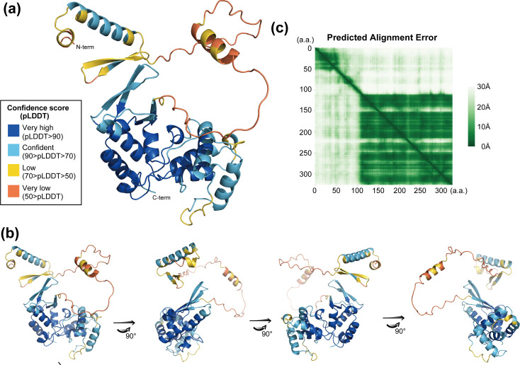
Fig 1 The ORF34 structure model predicted by AlphaFold2 and conserved cysteines on ORF34. The predicted ORF34 structure was visualized, and the predicted domain structure was analyzed. (a) The ORF34 structure was predicted using the deep learning algorithm AlphaFold2 (AF2). The illustration depicts the protein labeled with colors according to the pLDDT score of each amino acid residue. (b) Rotation of the predicted ORF34 structure model depicted in (a). (c) Prediction alignment error (PAE) plot of the predicted ORF34 structure. Each white-to-green pixel indicates a predicted distance of 30 to 0 Å, respectively, between amino acid residues.
Fig 1 The ORF34 structure model predicted by AlphaFold2 and conserved cysteines on ORF34. The predicted ORF34 structure was visualized, and the predicted domain structure was analyzed. (a) The ORF34 structure was predicted using the deep learning algorithm AlphaFold2 (AF2). The illustration depicts the protein labeled with colors according to the pLDDT score of each amino acid residue. (b) Rotation of the predicted ORF34 structure model depicted in (a). (c) Prediction alignment error (PAE) plot of the predicted ORF34 structure. Each white-to-green pixel indicates a predicted distance of 30 to 0 Å, respectively, between amino acid residues.CC BY · PubMed Central
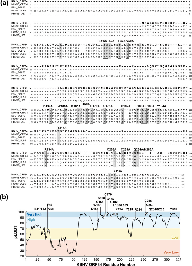
Fig 2 Conserved amino acid residues of ORF34 homolog and alanine-scanning mutants. Amino acid sequence alignment of KSHV ORF34. Herpesvirus homolog amino acid sequences were translated from nucleotide sequences found in the NCBI database [KSHV ORF34 (JSC-1-BAC16; accession number GQ994935), MHV68 ORF34 (strain WUMS; NC_001826), EBV BGLF3 (strain B95-8; V01555), CMV UL95 (strain Towne; FJ616285), and HHV6B U67 (strain japan-a1; KY239023)]. Raw data regarding alignment were obtained using Clustal Omega (EMBL-EBI; https://www.ebi.ac.uk/Tools/msa/clustalo/). Completely conserved amino acids between homologs are indicated by a gray background. Based on the alignment information, one to two conserved amino acid residues, except for proline and alanine, were substituted with alanine for ORF34 alanine-scanning mutants, as depicted. (b) The histogram of the pLDDT score is demonstrated by AF2 on each amino acid residue on ORF34 (1–327 aa), and the conserved amino acid residues are indicated with light-gray circles.
Fig 2 Conserved amino acid residues of ORF34 homolog and alanine-scanning mutants. Amino acid sequence alignment of KSHV ORF34. Herpesvirus homolog amino acid sequences were translated from nucleotide sequences found in the NCBI database [KSHV ORF34 (JSC-1-BAC16; accession number GQ994935), MHV68 ORF34 (strain WUMS; NC_001826), EBV BGLF3 (strain B95-8; V01555), CMV UL95 (strain Towne; FJ616285), and HHV6B U67 (strain japan-a1; KY239023)]. Raw data regarding alignment were obtained using Clustal Omega (EMBL-EBI; https://www.ebi.ac.uk/Tools/msa/clustalo/). Completely conserved amino acids between homologs are indicated by a gray background. Based on the alignment information, one to two conserved amino acid residues, except for proline and alanine, were substituted with alanine for ORF34 alanine-scanning mutants, as depicted. (b) The histogram of the pLDDT score is demonstrated by AF2 on each amino acid residue on ORF34 (1–327 aa), and the conserved amino acid residues are indicated with light-gray circles.CC BY · PubMed Central
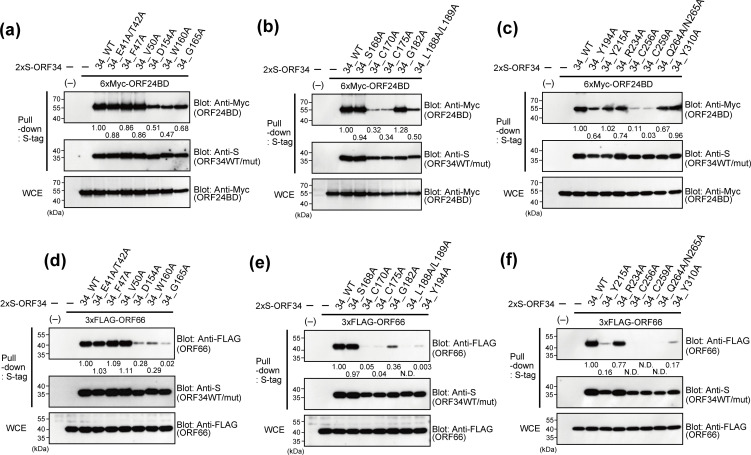
Fig 3 Physical interactions between ORF34 alanine-scanning mutants and vPIC components, ORF24 and ORF66. 293T cells were cotransfected with expression plasmids of the 2×S-ORF34 alanine-scanning mutant and 6×Myc-ORF24BD (a–c) or 3×FLAG-ORF66 (d–f). ORF24BD corresponds to the KSHV ORF24 N-terminal domain (1–400 aa) reported as having a domain interaction with ORF34 (28). Transfected cells were lysed, and cell lysates were subjected to pull-down assays using S-protein-agarose that captured the 2×S-ORF34 mutants. Obtained precipitates including 2×S-ORF34 mutants were probed with indicated antibodies to detect interactions. Whole-cell extracts (WCEs) were also probed as the input controls of prey proteins for each sample. The pull-down assay efficiency score of each interaction was indicated below the pull-down panel of prey proteins (6×Myc-ORF24BD or 3×FLAG-ORF66). The pull-down assay efficiency score of the sample co-transfected with ORF34 WT and ORF24BD/ORF66 was defined as 1.0. N.D. (not detected), indicating that a positive signal of the prey proteins was not detected.
Fig 3 Physical interactions between ORF34 alanine-scanning mutants and vPIC components, ORF24 and ORF66. 293T cells were cotransfected with expression plasmids of the 2×S-ORF34 alanine-scanning mutant and 6×Myc-ORF24BD (a–c) or 3×FLAG-ORF66 (d–f). ORF24BD corresponds to the KSHV ORF24 N-terminal domain (1–400 aa) reported as having a domain interaction with ORF34 (28). Transfected cells were lysed, and cell lysates were subjected to pull-down assays using S-protein-agarose that captured the 2×S-ORF34 mutants. Obtained precipitates including 2×S-ORF34 mutants were probed with indicated antibodies to detect interactions. Whole-cell extracts (WCEs) were also probed as the input controls of prey proteins for each sample. The pull-down assay efficiency score of each interaction was indicated below the pull-down panel of prey proteins (6×Myc-ORF24BD or 3×FLAG-ORF66). The pull-down assay efficiency score of the sample co-transfected with ORF34 WT and ORF24BD/ORF66 was defined as 1.0. N.D. (not detected), indicating that a positive signal of the prey proteins was not detected.CC BY · PubMed Central
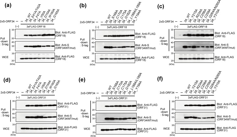
Fig 4 Physical interaction between ORF34 alanine-scanning mutants and vPIC components, ORF18 and ORF31. 293T cells were cotransfected with expression plasmids of the 2×S-ORF34 alanine-scanning mutant and 3×FLAG-ORF18 (a–c) or 3×FLAG-ORF31 (d–f). Transfected cells were lysed, and cell lysates were subjected to pull-down assays using S-protein-agarose that captured the 2×S-ORF34 mutants. Obtained precipitates including 2×S-ORF34 mutants were probed with the indicated antibodies to detect interactions. Whole-cell extracts (WCEs) were also probed as the input controls of prey proteins for each sample.
Fig 4 Physical interaction between ORF34 alanine-scanning mutants and vPIC components, ORF18 and ORF31. 293T cells were cotransfected with expression plasmids of the 2×S-ORF34 alanine-scanning mutant and 3×FLAG-ORF18 (a–c) or 3×FLAG-ORF31 (d–f). Transfected cells were lysed, and cell lysates were subjected to pull-down assays using S-protein-agarose that captured the 2×S-ORF34 mutants. Obtained precipitates including 2×S-ORF34 mutants were probed with the indicated antibodies to detect interactions. Whole-cell extracts (WCEs) were also probed as the input controls of prey proteins for each sample.CC BY · PubMed Central
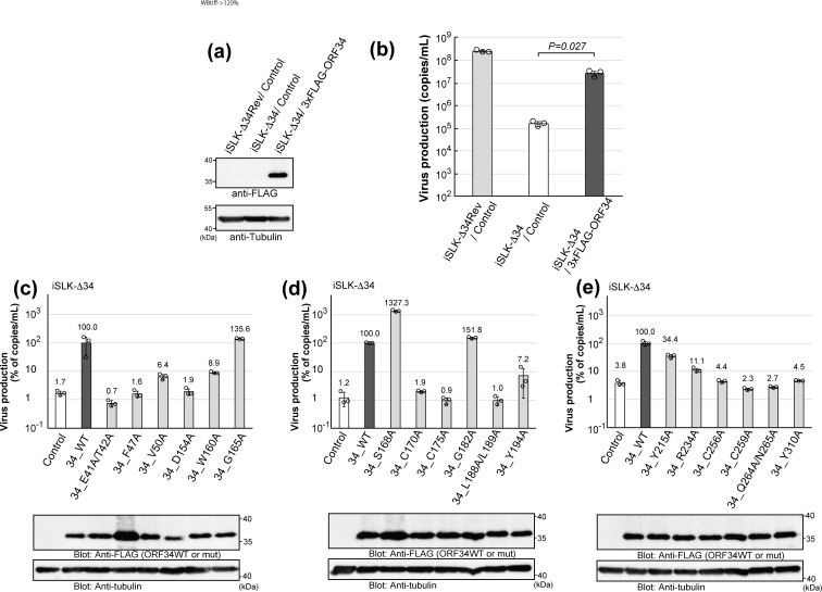
Fig 5 Complement abilities of ORF34 alanine-scanning mutants on viral production. Establishment of 3×FLAG-tagged ORF34-expressing iSLK-Δ34 stable cell lines and confirmation of the recovery by ORF34 WT in ΔORF34-virus production. (a) The Western blot showed exogenous 3×FLAG-tagged ORF34 WT expression in iSLK-Δ34 cells. (b) A complementation assay was performed to rescue virus production. (c–e) 3×FLAG-tagged ORF34 alanine-scanning mutant-expressing iSLK-Δ34 stable cell lines were investigated for their virus production recoveries, with results shown in the upper graph panels. The lower blot panels indicate the exogenous ORF34 WT/mutant expression under normal culture conditions. In the upper graph panels, the virus production of iSLK-Δ34/ORF34 WT cells was defined as 100%. Each stable iSLK-KSHV BAC-harboring cell line was activated by NaB (0.75 mM) and Dox (4 µg/mL) and cultured for 3 days, and the culture supernatant containing the encapsidated KSHV genome was harvested and quantified. Three independent samples were evaluated by real-time PCR and indicated as dots in each column. The error bars indicate standard deviations.
Fig 5 Complement abilities of ORF34 alanine-scanning mutants on viral production. Establishment of 3×FLAG-tagged ORF34-expressing iSLK-Δ34 stable cell lines and confirmation of the recovery by ORF34 WT in ΔORF34-virus production. (a) The Western blot showed exogenous 3×FLAG-tagged ORF34 WT expression in iSLK-Δ34 cells. (b) A complementation assay was performed to rescue virus production. (c–e) 3×FLAG-tagged ORF34 alanine-scanning mutant-expressing iSLK-Δ34 stable cell lines were investigated for their virus production recoveries, with results shown in the upper graph panels. The lower blot panels indicate the exogenous ORF34 WT/mutant expression under normal culture conditions. In the upper graph panels, the virus production of iSLK-Δ34/ORF34 WT cells was defined as 100%. Each stable iSLK-KSHV BAC-harboring cell line was activated by NaB (0.75 mM) and Dox (4 µg/mL) and cultured for 3 days, and the culture supernatant containing the encapsidated KSHV genome was harvested and quantified. Three independent samples were evaluated by real-time PCR and indicated as dots in each column. The error bars indicate standard deviations.CC BY · PubMed Central
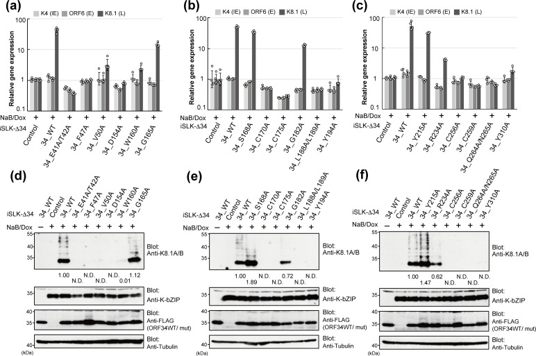
Fig 6 Complement abilities of ORF34 alanine-scanning mutants for viral gene transcription and protein expression. The mRNA and protein expression in lytic-induced iSLK-Δ34 cells complemented with ORF34 WT/alanine-scanning mutants. The cells were cultured for 3 days in the conditioned medium containing NaB and Dox to induce a lytic state. (a–c) Total RNA was extracted from cells and subjected to RT-qPCR. The mRNA expression of viral genes, which is representative of each step (K4 as IE gene, ORF6 as E gene, and K8.1 as L-gene), was normalized by GAPDH expression. The mRNA expression of iSLK-ΔORF34/control cells with NaB and Dox treatment was defined as 1.0. Four independent samples were evaluated and indicated as dots in each column. The error bars indicate standard deviations. (d–e) Total protein samples were prepared from each lytic-induced cell and subjected to Western blotting. The protein expression of the L-gene was analyzed by K8.1A/B blotting and the E gene by K-bZIP. The protein expression score, the band intensity, of iSLK-Δ34/ORF34 WT was defined as 1.0 N.D. (not detected), indicating that a positive signal of viral proteins was not detected.
Fig 6 Complement abilities of ORF34 alanine-scanning mutants for viral gene transcription and protein expression. The mRNA and protein expression in lytic-induced iSLK-Δ34 cells complemented with ORF34 WT/alanine-scanning mutants. The cells were cultured for 3 days in the conditioned medium containing NaB and Dox to induce a lytic state. (a–c) Total RNA was extracted from cells and subjected to RT-qPCR. The mRNA expression of viral genes, which is representative of each step (K4 as IE gene, ORF6 as E gene, and K8.1 as L-gene), was normalized by GAPDH expression. The mRNA expression of iSLK-ΔORF34/control cells with NaB and Dox treatment was defined as 1.0. Four independent samples were evaluated and indicated as dots in each column. The error bars indicate standard deviations. (d–e) Total protein samples were prepared from each lytic-induced cell and subjected to Western blotting. The protein expression of the L-gene was analyzed by K8.1A/B blotting and the E gene by K-bZIP. The protein expression score, the band intensity, of iSLK-Δ34/ORF34 WT was defined as 1.0 N.D. (not detected), indicating that a positive signal of viral proteins was not detected.CC BY · PubMed Central
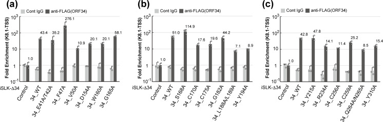
Fig 7 Recruitment of ORF34 alanine-scanning mutants to the ate gene transcription start site. The binding ability of the KSHV late gene TSS to ORF34 WT/alanine-scanning mutants was evaluated by ChIP. The iSLK-Δ34 cells complemented with ORF34 WT/alanine-scanning mutants were cultured for 3 days in the conditioned medium containing NaB and Dox to induce a lytic state. (a–c) A KSHV DNA fragment precipitated by control mouse IgG or anti-FLAG monoclonal antibody was subjected to RT-qPCR and normalized by input DNA subjected to ChIP. Each fold enrichment of iSLK-ΔORF34/control cells by control IgG or anti-FLAG antibodies was defined as 1.0. Three independent samples were evaluated and indicated as dots in each column. The error bars indicate standard deviation.
Fig 7 Recruitment of ORF34 alanine-scanning mutants to the ate gene transcription start site. The binding ability of the KSHV late gene TSS to ORF34 WT/alanine-scanning mutants was evaluated by ChIP. The iSLK-Δ34 cells complemented with ORF34 WT/alanine-scanning mutants were cultured for 3 days in the conditioned medium containing NaB and Dox to induce a lytic state. (a–c) A KSHV DNA fragment precipitated by control mouse IgG or anti-FLAG monoclonal antibody was subjected to RT-qPCR and normalized by input DNA subjected to ChIP. Each fold enrichment of iSLK-ΔORF34/control cells by control IgG or anti-FLAG antibodies was defined as 1.0. Three independent samples were evaluated and indicated as dots in each column. The error bars indicate standard deviation.CC BY · PubMed Central
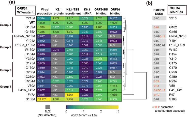
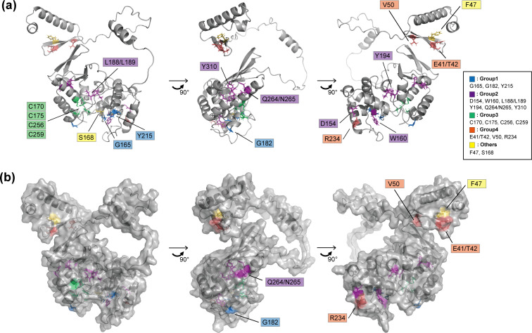
Fig 8 Summary of the effects of ORF34 alanine-scanning mutants on vPIC function. (a) The results (Fig. 5 to 7) were summarized and visualized on the heatmap with a dendrogram, highlighting the hierarchical clustering. Each score of ORF34 WT or iSLK-Δ34/WT was defined as 1.0. The scores are indicated on each panel with the color gradient (dark blue [0.0], green [1.0], and yellow [>3.0]) as indicated by the lower bar. N.D. (not detected) in each experiment is indicated by the gray panel. Groups were classified according to the visualized dendrogram. (b) Based on the ORF34 structure model (Fig. 1a), the prediction scores of protein surface exposure, relative solvent-accessible surface area (relative SASA), were listed for each mutated site. The relative SASA score colored with orange [>0.1] indicates that the amino acid residue has a high possibility of being exposed on the protein surface.
Fig 8 Summary of the effects of ORF34 alanine-scanning mutants on vPIC function. (a) The results (Fig. 5 to 7) were summarized and visualized on the heatmap with a dendrogram, highlighting the hierarchical clustering. Each score of ORF34 WT or iSLK-Δ34/WT was defined as 1.0. The scores are indicated on each panel with the color gradient (dark blue [0.0], green [1.0], and yellow [>3.0]) as indicated by the lower bar. N.D. (not detected) in each experiment is indicated by the gray panel. Groups were classified according to the visualized dendrogram. (b) Based on the ORF34 structure model (Fig. 1a), the prediction scores of protein surface exposure, relative solvent-accessible surface area (relative SASA), were listed for each mutated site. The relative SASA score colored with orange [>0.1] indicates that the amino acid residue has a high possibility of being exposed on the protein surface.CC BY · PubMed Central
Fig 9 Overview of the predicted ORF34 structure and mutated residues. Rotated ORF34 structures with caption based on the predicted structure (Fig. 1a and b) (a) In the illustration model, all the mutated amino acid residues are highlighted with colors according to the grouping (Fig. 8a) and captioned. (b) In the surface illustration model based on (a), only the mutated amino acid residues expected to be exposed on the protein surface (Fig. 8b) are captioned.
Fig 9 Overview of the predicted ORF34 structure and mutated residues. Rotated ORF34 structures with caption based on the predicted structure (Fig. 1a and b) (a) In the illustration model, all the mutated amino acid residues are highlighted with colors according to the grouping (Fig. 8a) and captioned. (b) In the surface illustration model based on (a), only the mutated amino acid residues expected to be exposed on the protein surface (Fig. 8b) are captioned.CC BY · PubMed Central
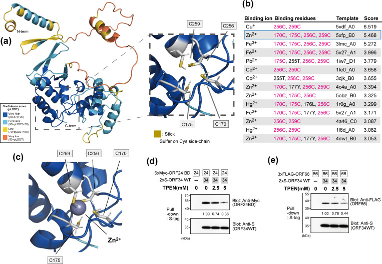
Fig 10 The prediction of metal cation binding to ORF34 and its influence on the binding ability of ORF34. (a) The table organizes the binding residues, templates, and binding scores of 18 different metal ions predicted by the metal ion-binding site prediction and modeling server, MIB2. When multiple copies of identical binding residues were present for a single metal ion, the template with the highest binding score was noted. If any new binding residues were present among previously recorded ones, the template was still recorded. Prediction binding scores higher than 3.0 are listed. Conserved cysteine residues (C170, C175, C256, and C256) were highlighted with magenta. (b) Predicted model of ORF34 structure binding to ions. The illustration model of ORF34 was visualized according to Fig. 7a (right panel), and the binding zinc ion (Zn2+) is shown as a gray sphere. Heavy metal cation chelation influences the binding abilities of ORF34 to ORF24BD (c) and ORF66 (d). Pull-down assays using S-tagged ORF34 binding beads as bait in the presence of cell extract overexpressed prey proteins (6×Myc-ORF24BD or 3×FLAG-ORF66) and the heavy metal chelator TPEN. The pull-down efficiency score is indicated below the pull-down panel of the prey protein. The score of the sample coincubated with 2×S-ORF34 WT binding beads as bait and 6×Myc-ORF24BD or 3×FLAG-ORF66 as prey was defined as 1.0.
Fig 10 The prediction of metal cation binding to ORF34 and its influence on the binding ability of ORF34. (a) The table organizes the binding residues, templates, and binding scores of 18 different metal ions predicted by the metal ion-binding site prediction and modeling server, MIB2. When multiple copies of identical binding residues were present for a single metal ion, the template with the highest binding score was noted. If any new binding residues were present among previously recorded ones, the template was still recorded. Prediction binding scores higher than 3.0 are listed. Conserved cysteine residues (C170, C175, C256, and C256) were highlighted with magenta. (b) Predicted model of ORF34 structure binding to ions. The illustration model of ORF34 was visualized according to Fig. 7a (right panel), and the binding zinc ion (Zn2+) is shown as a gray sphere. Heavy metal cation chelation influences the binding abilities of ORF34 to ORF24BD (c) and ORF66 (d). Pull-down assays using S-tagged ORF34 binding beads as bait in the presence of cell extract overexpressed prey proteins (6×Myc-ORF24BD or 3×FLAG-ORF66) and the heavy metal chelator TPEN. The pull-down efficiency score is indicated below the pull-down panel of the prey protein. The score of the sample coincubated with 2×S-ORF34 WT binding beads as bait and 6×Myc-ORF24BD or 3×FLAG-ORF66 as prey was defined as 1.0.CC BY · PubMed Central

Verification

Claims the paper makes

The claims and statistics of this paper haven’t been checked yet.

Run the check — free, no account needed. It reads the open-access text and checks the sources it cites.

A few free checks a day · papers already verified open instantly · results are public

Self-check

Percentages, sums and reported differences re-computed from the paper’s own numbers.

The self-check needs the paper’s full text.

Citations

Cites

Research works printed but not linked: 54

  1. Pellett PE , Davison, A.J. , Eberle, R. , Ehlers, B. , Hayward, G.S. , Lacoste, V. , Minson, A.C. , Nicholas, J. , Roizman, B. , Studdert, M.J. and Wang, F. 2011. Order Herpesvirales . In King A , Lefkowitz E. , Adams, M.J. , Carstens, E.B. (ed), Virus Taxonomy: Ninth Report of the International Committee on Taxonomy of Viruses. Elsevier.
  2. Pellett PE , Davison, A.J. , Eberle, R. , Ehlers, B. , Hayward, G.S. , Lacoste, V. , Minson, A.C. , Nicholas, J. , Roizman, B. , Studdert, M.J. , Wang, F. 2011. Family Herpesviridae , p 81-92. In King A , Lefkowitz E. , Adams, M.J. , Carstens, E.B. (ed), Virus Taxonomy: Ninth Report of the International Committee on Taxonomy of Viruses. Elsevier.
  3. 2022, Fields Virology, 2, 212
  4. 10.1111/febs.16206 · DOI
  5. 2022, Fields Virology, 2, 324
  6. 2022, Fields Virology, 2, 513
  7. 2019, Kaposi sarcoma, Nat Rev Dis Primers, 5, 9, 10.1038/s41572-019-0060-9 · DOI
  8. 10.1182/blood.2020005470 · DOI
  9. 10.1016/j.canlet.2009.07.004 · DOI
  10. 10.1371/journal.ppat.1003847 · DOI
  11. 10.1128/JVI.76.10.5000-5013.2002 · DOI
  12. 10.1128/JVI.76.7.3168-3178.2002 · DOI
  13. 10.1073/pnas.0408192102 · DOI
  14. 10.1073/pnas.1816183115 · DOI
  15. 10.1172/JCI136225 · DOI
  16. 10.1371/journal.ppat.1000935 · DOI
  17. 10.1371/journal.ppat.1001013 · DOI
  18. 10.3390/v5051346 · DOI
  19. 10.1128/JVI.75.2.891-902.2001 · DOI
  20. 10.3389/fmicb.2012.00030 · DOI
  21. 10.1016/s0065-230x(06)97004-3 · DOI
  22. 10.1073/pnas.95.18.10866 · DOI
  23. 10.1007/82_2017_31 · DOI
  24. 10.1016/j.semcdb.2022.12.001 · DOI
  25. 10.1371/journal.ppat.1007774 · DOI
  26. Gruffat H , Marchione R , Manet E . 2016. Herpesvirus Late Gene Expression: A Viral-Specific Pre-initiation Complex Is Key. Front Microbiol 7. · DOI
  27. 10.1038/s41598-017-00401-7 · DOI
  28. 10.1128/jvi.02157-15 · DOI
  29. 10.1128/JVI.00272-15 · DOI
  30. Watanabe T , Nishimura M , Izumi T , Kuriyama K , Iwaisako Y , Hosokawa K , Takaori-Kondo A , Fujimuro M . 2020. Kaposi’s Sarcoma-Associated Herpesvirus ORF66 Is Essential for Late Gene Expression and Virus Production via Interaction with ORF34. J Virol 94. · DOI
  31. Didychuk AL , Castaneda AF , Kushnir LO , Huang CJ , Glaunsinger BA . 2020. Conserved CxnC Motifs in Kaposi’s Sarcoma-Associated Herpesvirus ORF66 Are Required for Viral Late Gene Expression and Are Essential for Its Interaction with ORF34. J Virol 94. · DOI
  32. 10.1128/JVI.00793-14 · DOI
  33. 2019, The Interaction between ORF18 and ORF30 Is Required for Late Gene Expression in Kaposi’s Sarcoma-Associated Herpesvirus, J Virol, 93, e01488
  34. 10.1016/j.molcel.2014.11.026 · DOI
  35. 2022, Biomolecular Fluorescence Complementation Profiling and Artificial Intelligence Structure Prediction of the Kaposi’s Sarcoma-Associated Herpesvirus ORF18 and ORF30 Interaction, Int J Mol Sci, 23, 9647, 10.3390/ijms23179647 · DOI
  36. 2022, MIB2: metal ion-binding site prediction and modeling server, Bioinformatics, 38, 4428, 10.1093/bioinformatics/btac534 · DOI
  37. 2022, Identification of Iron-Sulfur (Fe-S) Cluster and Zinc (Zn) Binding Sites Within Proteomes Predicted by DeepMind’s AlphaFold2 Program Dramatically Expands the Metalloproteome, J Mol Biol, 434, 167377, 10.1016/j.jmb.2021.167377 · DOI · Paper page
  38. 10.1074/jbc.M404722200 · DOI
  39. 10.1110/ps.9.9.1743 · DOI
  40. 10.1038/365277a0 · DOI
  41. 10.1016/S0969-2126(98)00058-6 · DOI
  42. 10.1093/jb/mvi138 · DOI
  43. 10.1042/bj20031706 · DOI
  44. 10.1093/nar/gkm363 · DOI
  45. 10.1093/bioinformatics/btu744 · DOI
  46. 10.1371/journal.ppat.1007980 · DOI
  47. Sato Y , Watanabe T , Suzuki C , Abe Y , Masud H , Inagaki T , Yoshida M , Suzuki T , Goshima F , Adachi J , Tomonaga T , Murata T , Kimura H . 2019. S-Like-Phase Cyclin-Dependent Kinases Stabilize the Epstein-Barr Virus BDLF4 Protein To Temporally Control Late Gene Transcription. J Virol 93. · DOI
  48. 10.1371/journal.pone.0039252 · DOI
  49. 10.1016/j.bbrc.2014.01.017 · DOI
  50. 10.1016/j.bbrc.2014.07.068 · DOI
  51. 10.1128/JVI.76.12.6213-6223.2002 · DOI
  52. 2017, Quantitative Analysis of the KSHV Transcriptome Following Primary Infection of Blood and Lymphatic Endothelial Cells, 6, 11
  53. 10.1242/jcs.168740 · DOI
  54. 10.1080/10618600.2018.1473780 · DOI

Metadata

Authors
Watanabe, T., McGraw, A., Narayan, K., Tibebe, H., Kuriyama, K., Nishimura, M., Izumi, T., Fujimuro, M., & Ohno, S.
Affiliations
  • University of the Ryukyus (JP)
  • American University (US)
  • Kyoto Pharmaceutical University (JP)
Corresponding author
  • Watanabe, Tadashi
  • Ohno, Shinji
Venue
Journal of Virology
Publisher
openRxiv
Published
2023-03-09
Volume
98
Issue
8
Pages
e0100024-e0100024
ISSN
0022-538X (ISSN-L)
OpenAlex
W4401117881
Cited by
4
References
1
Citations by year
2025: 4
FWCI
0.68
Type
journal article
Language
en
Open access
Licence
cc-by
Locations
  • Journal of Virology (publishedVersion, cc-by)
  • bioRxiv (Cold Spring Harbor Laboratory) (acceptedVersion, cc-by-nc-nd)
  • PubMed Central (publishedVersion, cc-by)
  • PubMed (publishedVersion)
  • PubMed Central (submittedVersion, cc-by-nc-nd)
  • Institutional Repositories DataBase (IRDB) (submittedVersion, cc-by-nc-nd)
  • University oh the Ryukyus Repository (submittedVersion, cc-by-nc-nd)
Funders
  • Northwestern University
  • Cleveland Clinic
  • Takeda Science Foundation
  • American University
  • Ministry of Education, Culture, Sports, Science and Technology (JP21K08509, JP19K07669)
  • University of the Ryukyus (20SP04102)
  • Yakult Honsha
  • National Institutes of Health
  • Center for AIDS Research, University of Washington (P30 AI117970)
  • Japan Society for the Promotion of Science (19K07669, 21K08509)
  • National Institute on Aging
  • National Heart, Lung, and Blood Institute
  • National Institute of Mental Health
  • National Institute on Drug Abuse
  • National Cancer Institute
  • National Institute of Nursing Research
  • National Institute of Allergy and Infectious Diseases (R15AI172610, P30AI117970)
  • National Institute of Diabetes and Digestive and Kidney Diseases
  • National Institute of Dental and Craniofacial Research
  • National Institute on Minority Health and Health Disparities
Topics
  • Health Sciences › Medicine › Oncology › Viral-associated cancers and disorders
  • Health Sciences › Medicine › Infectious Diseases › Herpesvirus Infections and Treatments
  • Life Sciences › Biochemistry, Genetics and Molecular Biology › Molecular Biology › Genomics and Chromatin Dynamics
Keywords
Kaposi's sarcoma-associated herpesvirus, conserved cysteine residues, metal cation coordination, late gene transcription, viral production, viral replication

About these checks

Registry, retraction, Cites and statistics results are computed, not judged, with one exception: a printed reference that no index lists is read once by a model before it counts as a citation error. Claims are extracted by a model and kept only when their quote appears verbatim in the text; each cited source is then checked, with a model’s help, for whether it says what the claim relies on. Those verdicts can be wrong — read the claim’s quote and the cited source before relying on one.

Statistics and arithmetic are recomputed, not judged.

Confidence bands are shown only for fields calibrated on our annotated test set. None is calibrated yet, so no band is shown.

What counts as a claim ›How we measure confidence ›How we check this ›

Are you an author, or do you see a problem with this page? Request a review