PreprintCited by 16RNA and protein synthesis mechanismsCellular transport and secretionAdvanced Electron Microscopy Techniques and Applications

The DOI matches its Crossref record; 1 of the 1 references it cites were found in a registry or an index.

Exploring the molecular composition of the multipass translocon in its native membrane environment

Open access
Gemmer, Max, Chaillet, Marten L., Förster, Friedrich·2023·Life Science Alliance

Preprint — published as 10.26508/lsa.202302496

Abstract

Multispanning membrane proteins are inserted into the endoplasmic reticulum membrane by the ribosome-bound multipass translocon machinery. Based on cryo-electron tomography and extensive subtomogram analysis, we reveal the composition and arrangement of multipass translocon components in their native membrane environment. The intramembrane chaperone complex PAT and the translocon associated protein (TRAP) complex associate substoichiometrically with the multipass translocon in a translation-dependent manner. While PAT is preferentially recruited to active complexes, TRAP primarily associates with inactive translocons. The subtomogram average of the TRAP-multipass translocon reveals intermolecular contacts between the luminal domains of TRAP and an unknown subunit of the BOS complex. AlphaFold modeling suggests this protein is NOMO, bridging the luminal domains of nicalin and TRAPα. Collectively, our results visualize the interplay of accessory factors associated with multipass membrane protein biogenesis under near-native conditions.

Overview

The claims and statistics of this paper haven’t been checked yet.

Run the check — free, no account needed. It reads the open-access text and checks the sources it cites.

A few free checks a day · papers already verified open instantly · results are public

Integrity signals

Registry

Crossref · 10.1101/2023.11.28.569136

Cites

1 of 1 references verified0 citation errors · 0 not checkable

Statistics

Figures

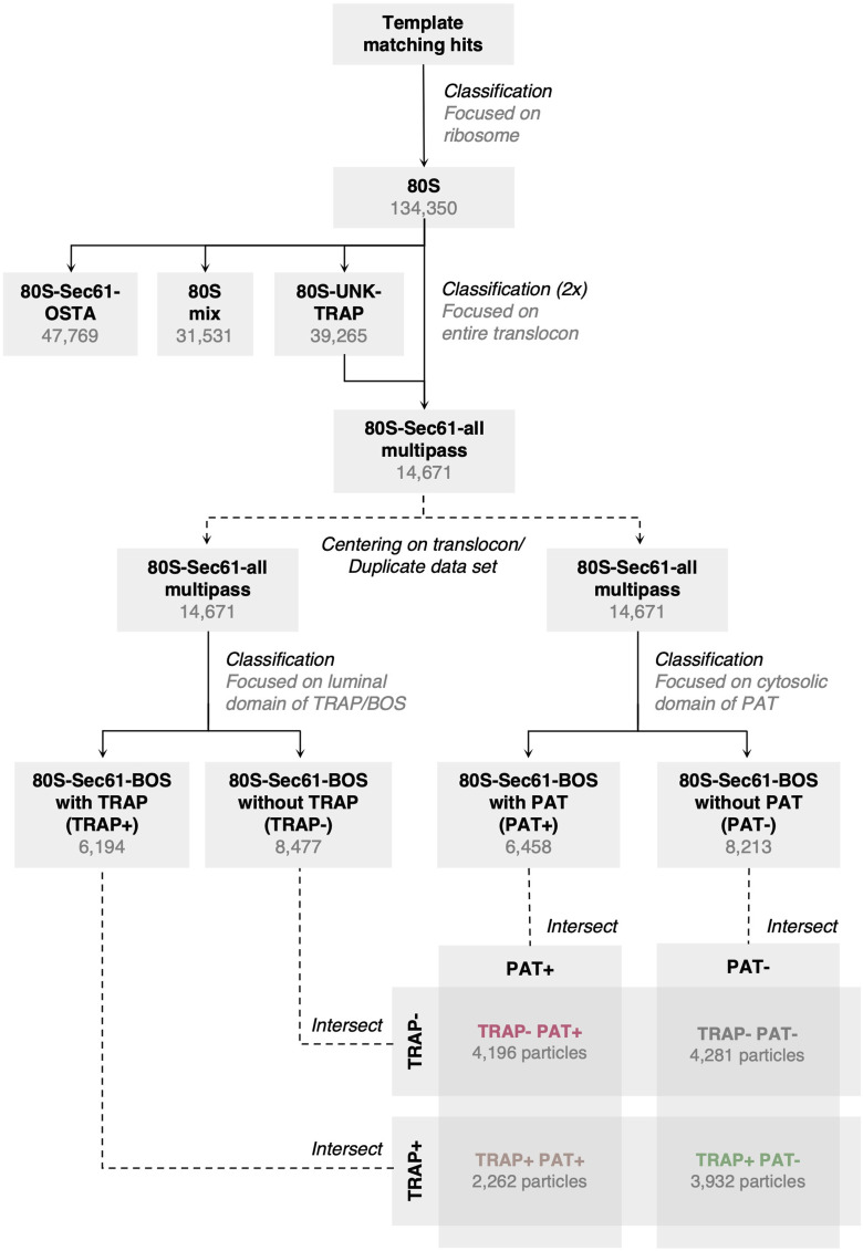
Figure S1. Classification overview of multipass translocon (MPT)-associated ribosomes. All 3D classification steps were performed in relion without alignment using subtomograms with a pixel size of 6.9 Å. After removing false positives after template matching, all well-aligned 80S ribosomes have been subjected to two rounds of classification focused on the entire translocon complex (Gemmer et al, 2023), yielding a population of ribosome-bound MPT. MPT particles were then either sorted into translocon-associated protein (TRAP)-containing and TRAP-lacking classes by classification focused on TRAP/back-of-Sec61, or into PAT-containing and PAT-lacking classes by classification focused on PAT, in two independent parallel rounds of classification. Afterwards, the classes TRAP−/PAT−, TRAP+/PAT−, TRAP−/PAT+, and TRAP+/PAT+ were generated by intersection of the particle sets obtained from the previous classification. The translocon-centered reconstruction of the TRAP-containing class (TRAP+, i.e., TRAP+/PAT− plus TRAP+/PAT+) was further aligned by focusing on the luminal domains of TRAP and back-of-Sec61. All classes (TRAP−/PAT−, TRAP+/PAT−, TRAP−/PAT+, and TRAP+/PAT+) were centered on the ribosome again and averaged.
Figure S1. Classification overview of multipass translocon (MPT)-associated ribosomes. All 3D classification steps were performed in relion without alignment using subtomograms with a pixel size of 6.9 Å. After removing false positives after template matching, all well-aligned 80S ribosomes have been subjected to two rounds of classification focused on the entire translocon complex (Gemmer et al, 2023), yielding a population of ribosome-bound MPT. MPT particles were then either sorted into translocon-associated protein (TRAP)-containing and TRAP-lacking classes by classification focused on TRAP/back-of-Sec61, or into PAT-containing and PAT-lacking classes by classification focused on PAT, in two independent parallel rounds of classification. Afterwards, the classes TRAP−/PAT−, TRAP+/PAT−, TRAP−/PAT+, and TRAP+/PAT+ were generated by intersection of the particle sets obtained from the previous classification. The translocon-centered reconstruction of the TRAP-containing class (TRAP+, i.e., TRAP+/PAT− plus TRAP+/PAT+) was further aligned by focusing on the luminal domains of TRAP and back-of-Sec61. All classes (TRAP−/PAT−, TRAP+/PAT−, TRAP−/PAT+, and TRAP+/PAT+) were centered on the ribosome again and averaged.CC BY · PubMed Central
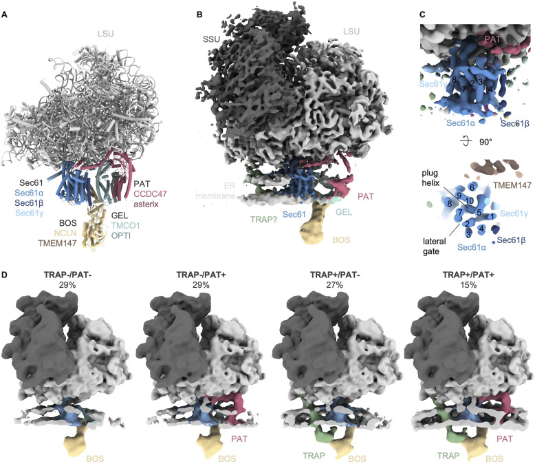
Figure 1. Structure and composition of the multipass translocon (MPT) in the native ER membrane. (A) Structure of the ribosome-associated MPT (PDB-7TUT) determined by Smalinskaitė et al (2022). (B) Cryo-ET reconstruction of the heterogenous ribosome-associated MPT-population (14,671 particles). Ribosome and Sec61 were filtered to 8 Å and the accessory factors and ER membrane area to 20 Å. (B, C) Close-up front (top panel) and top (bottom panel) view of Sec61 from the MPT population from (B). Numbering of transmembrane helixs of Sec61α is indicated. (D) Reconstructions of different MPT populations. Reconstructions were filtered to 20 Å resolution. The ER membrane was clipped for visual clarity.
Figure 1. Structure and composition of the multipass translocon (MPT) in the native ER membrane. (A) Structure of the ribosome-associated MPT (PDB-7TUT) determined by Smalinskaitė et al (2022). (B) Cryo-ET reconstruction of the heterogenous ribosome-associated MPT-population (14,671 particles). Ribosome and Sec61 were filtered to 8 Å and the accessory factors and ER membrane area to 20 Å. (B, C) Close-up front (top panel) and top (bottom panel) view of Sec61 from the MPT population from (B). Numbering of transmembrane helixs of Sec61α is indicated. (D) Reconstructions of different MPT populations. Reconstructions were filtered to 20 Å resolution. The ER membrane was clipped for visual clarity.CC BY · PubMed Central
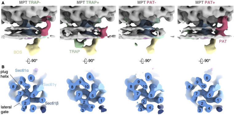
Figure S2. Sec61 conformational states of different multipass translocon (MPT) populations. (A) Reconstructions of all WT MPT populations which lack translocon-associated protein (TRAP) (TRAP−, i.e., TRAP−/PAT− plus TRAP−/PAT+) and contain TRAP (TRAP+, i.e., TRAP+/PAT− plus TRAP+/PAT+), and all WT MPT populations which lack PAT (PAT−, i.e., TRAP−/PAT− plus TRAP+/PAT−) and contain PAT (PAT+, i.e., TRAP−/PAT+ plus TRAP+/PAT+). MPT populations are filtered to 20 Å. (B) Top view of the corresponding map of Sec61 filtered to 8 Å. The membrane resides in the paper plane. The complex was clipped in the center of the membrane plane for visual clarity. Transmembrane helix numbering is indicated. The summed density fits the laterally closed Sec61 atomic model (PDB-7TUT) better than an open model (PDB-3CJ2) (correlation coefficient 0.8541 versus closed 0.8815 when filtered to 10 Å, models not shown).
Figure S2. Sec61 conformational states of different multipass translocon (MPT) populations. (A) Reconstructions of all WT MPT populations which lack translocon-associated protein (TRAP) (TRAP−, i.e., TRAP−/PAT− plus TRAP−/PAT+) and contain TRAP (TRAP+, i.e., TRAP+/PAT− plus TRAP+/PAT+), and all WT MPT populations which lack PAT (PAT−, i.e., TRAP−/PAT− plus TRAP+/PAT−) and contain PAT (PAT+, i.e., TRAP−/PAT+ plus TRAP+/PAT+). MPT populations are filtered to 20 Å. (B) Top view of the corresponding map of Sec61 filtered to 8 Å. The membrane resides in the paper plane. The complex was clipped in the center of the membrane plane for visual clarity. Transmembrane helix numbering is indicated. The summed density fits the laterally closed Sec61 atomic model (PDB-7TUT) better than an open model (PDB-3CJ2) (correlation coefficient 0.8541 versus closed 0.8815 when filtered to 10 Å, models not shown).CC BY · PubMed Central
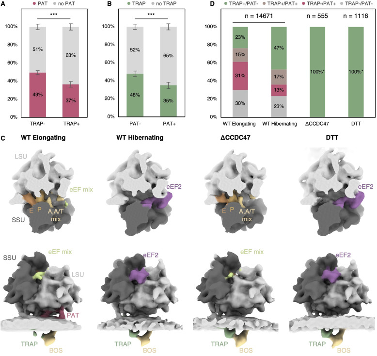
Figure 2. Recruitment behavior of PAT and translocon-associated protein (TRAP). (A) Relative abundance of PAT (magenta) in the TRAP-lacking (TRAP−) or -containing (TRAP+) multipass translocon (MPT) population resulting from subtomogram classification of ribosome–translocon complexes on ER-derived vesicles. The distributions differ significantly with P = 7.14 × 10⁻¹². (B) Relative abundance of TRAP (green) in the PAT-lacking (PAT−) or PAT-containing (PAT+) MPT subtomogram-average population with P = 8.52 × 10⁻¹¹. (C) Reconstructions of MPT-associated ribosomes obtained from HEK293F under unperturbed conditions (WT) of elongating and hibernating classes, ΔCCDC47, and DTT-treated samples. Top row: top views of the ribosomal binding cleft and elongation factor binding site. The ribosome was clipped for clarity. Bottom row: corresponding front views of the ribosome-associated translocon populations. The ER membrane was again clipped for clarity. The subtomogram averages represent the entire multipass population in each class (hibernating and elongating) or sample (ΔCCDC47 and DTT). Note the varying occupancy of TRAP and PAT in different classes. (C, D) Distribution of TRAP and PAT in the multipass populations from (C) color-coded as indicated. (*) PAT-containing or TRAP-lacking classes were not detected in ΔCCDC47 and DTT-treated samples.
Figure 2. Recruitment behavior of PAT and translocon-associated protein (TRAP). (A) Relative abundance of PAT (magenta) in the TRAP-lacking (TRAP−) or -containing (TRAP+) multipass translocon (MPT) population resulting from subtomogram classification of ribosome–translocon complexes on ER-derived vesicles. The distributions differ significantly with P = 7.14 × 10⁻¹². (B) Relative abundance of TRAP (green) in the PAT-lacking (PAT−) or PAT-containing (PAT+) MPT subtomogram-average population with P = 8.52 × 10⁻¹¹. (C) Reconstructions of MPT-associated ribosomes obtained from HEK293F under unperturbed conditions (WT) of elongating and hibernating classes, ΔCCDC47, and DTT-treated samples. Top row: top views of the ribosomal binding cleft and elongation factor binding site. The ribosome was clipped for clarity. Bottom row: corresponding front views of the ribosome-associated translocon populations. The ER membrane was again clipped for clarity. The subtomogram averages represent the entire multipass population in each class (hibernating and elongating) or sample (ΔCCDC47 and DTT). Note the varying occupancy of TRAP and PAT in different classes. (C, D) Distribution of TRAP and PAT in the multipass populations from (C) color-coded as indicated. (*) PAT-containing or TRAP-lacking classes were not detected in ΔCCDC47 and DTT-treated samples.CC BY · PubMed Central
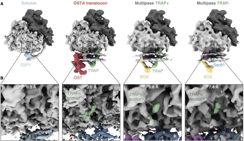
Figure 3. Association of the TRAPα anchor domain with different ribosome populations. (A) Back side of the ribosome associated with different soluble or ER translocon variants. Reconstructions of soluble and oligosaccharyltransferase A translocon-bound ribosomes are filtered to 12 Å and multipass-bound ribosomes to 20 Å. (B) Close-up views of the TRAPα anchor binding site. Reconstructions are filtered to their overall resolution as indicated.
Figure 3. Association of the TRAPα anchor domain with different ribosome populations. (A) Back side of the ribosome associated with different soluble or ER translocon variants. Reconstructions of soluble and oligosaccharyltransferase A translocon-bound ribosomes are filtered to 12 Å and multipass-bound ribosomes to 20 Å. (B) Close-up views of the TRAPα anchor binding site. Reconstructions are filtered to their overall resolution as indicated.CC BY · PubMed Central
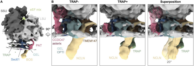
Figure 4. Orientation of the back-of-Sec61 complex in different populations. (A) Overall structure of the ribosome-bound multipass translocon filtered to 20 Å. (B) Close-up side views of the translocon-associated protein (TRAP)-lacking (TRAP−) and TRAP-containing (TRAP+) multipass translocon, and their superposition with TRAP in transparent. The longitudinal axis of back-of-Sec61 is indicated as dashed line.
Figure 4. Orientation of the back-of-Sec61 complex in different populations. (A) Overall structure of the ribosome-bound multipass translocon filtered to 20 Å. (B) Close-up side views of the translocon-associated protein (TRAP)-lacking (TRAP−) and TRAP-containing (TRAP+) multipass translocon, and their superposition with TRAP in transparent. The longitudinal axis of back-of-Sec61 is indicated as dashed line.CC BY · PubMed Central
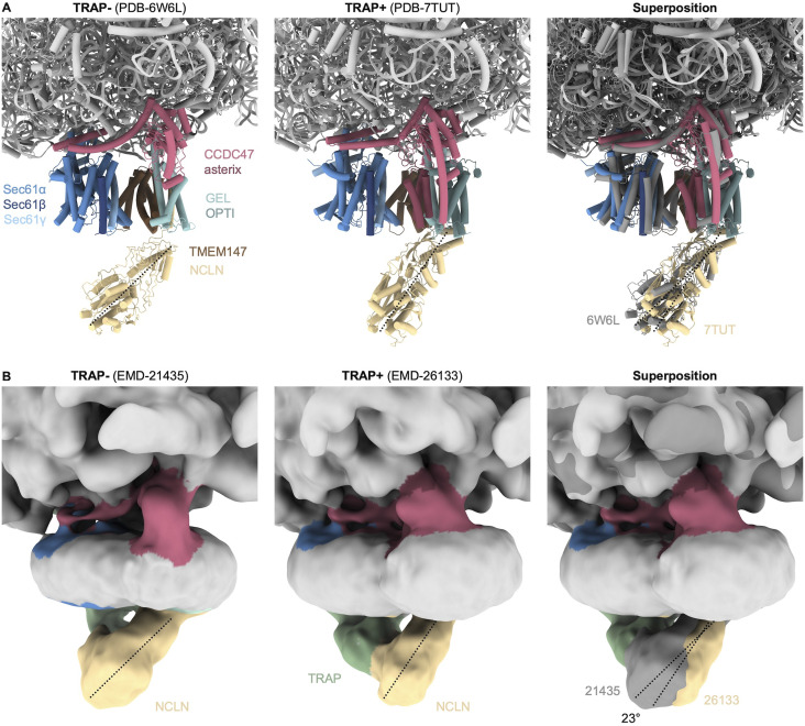
Figure S3. Orientation of the back-of-Sec61 (BOS) complex in different structures. (A) Close-up side views of the translocon-associated protein (TRAP)-lacking (TRAP−, 6W6L) and TRAP-containing (TRAP+, PDB-7TUT) multipass translocon, and their superposition with TRAP in dark grey. (A, B) Cryo-EM reconstructions corresponding to the structures from (A). Note that although a fragmented density of TRAP is present in the micelle of the TRAP reconstruction (EMD-21435), it is disconnected from Sec61 and BOS. The longitudinal axis of BOS is indicated as dashed line. The indicated rotation angle was determined in ChimeraX.
Figure S3. Orientation of the back-of-Sec61 (BOS) complex in different structures. (A) Close-up side views of the translocon-associated protein (TRAP)-lacking (TRAP−, 6W6L) and TRAP-containing (TRAP+, PDB-7TUT) multipass translocon, and their superposition with TRAP in dark grey. (A, B) Cryo-EM reconstructions corresponding to the structures from (A). Note that although a fragmented density of TRAP is present in the micelle of the TRAP reconstruction (EMD-21435), it is disconnected from Sec61 and BOS. The longitudinal axis of BOS is indicated as dashed line. The indicated rotation angle was determined in ChimeraX.CC BY · PubMed Central
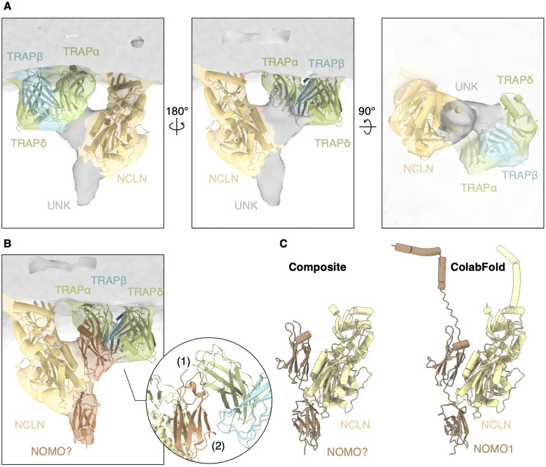
Figure 5. Translocon-associated protein (TRAP) interacts with the NCLN–nodal modulator (NOMO)–TMEM147 complex. (A) Recentered and locally refined reconstruction of the TRAP-containing multipass translocon. Luminal domains of TRAP (8B6L, green) and NCLN (AF-Q969V3, yellow) fit well into the density map. Two unidentified densities (UNK, grey) have a molecular weight of 10 kD each. (B) Molecular model of the C-terminal Ig-like domain 11–12 of NOMO1 (AF-Q15155) placed into the density map of the unidentified densities. The inset shows a close-up view of the putative interaction site between NOMO and TRAP. NOMO residues 1,068–1,072 and 1,117–1,120 likely interact with TRAPα residues 90–94 and 191–195 (1), and NOMO 1,084–1,089 with TRAPβ 51–53 and 83–86 (2). (B, C) Comparison of our model based on the reconstruction from (B) and the ColabFold prediction model of the NCLN–NOMO1 complex. NOMO1 domains 1–9 were removed for clarity.
Figure 5. Translocon-associated protein (TRAP) interacts with the NCLN–nodal modulator (NOMO)–TMEM147 complex. (A) Recentered and locally refined reconstruction of the TRAP-containing multipass translocon. Luminal domains of TRAP (8B6L, green) and NCLN (AF-Q969V3, yellow) fit well into the density map. Two unidentified densities (UNK, grey) have a molecular weight of 10 kD each. (B) Molecular model of the C-terminal Ig-like domain 11–12 of NOMO1 (AF-Q15155) placed into the density map of the unidentified densities. The inset shows a close-up view of the putative interaction site between NOMO and TRAP. NOMO residues 1,068–1,072 and 1,117–1,120 likely interact with TRAPα residues 90–94 and 191–195 (1), and NOMO 1,084–1,089 with TRAPβ 51–53 and 83–86 (2). (B, C) Comparison of our model based on the reconstruction from (B) and the ColabFold prediction model of the NCLN–NOMO1 complex. NOMO1 domains 1–9 were removed for clarity.CC BY · PubMed Central
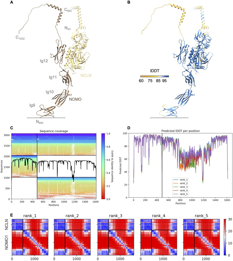
Figure S4. Colabfold model prediction of the NCLN–NOMO1 complex. (A) Colabfold prediction model of NCLN (full-length) in complex with NOMO1 (aa800-aa1222; Ig9-C-terminus) color-coded according to chain as indicated. Ig-like domains 1–8 were removed for clarity. N- and C-termini, corresponding residue numbering, and NOMO1 domain numbering is indicated. Signal peptides were removed before prediction. (A, B) Prediction model as in (A) color-coded according to predicted local distance difference test score. (C) Sequence coverage obtained by sequence alignments generated by MMseqs2. (D) Predicted local distance difference test scores per position of five model predictions. (E) Predicted aligned error of five model predictions of the NCLN–NOMO1 complex.
Figure S4. Colabfold model prediction of the NCLN–NOMO1 complex. (A) Colabfold prediction model of NCLN (full-length) in complex with NOMO1 (aa800-aa1222; Ig9-C-terminus) color-coded according to chain as indicated. Ig-like domains 1–8 were removed for clarity. N- and C-termini, corresponding residue numbering, and NOMO1 domain numbering is indicated. Signal peptides were removed before prediction. (A, B) Prediction model as in (A) color-coded according to predicted local distance difference test score. (C) Sequence coverage obtained by sequence alignments generated by MMseqs2. (D) Predicted local distance difference test scores per position of five model predictions. (E) Predicted aligned error of five model predictions of the NCLN–NOMO1 complex.CC BY · PubMed Central
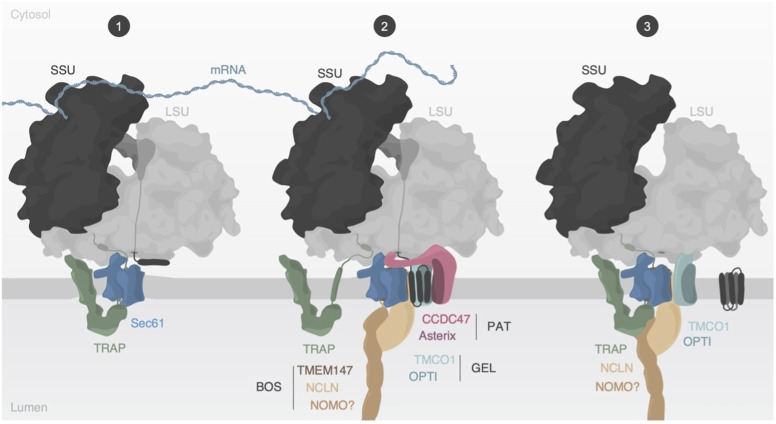
Figure 6. Model of multipass membrane protein biogenesis. (1) Synthesis of multispanning membrane proteins is facilitated by the ER translocon-bound ribosome. The translocon-associated protein (TRAP) complex engages with the translocon during the early stage. (2) Transmembrane helices of the nascent substrate are inserted into the cavity behind Sec61, whereas Sec61’s lateral gate and pore remain closed. The PAT complex engages with exposed hydrophilic areas of substrate proteins and the TRAP complex partially dissociates from the translocon likely to avoid TRAP-assisted insertion into Sec61’s lateral gate. The TRAPα anchor domain may remain bound to the ribosome to retain TRAP in proximity to the translocon. (3) After substrate folding and release, PAT dissociates from the ribosome and translocon, whereas TRAP is recruited again and interacts with the back-of-Sec61 complex in the ER lumen.
Figure 6. Model of multipass membrane protein biogenesis. (1) Synthesis of multispanning membrane proteins is facilitated by the ER translocon-bound ribosome. The translocon-associated protein (TRAP) complex engages with the translocon during the early stage. (2) Transmembrane helices of the nascent substrate are inserted into the cavity behind Sec61, whereas Sec61’s lateral gate and pore remain closed. The PAT complex engages with exposed hydrophilic areas of substrate proteins and the TRAP complex partially dissociates from the translocon likely to avoid TRAP-assisted insertion into Sec61’s lateral gate. The TRAPα anchor domain may remain bound to the ribosome to retain TRAP in proximity to the translocon. (3) After substrate folding and release, PAT dissociates from the ribosome and translocon, whereas TRAP is recruited again and interacts with the back-of-Sec61 complex in the ER lumen.CC BY · PubMed Central

Verification

Claims the paper makes

The claims and statistics of this paper haven’t been checked yet.

Run the check — free, no account needed. It reads the open-access text and checks the sources it cites.

A few free checks a day · papers already verified open instantly · results are public

Self-check

Percentages, sums and reported differences re-computed from the paper’s own numbers.

The self-check needs the paper’s full text.

Citations

Cites

Research works printed but not linked: 42

  1. M. Gemmer & F. Forster . A clearer picture of the ER translocon complex. J Cell Sci 133 (2020). · DOI
  2. 10.1146/annurev-biophys-050511-102312 · DOI
  3. A. M. Liaci & F. Forster . Take Me Home, Protein Roads: Structural Insights into Signal Peptide Interactions during ER Translocation. Int J Mol Sci 22 (2021). · DOI
  4. 2022, The mechanisms of integral membrane protein biogenesis, Nat Rev Mol Cell Biol, 23, 107, 10.1038/s41580-021-00413-2 · DOI
  5. 2019, Cryo-EM is uncovering the mechanism of eukaryotic protein N-glycosylation, FEBS J, 286, 1638, 10.1111/febs.14705 · DOI
  6. S. Lang , S. Pfeffer , P. H. Lee , A. Cavalie , V. Helms , F. Förster & R. Zimmermann . An Update on Sec61 Channel Functions, Mechanisms, and Related Diseases. Frontiers in physiology 8, 887 (2017). · DOI
  7. 10.1038/ncomms9403 · DOI
  8. 10.1083/jcb.200210095 · DOI
  9. 10.1038/s41467-018-06188-z · DOI
  10. 10.1016/j.sbi.2011.08.005 · DOI
  11. 10.1126/science.aar5140 · DOI
  12. 10.1016/j.celrep.2017.12.006 · DOI · Paper page
  13. 10.1016/j.cell.2008.06.025 · DOI
  14. 10.1016/j.molcel.2020.08.012 · DOI
  15. 10.1126/science.aao3099 · DOI
  16. 10.1016/j.cell/2018.10.009 · DOI
  17. 10.1126/science.abb5008 · DOI
  18. M. J. Shurtleff , D. N. Itzhak , J. A. Hussmann , N. T. Schirle Oakdale , E. A. Costa , M. Jonikas , J. Weibezahn , K. D. Popova , C. H. Jan , P. Sinitcyn , S. S. Vembar , H. Hernandez , J. Cox , A. L. Burlingame , J. L. Brodsky , A. Frost , G. H. Borner & J. S. Weissman . The ER membrane protein complex interacts cotranslationally to enable biogenesis of multipass membrane proteins. Elife 7 (2018). · DOI
  19. P. T. McGilvray , S. A. Anghel , A. Sundaram , F. Zhong , M. J. Trnka , J. R. Fuller , H. Hu , A. L. Burlingame & R. J. Keenan . An ER translocon for multi-pass membrane protein biogenesis. Elife 9 (2020). · DOI
  20. 10.1038/s41586-022-05336-2 · DOI
  21. 10.1038/s41586-022-05330-8 · DOI
  22. A. J. O. Lewis & R. S. Hegde . A unified evolutionary origin for the ubiquitous protein transporters SecY and YidC. BMC Biol. 19, 266 (2021). · DOI
  23. 2020, An intramembrane chaperone complex facilitates membrane protein biogenesis, Nature, 584, 630, 10.1038/s41586-020-2624-y · DOI
  24. 10.1091/mbc.E02-04-0198 · DOI
  25. 10.1074/jbc.M611033200 · DOI
  26. 10.1074/jbc.M110.132548 · DOI
  27. 10.1038/sj.emboj.7600307 · DOI
  28. 10.1038/emboj.2009.204 · DOI
  29. 10.1074/jbc.M113.533695 · DOI
  30. A. Christodoulou , G. Maimaris , A. Makrigiorgi , E. Charidemou , C. Luchtenborg , A. Ververis , R. Georgiou , C. W. Lederer , C. Haffner , B. Brugger & N. Santama . TMEM147 interacts with lamin B receptor, regulates its localization and levels, and affects cholesterol homeostasis. J Cell Sci 133 (2020). · DOI
  31. 10.1038/s41586-022-05638-5 · DOI
  32. 10.1126/science.1209038 · DOI
  33. 10.1038/sj.embor.7400933 · DOI
  34. M. Jaskolowski , A. Jomaa , M. Gamerdinger , S. Shrestha , M. Leibundgut , E. Deuerling & N. Ban . Molecular basis of the TRAP complex function in ER protein biogenesis. bioRxiv, 2022.2010.2004.510795 (2022). · DOI · Paper page
  35. 10.1016/j.bbamcr.2013.08.018 · DOI
  36. 2021, Nodal modulator (NOMO) is required to sustain endoplasmic reticulum morphology, J. Biol. Chem, 297, 100937, 10.1016/j.jbc.2021.100937 · DOI
  37. 10.1093/NAR/GKAB1061 · DOI · Paper page
  38. 10.1038/S41592-022-01488-1 · DOI · Paper page
  39. 10.1124/mol.110.067363 · DOI
  40. 10.1016/j.jsb.2012.09.006 · DOI · Paper page
  41. 2021, Multi-particle cryo-EM refinement with M visualizes ribosome-antibiotic complex at 3.5 A in cells, Nat Methods, 18, 186, 10.1038/s41592-020-01054-7 · DOI
  42. R. Evans , M. O’Neill , A. Pritzel , N. Antropova , A. Senior , T. Green , A. Žídek , R. Bates , S. Blackwell , J. Yim , O. Ronneberger , S. Bodenstein , M. Zielinski , A. Bridgland , A. Potapenko , A. Cowie , K. Tunyasuvunakool , R. Jain , E. Clancy , P. Kohli , J. Jumper & D. Hassabis . Protein complex prediction with AlphaFold-Multimer. bioRxiv, 2021.2010.2004.463034 (2021). · DOI · Paper page

Metadata

Authors
Gemmer, M., Chaillet, M. L., & Förster, F.
Affiliations
Utrecht University (NL)
Corresponding author
Förster, Friedrich
Venue
Life Science Alliance
Publisher
openRxiv
Published
2023-11-29
Volume
7
Issue
8
Pages
e202302496-e202302496
ISSN
2575-1077 (ISSN-L)
OpenAlex
W4399566271
Cited by
16
References
1
Citations by year
2026: 4 · 2025: 7 · 2024: 4 · 2023: 1
FWCI
2.04
Type
journal article
Language
en
Open access
Licence
cc-by
Locations
  • Life Science Alliance (publishedVersion, cc-by)
  • bioRxiv (Cold Spring Harbor Laboratory) (acceptedVersion, cc-by-nc-nd)
  • PubMed Central (publishedVersion, cc-by)
  • PubMed (publishedVersion)
  • DOAJ (DOAJ: Directory of Open Access Journals) (submittedVersion)
  • Utrecht University Repository (Utrecht University) (submittedVersion, other-oa)
Funders
  • European Commission (724425, HORIZON2020)
  • Nederlandse Organisatie voor Wetenschappelijk Onderzoek (724.016.001, 184.034.014, 724.016.001, 184.034.014, 741.018.201, 741.018.201)
  • European Research Council (724425)
Topics
  • Life Sciences › Biochemistry, Genetics and Molecular Biology › Molecular Biology › RNA and protein synthesis mechanisms
  • Life Sciences › Biochemistry, Genetics and Molecular Biology › Cell Biology › Cellular transport and secretion
  • Life Sciences › Biochemistry, Genetics and Molecular Biology › Structural Biology › Advanced Electron Microscopy Techniques and Applications
Keywords
endoplasmic reticulum membrane, TRAP complex, Sec61, protein translocation, membrane protein insertion, cryo-electron tomography, protein complex composition
MeSH terms
SEC Translocation Channels, Endoplasmic Reticulum, Membrane Proteins, Models, Molecular, Ribosomes, Protein Biosynthesis, Molecular Chaperones, Cryoelectron Microscopy, Protein Transport

About these checks

Registry, retraction, Cites and statistics results are computed, not judged, with one exception: a printed reference that no index lists is read once by a model before it counts as a citation error. Claims are extracted by a model and kept only when their quote appears verbatim in the text; each cited source is then checked, with a model’s help, for whether it says what the claim relies on. Those verdicts can be wrong — read the claim’s quote and the cited source before relying on one.

Statistics and arithmetic are recomputed, not judged.

Confidence bands are shown only for fields calibrated on our annotated test set. None is calibrated yet, so no band is shown.

What counts as a claim ›How we measure confidence ›How we check this ›

Are you an author, or do you see a problem with this page? Request a review