PreprintCited by 6Photosynthetic Processes and MechanismsMetalloenzymes and iron-sulfur proteinsMetal-Catalyzed Oxygenation Mechanisms

The DOI matches its Crossref record; 1 of the 1 references it cites were found in a registry or an index.

Physcomitrium patens flavodiiron proteins form a redox-dependent heterocomplex

Open access

Preprint — published as 10.1016/j.jbc.2024.107643

Abstract

Flavodiiron proteins (FLVs) catalyze the reduction of oxygen to water by exploiting electrons from Photosystem I (PSI). In several photosynthetic organisms such as cyanobacteria, green algae, mosses and gymnosperms, FLV-dependent electron flow protects PSI from over-reduction and consequent damage especially under fluctuating light conditions. In this work we investigated biochemical and structural properties of FLVA and FLVB from the model moss Physcomitrium patens . The two proteins, expressed and purified from Escherichia coli , bind both iron and flavin cofactors and show NAD(P)H oxidase activity as well as oxygen reductase capacities. Moreover, the co-expression of both FLVA and FLVB, coupled to a tandem affinity purification procedure with two different affinity tags, enabled the isolation of the stable and catalytically active FLVA/B hetero multimer protein complex, that has never been isolated and characterized so far. The multimeric organization was shown to be stabilized by inter-subunit disulfide bonds. This investigation provides valuable new information on the biochemical properties of FLVs, with new insights into their in vivo role and regulation.

Overview

The claims and statistics of this paper haven’t been checked yet.

Run the check — free, no account needed. It reads the open-access text and checks the sources it cites.

A few free checks a day · papers already verified open instantly · results are public

Integrity signals

Registry

Crossref · 10.1101/2024.01.29.577648

Cites

1 of 1 references verified0 citation errors · 0 not checkable

Statistics

Figures

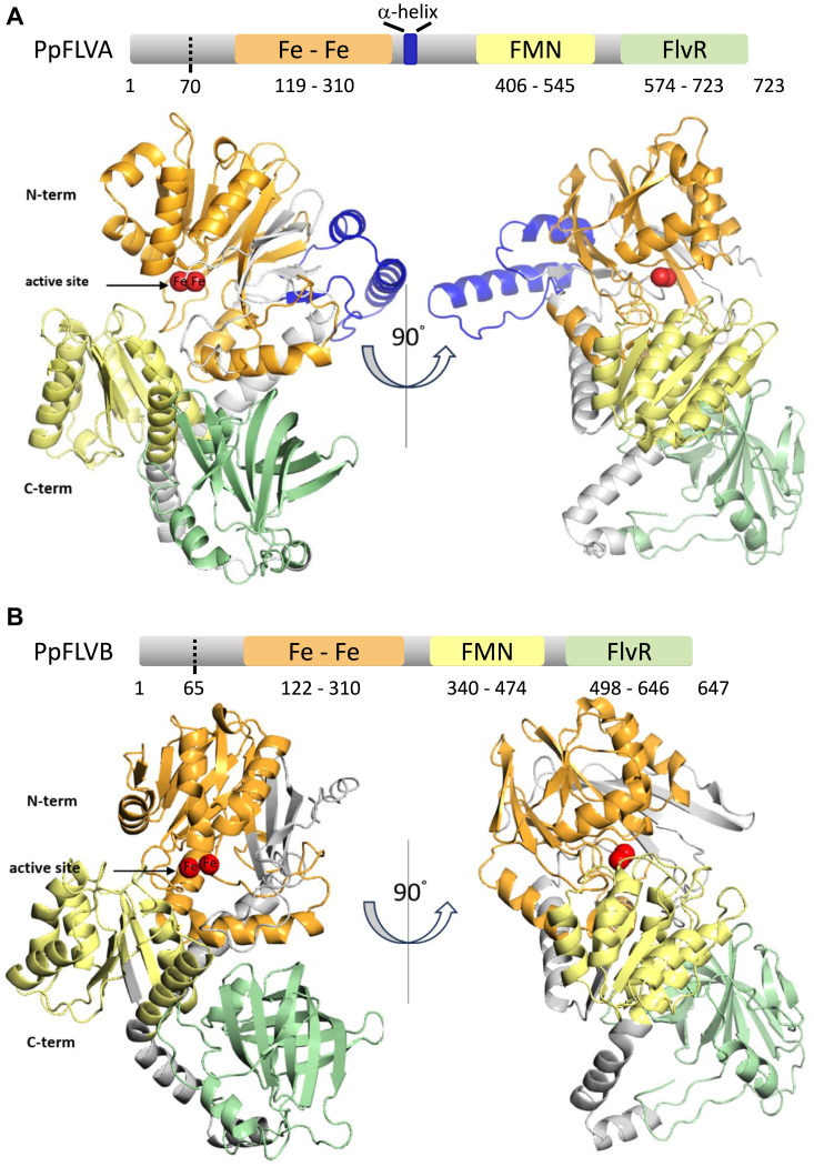
Figure 1 Predicted structure analysis of Physcomitrium patens FLVA and FLVB.A, schematic representation and AlphaFold model of FLVA (A9RYP3) and (B) FLVB (A9RQ00) structures. Dotted lines indicate the predicted cleavage sites for FLVA (position 70) and FLVB (position 65) chloroplast target peptides. The N-terminal metallo-β-lactamase domain contains a diiron center (Fe-Fe), the flavodoxin-like domain harbors a flavin mononucleotide (FMN), and the C-terminal domain is a flavin reductase-like domain (FlvR). The three domains are shown respectively in orange, yellow, and green. FLVA-specific residues (I313-K364) are shown in blue. Iron atoms (Fe) are represented as red spheres present in the active site.
Figure 1 Predicted structure analysis of Physcomitrium patens FLVA and FLVB.A, schematic representation and AlphaFold model of FLVA (A9RYP3) and (B) FLVB (A9RQ00) structures. Dotted lines indicate the predicted cleavage sites for FLVA (position 70) and FLVB (position 65) chloroplast target peptides. The N-terminal metallo-β-lactamase domain contains a diiron center (Fe-Fe), the flavodoxin-like domain harbors a flavin mononucleotide (FMN), and the C-terminal domain is a flavin reductase-like domain (FlvR). The three domains are shown respectively in orange, yellow, and green. FLVA-specific residues (I313-K364) are shown in blue. Iron atoms (Fe) are represented as red spheres present in the active site.CC BY-NC-ND · PubMed Central
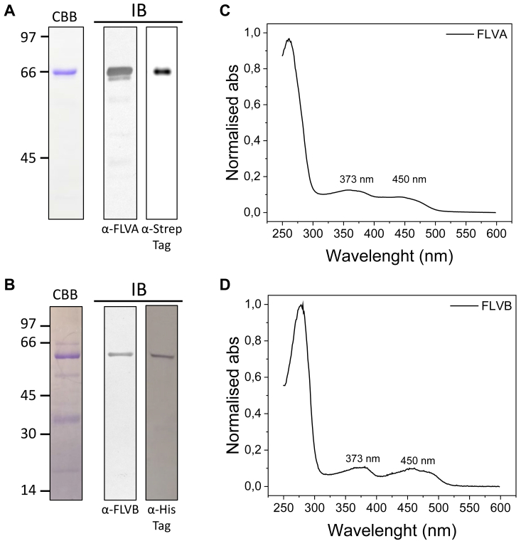
Figure 2 FLVA and FLVB biochemical properties. FLVA and FLVB biochemical properties. Coomassie Brilliant Blue-stained SDS-PAGE (CBB) of purified FLVA (A) and FLVB (B) and immunoblot analysis (IB) performed with antisera anti-FLVA and anti-Strep-Tag (A) and anti-FLVB and anti-His-Tag. On the left side, the apparent molecular weight of the ladder is reported in kDa. UV-visible absorption spectra of purified FLVA (C) and FLVB (D). Absorption spectra of approximately 5 μM protein in a solution containing 30 mM Tris and 300 mM NaCl at pH 8.0 were recorded at room temperature.
Figure 2 FLVA and FLVB biochemical properties. FLVA and FLVB biochemical properties. Coomassie Brilliant Blue-stained SDS-PAGE (CBB) of purified FLVA (A) and FLVB (B) and immunoblot analysis (IB) performed with antisera anti-FLVA and anti-Strep-Tag (A) and anti-FLVB and anti-His-Tag. On the left side, the apparent molecular weight of the ladder is reported in kDa. UV-visible absorption spectra of purified FLVA (C) and FLVB (D). Absorption spectra of approximately 5 μM protein in a solution containing 30 mM Tris and 300 mM NaCl at pH 8.0 were recorded at room temperature.CC BY-NC-ND · PubMed Central
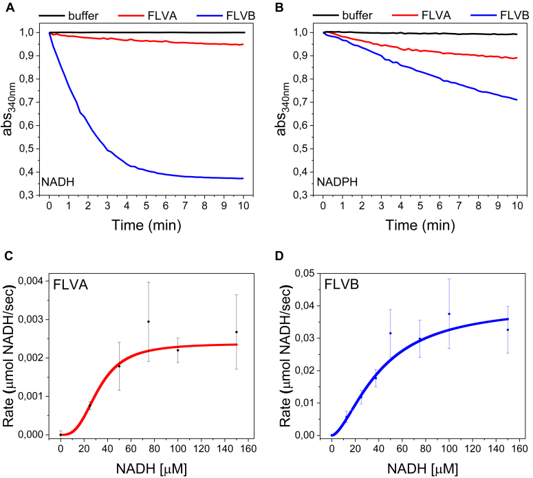
Figure 3 Kinetics of FLVA and FLVB NAD(P)H oxidase activity. FLVA and FLVB activity were tested in the presence of either 150 μM NADH (A) or NADPH (B). Red lines correspond to 7 μM FLVA and blue lines correspond to 7 μM FLVB. Black lines correspond to the negative control in the absence of FLV proteins. Traces were shifted to OD340nm equal to 1 as a starting point immediately after FLV protein addition. Absorption spectra were recorded at room temperature. C-D, Hill fitting kinetics and fitted values for purified FLVA and FLVB in the presence of NADH. The solid line indicates Hill fitting curve of FLVA (C) or FLVB (D). n = 3 reactions were performed in the presence of FLVA and different concentrations of NADH, n = 4 reactions were performed in the presence of FLVB and different concentrations of NADH. Mean values are represented ± SEM.
Figure 3 Kinetics of FLVA and FLVB NAD(P)H oxidase activity. FLVA and FLVB activity were tested in the presence of either 150 μM NADH (A) or NADPH (B). Red lines correspond to 7 μM FLVA and blue lines correspond to 7 μM FLVB. Black lines correspond to the negative control in the absence of FLV proteins. Traces were shifted to OD340nm equal to 1 as a starting point immediately after FLV protein addition. Absorption spectra were recorded at room temperature. C-D, Hill fitting kinetics and fitted values for purified FLVA and FLVB in the presence of NADH. The solid line indicates Hill fitting curve of FLVA (C) or FLVB (D). n = 3 reactions were performed in the presence of FLVA and different concentrations of NADH, n = 4 reactions were performed in the presence of FLVB and different concentrations of NADH. Mean values are represented ± SEM.CC BY-NC-ND · PubMed Central
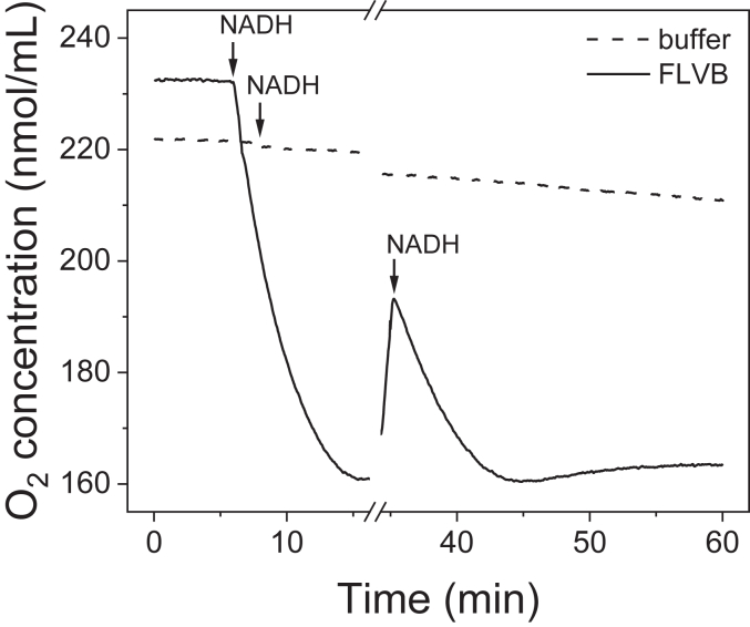
Figure 4 High-resolution respirometry of P. patens FLVB performed in an Oroboros Oxygraph-2k. The measurement started with sample buffer (Tris 30 mM and NaCl 300 mM) either containing 6 μM FLVB (solid line) or not (dashed line). After about 7.5 min 150 μM NADH was added to both samples. To verify protein stability, 70 μM NADH was added again after about 35 min.
Figure 4 High-resolution respirometry of P. patens FLVB performed in an Oroboros Oxygraph-2k. The measurement started with sample buffer (Tris 30 mM and NaCl 300 mM) either containing 6 μM FLVB (solid line) or not (dashed line). After about 7.5 min 150 μM NADH was added to both samples. To verify protein stability, 70 μM NADH was added again after about 35 min.CC BY-NC-ND · PubMed Central
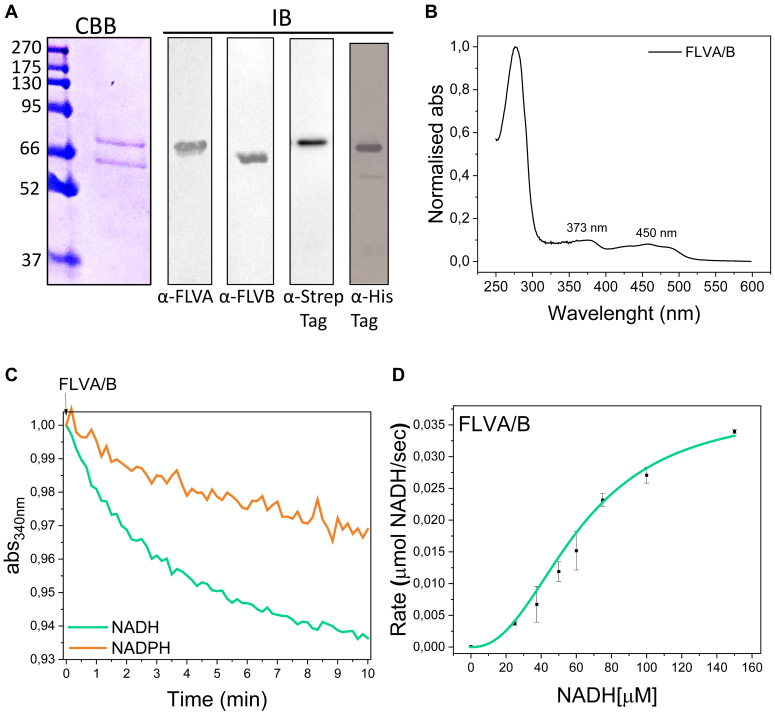
Figure 5 FLVA/FLVB heterocomplex biochemical and functional properties.A, representative Coomassie Brilliant Blue-stained SDS-polyacrylamide gel and Western blot analysis of purified heterocomplex using α-FLVA and α-FLVB, α-StrepTag, α-HisTag antibodies. On the left side, the apparent molecular weight of the ladder is reported in kDa. B, the absorption spectrum of approximately 5 μM FLVA/B in a solution containing 30 mM Tris and 300 mM NaCl at pH 8.0 was recorded at room temperature. C, NAD(P)H oxidase activity of 1 μM FLVA/B heterocomplex in the presence of 150 μM NADH (green line) and 150 μM NADPH (orange line). Traces were shifted to OD340nm equal to 1 as the starting point immediately after protein adding. Absorption spectra were recorded at room temperature. D, Hill fitting Kinetics and fitted values for purified FLVA/B in the presence of NADH. The solid line indicates the Hill fitting curve. n = 3 Values are expressed ± SEM.
Figure 5 FLVA/FLVB heterocomplex biochemical and functional properties.A, representative Coomassie Brilliant Blue-stained SDS-polyacrylamide gel and Western blot analysis of purified heterocomplex using α-FLVA and α-FLVB, α-StrepTag, α-HisTag antibodies. On the left side, the apparent molecular weight of the ladder is reported in kDa. B, the absorption spectrum of approximately 5 μM FLVA/B in a solution containing 30 mM Tris and 300 mM NaCl at pH 8.0 was recorded at room temperature. C, NAD(P)H oxidase activity of 1 μM FLVA/B heterocomplex in the presence of 150 μM NADH (green line) and 150 μM NADPH (orange line). Traces were shifted to OD340nm equal to 1 as the starting point immediately after protein adding. Absorption spectra were recorded at room temperature. D, Hill fitting Kinetics and fitted values for purified FLVA/B in the presence of NADH. The solid line indicates the Hill fitting curve. n = 3 Values are expressed ± SEM.CC BY-NC-ND · PubMed Central
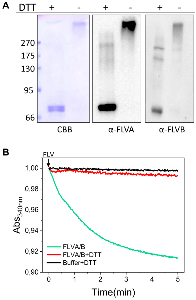
Figure 6 Redox dependence of flavodiiron proteins heterocomplex.A, SDS-PAGE and Western blotting of purified FLVA/B heterocomplex in the presence or absence of reducing agent. On the left side, the apparent molecular weight of the ladder is reported in kDa. The samples were incubated at room temperature in the presence or absence of DTT as indicated on the top of each lane. Prior to SDS-PAGE all samples were heated at 100 °C. B, FLVA/B heterocomplex NADH oxidase activity in the presence (red line) or absence (green line) of reducing agent. 4 μM of purified enzyme and the equivalent volume of protein buffer (black line) were incubated at room temperature for ∼1 h with DTT prior to activity test. 150 μM NADH without adding FLV and DTT was also monitored as a control (black line). Traces were shifted to OD340nm equal to 1 as the starting point immediately after protein adding. Absorption spectra were recorded at room temperature.
Figure 6 Redox dependence of flavodiiron proteins heterocomplex.A, SDS-PAGE and Western blotting of purified FLVA/B heterocomplex in the presence or absence of reducing agent. On the left side, the apparent molecular weight of the ladder is reported in kDa. The samples were incubated at room temperature in the presence or absence of DTT as indicated on the top of each lane. Prior to SDS-PAGE all samples were heated at 100 °C. B, FLVA/B heterocomplex NADH oxidase activity in the presence (red line) or absence (green line) of reducing agent. 4 μM of purified enzyme and the equivalent volume of protein buffer (black line) were incubated at room temperature for ∼1 h with DTT prior to activity test. 150 μM NADH without adding FLV and DTT was also monitored as a control (black line). Traces were shifted to OD340nm equal to 1 as the starting point immediately after protein adding. Absorption spectra were recorded at room temperature.CC BY-NC-ND · PubMed Central
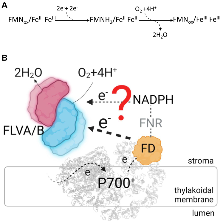
Figure 7 Proposed model describing P. patens flavodiiron protein catalytic activity.A, overall proposed reaction scheme. Four electrons are necessary for the complete reduction of O2 to water, two electrons reduce the FMN moiety, while the other two the Fe-Fe center. B, FLVA and FLVB form stable bound heterotetramer in the stroma of P. patens chloroplasts. The dashed line indicates electron flow. Electrons coming from Fd and NADPH pool could participate in FLV oxygen reduction reaction.
Figure 7 Proposed model describing P. patens flavodiiron protein catalytic activity.A, overall proposed reaction scheme. Four electrons are necessary for the complete reduction of O2 to water, two electrons reduce the FMN moiety, while the other two the Fe-Fe center. B, FLVA and FLVB form stable bound heterotetramer in the stroma of P. patens chloroplasts. The dashed line indicates electron flow. Electrons coming from Fd and NADPH pool could participate in FLV oxygen reduction reaction.CC BY-NC-ND · PubMed Central

Verification

Claims the paper makes

The claims and statistics of this paper haven’t been checked yet.

Run the check — free, no account needed. It reads the open-access text and checks the sources it cites.

A few free checks a day · papers already verified open instantly · results are public

Self-check

Percentages, sums and reported differences re-computed from the paper’s own numbers.

The self-check needs the paper’s full text.

Citations

Cites

Show all 1 references

Research works printed but not linked: 37

  1. 10.1007/BF01101690 · DOI
  2. 2016, Photodamage of iron-sulphur clusters in photosystem I induces non-photochemical energy dissipation, Nat Plants, 2, 16035, 10.1038/nplants.2016.35 · DOI
  3. 2019, Consequences of photosystem-I damage and repair on photosynthesis and carbon use in Arabidopsis thaliana, Plant J, 97, 1061, 10.1111/tpj.14177 · DOI
  4. 10.1098/rstb.2000.0703 · DOI
  5. 10.1104/pp.114.238238 · DOI
  6. 10.1111/nph.15372 · DOI
  7. 10.1016/S0960-9822(03)00046-0 · DOI
  8. 10.1073/pnas.1407327111 · DOI
  9. 10.7554/eLife.45766 · DOI
  10. 10.1104/pp.17.00421 · DOI
  11. 10.1104/pp.16.01038 · DOI
  12. 10.1073/pnas.1606685113 · DOI
  13. 2023, Flavodiiron-mediated O2 photoreduction at photosystem I acceptor-side provides photoprotection to conifer thylakoids in early spring, Nat Commun, 14, 3210, 10.1038/s41467-023-38938-z · DOI
  14. 10.1093/femsle/fnx267 · DOI
  15. 10.1016/j.freeradbiomed.2019.01.051 · DOI
  16. 10.1016/S0006-291X(02)00434-5 · DOI
  17. 2019, The oxygen reduction reaction catalyzed by Synechocystis sp. PCC 6803 flavodiiron proteins, Sustainable Energy Fuels, 3, 3191, 10.1039/C9SE00523D · DOI
  18. 2020, Identification of the electron donor to flavodiiron proteins in Synechocystis sp. PCC 6803 by in vivo spectroscopy, Biochim Biophys Acta Bioenerg, 1861, 148256, 10.1016/j.bbabio.2020.148256 · DOI
  19. 10.1186/s13068-019-1618-1 · DOI
  20. 2019, Analysis of a new flavodiiron core structural arrangement in Flv1-ΔFlR protein from Synechocystis sp. PCC6803, Journal of Structural Biology, 205, 91, 10.1016/j.jsb.2018.11.004 · DOI
  21. 2015, Cyanobacterial Oxygenic Photosynthesis is Protected by Flavodiiron Proteins, Life (Basel, 5, 716
  22. 10.1042/BCJ20180648 · DOI
  23. 10.1074/jbc.M111.223289 · DOI
  24. 10.1073/pnas.1221194110 · DOI
  25. 2022, Coral symbionts evolved a functional polycistronic flavodiiron gene, Photosynth Res, 151, 113, 10.1007/s11120-021-00867-7 · DOI
  26. 2016, Artificial remodelling of alternative electron flow by flavodiiron proteins in Arabidopsis, Nat Plants, 2, 16012, 10.1038/nplants.2016.12 · DOI
  27. 10.1104/pp.17.01335 · DOI
  28. 10.1046/j.1432-1327.1998.2540325.x · DOI
  29. 10.1038/s41598-018-28453-3 · DOI
  30. 10.1105/tpc.111.094417 · DOI
  31. 10.1016/j.tplants.2021.03.008 · DOI
  32. 2016, Structure of Escherichia coli Flavodiiron Nitric Oxide Reductase, Journal of Molecular Biology, 428, 4686, 10.1016/j.jmb.2016.10.008 · DOI
  33. 10.1042/BST0390216 · DOI
  34. 10.1074/jbc.M705605200 · DOI
  35. 2015, Dioxygen and nitric oxide scavenging by Treponema denticola flavodiiron protein: a mechanistic paradigm for catalysis, J Biol Inorg Chem, 20, 603, 10.1007/s00775-015-1248-4 · DOI
  36. 10.1021/ac60289a016 · DOI
  37. 10.1016/j.plantsci.2016.08.003 · DOI

Metadata

Authors
Beraldo, C., Traverso, E., Boschin, M., Cendron, L., Morosinotto, T., & Alboresi, A.
Affiliations
University of Padua (IT)
Corresponding author
Alboresi, Alessandro
Venue
Journal of Biological Chemistry
Publisher
openRxiv
Published
2024-01-29
Volume
300
Issue
9
Pages
107643-107643
ISSN
0021-9258 (ISSN-L)
OpenAlex
W4401438711
Cited by
6
References
1
Citations by year
2026: 1 · 2025: 5
FWCI
0.82
Type
journal article
Language
en
Open access
Licence
cc-by-nc-nd
Locations
  • Journal of Biological Chemistry (publishedVersion, cc-by-nc-nd)
  • bioRxiv (Cold Spring Harbor Laboratory) (acceptedVersion, cc-by-nc-nd)
  • PubMed Central (publishedVersion, cc-by-nc-nd)
  • PubMed (publishedVersion)
  • Padua Research Archive (University of Padova) (submittedVersion, cc-by)
Funders
  • European Commission (H2020, CN00000033, 859770)
  • Ministero dell’Istruzione, dell’Università e della Ricerca
  • Horizon 2020 Framework Programme (859770)
  • Ministero dell'Istruzione e del Merito
Topics
  • Life Sciences › Biochemistry, Genetics and Molecular Biology › Molecular Biology › Photosynthetic Processes and Mechanisms
  • Life Sciences › Biochemistry, Genetics and Molecular Biology › Biochemistry › Metalloenzymes and iron-sulfur proteins
  • Physical Sciences › Chemistry › Inorganic Chemistry › Metal-Catalyzed Oxygenation Mechanisms
Keywords
flavodiiron proteins, Physcomitrella patens, photosystem I, oxygen reduction reaction, protein oligomerization, tandem affinity purification, photosynthetic electron transport
MeSH terms
Escherichia coli, Iron, Plant Proteins, Bryopsida, Photosystem I Protein Complex, Protein Multimerization

About these checks

Registry, retraction, Cites and statistics results are computed, not judged, with one exception: a printed reference that no index lists is read once by a model before it counts as a citation error. Claims are extracted by a model and kept only when their quote appears verbatim in the text; each cited source is then checked, with a model’s help, for whether it says what the claim relies on. Those verdicts can be wrong — read the claim’s quote and the cited source before relying on one.

Statistics and arithmetic are recomputed, not judged.

Confidence bands are shown only for fields calibrated on our annotated test set. None is calibrated yet, so no band is shown.

What counts as a claim ›How we measure confidence ›How we check this ›

Are you an author, or do you see a problem with this page? Request a review

How to cite

Cite CoChat for the checks on this page, and the paper for its own findings.

This page
CoChat. (2026). Citation check: Physcomitrium patens flavodiiron proteins form a redox-dependent heterocomplex. https://app.cochat.ai/papers/10.1101/2024.01.29.577648
The paper
Beraldo, C., Traverso, E., Boschin, M., Cendron, L., Morosinotto, T., & Alboresi, A. (2024). Physcomitrium patens flavodiiron proteins form a redox-dependent heterocomplex. Journal of Biological Chemistry. https://doi.org/10.1101/2024.01.29.577648