Student or educator? Sign up with your school email and get 6 months of Pro free.
PreprintCited by 2RNA and protein synthesis mechanismsDNA and Nucleic Acid ChemistryRNA Research and Splicing

The DOI matches its Crossref record; 1 of the 1 references it cites were found in a registry or an index.

The impact of RNA chemical probing reagents on RNA binding proteins

Open access

Preprint — published as 10.1016/j.jbc.2025.110468 · Paper page

Abstract

RNA chemical probing experiments are a broadly used method for revealing the structure of RNA, as well as for identifying protein binding sites. This is beneficial for expanding our understanding of biological processes governed by protein-RNA complex interactions, as well as facilitating the identification of complex inhibiting molecules. The reagents commonly used in chemical probing experiments are highly reactive, methylating or acylating flexible RNA nucleotides. The highly reactive nature of the chemical probes means that they can also react with nucleophilic amino acid side chains, and subsequently affect protein-RNA binding events. We combine molecular dynamics (MD) simulations, matrix-assisted laser desorption ionization mass spectrometry (MALDI-MS), and nuclear magnetic resonance (NMR) experiments to show that commonly used RNA chemical probes react with protein amino acids, and demonstrate that this effect alters protein-RNA binding interactions through binding shift assays. We discuss the implications of this phenomenon in elucidating the protein-RNA interaction interface using chemical probing experiments.

Overview

The claims and statistics of this paper haven’t been checked yet.

Run the check — free, no account needed. It reads the open-access text and checks the sources it cites.

A few free checks a day · papers already verified open instantly · results are public

Integrity signals

Registry

Crossref · 10.1101/2024.03.21.586119

Cites

1 of 1 references verified0 citation errors · 0 not checkable

Statistics

Figures

Figure 1 Mechanisms of chemical probing experiments.A, chemical probes modify flexible or single-stranded nucleotides. When protein is present in solution. B, the protein can prevent binding by the probe. C, the probe can prevent binding of the protein. D, the probe can bind to the protein to prevent an interaction. E, the interaction site can be doubly modified, preventing interaction.
Figure 1 Mechanisms of chemical probing experiments.A, chemical probes modify flexible or single-stranded nucleotides. When protein is present in solution. B, the protein can prevent binding by the probe. C, the probe can prevent binding of the protein. D, the probe can bind to the protein to prevent an interaction. E, the interaction site can be doubly modified, preventing interaction.CC BY-NC-ND · PubMed Central
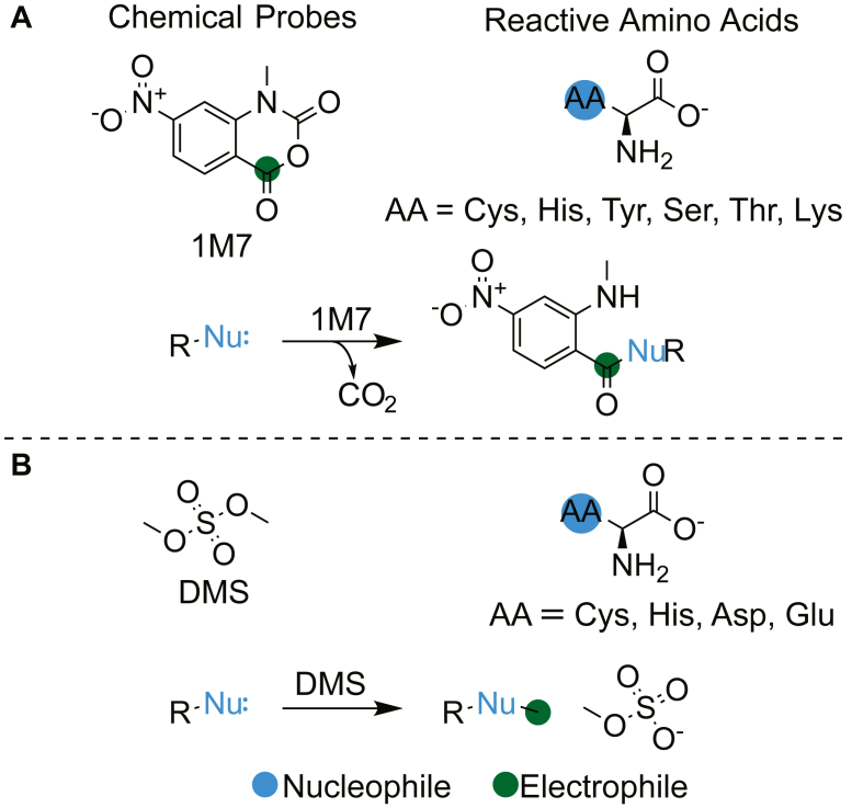
Figure 2 Nucleophilic amino acids that react with RNA-modifying probes.A, 1M7 acylates Cys, His, Tyr, Ser, Thr and Lys residues. B, DMS methylates Cys, His, Asp, and Glu residues.
Figure 2 Nucleophilic amino acids that react with RNA-modifying probes.A, 1M7 acylates Cys, His, Tyr, Ser, Thr and Lys residues. B, DMS methylates Cys, His, Asp, and Glu residues.CC BY-NC-ND · PubMed Central
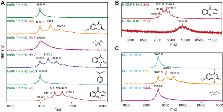
Figure 3 MALDI MS of proteins treated with RNA chemical probing reagents.A, MALDI MS of hnRNP K KH3 (top) treated with 1M7, DMS, NMIA, BzCN, 2A3. Single, double and multiple modifications are observed for 1M7, NMIA, and 2A3. B, increasing the 2A3 concentration from 18 mM (top) to 36 mM (bottom) leads to heavily modified protein species with the unmodified variant no longer observed. C, MALDI MS of SHARP RRM1 (top) treated with 1M7 (middle) and DMS (bottom).
Figure 3 MALDI MS of proteins treated with RNA chemical probing reagents.A, MALDI MS of hnRNP K KH3 (top) treated with 1M7, DMS, NMIA, BzCN, 2A3. Single, double and multiple modifications are observed for 1M7, NMIA, and 2A3. B, increasing the 2A3 concentration from 18 mM (top) to 36 mM (bottom) leads to heavily modified protein species with the unmodified variant no longer observed. C, MALDI MS of SHARP RRM1 (top) treated with 1M7 (middle) and DMS (bottom).CC BY-NC-ND · PubMed Central
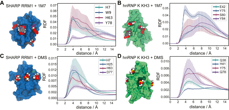
Figure 4 Analysis of the MD simulations of the RRM domain of SHARP, and the KH3 domain of hnRNP K with 1M7 and DMS.A, simulation structure of the RRM1 domain of SHARP (blue) with 1M7, and contours representing regions of high 1M7 density (magenta). B, simulation structure of the third KH domain of hnRNP K (green) with 1M7, and contours representing the regions of high 1M7 density (blue). C, simulation structure of the RRM1 domain of SHARP (blue) with DMS, and contours representing regions of high DMS density (green). D, simulation structure of the third KH domain of hnRNP K (green) and contours representing regions of high DMS density (blue). The plots represent the radial distribution function (RDF) of DMS or 1M7 to specific amino acids, averaged across the three simulation trials and reported with standard deviations.
Figure 4 Analysis of the MD simulations of the RRM domain of SHARP, and the KH3 domain of hnRNP K with 1M7 and DMS.A, simulation structure of the RRM1 domain of SHARP (blue) with 1M7, and contours representing regions of high 1M7 density (magenta). B, simulation structure of the third KH domain of hnRNP K (green) with 1M7, and contours representing the regions of high 1M7 density (blue). C, simulation structure of the RRM1 domain of SHARP (blue) with DMS, and contours representing regions of high DMS density (green). D, simulation structure of the third KH domain of hnRNP K (green) and contours representing regions of high DMS density (blue). The plots represent the radial distribution function (RDF) of DMS or 1M7 to specific amino acids, averaged across the three simulation trials and reported with standard deviations.CC BY-NC-ND · PubMed Central
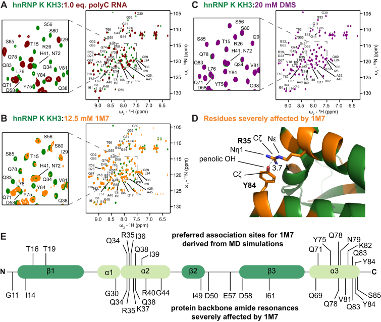
Figure 5 Backbone HMQC spectra recorded at 298K demonstrate binding of hnRNP K KH3 by RNA and interactions with chemical probes. Assignments refer to apo protein since CSPs and line broadening impede transfer of the assignments to bound/modified protein for many residues. Overlays show hnRNP K KH3 backbone spectrum (green) upon addition of (A) 1.0 eq. of 15 nt polyC RNA, (B) 12.5 mM 1M7, and (C) 20 mM DMS. D, residues severely affected by 1M7 (orange) mapped the X-ray structure of hnRNP K KH3. A stacking interaction between Arg35 and Tyr84 potentially catalyzes the reaction. The full structure is shown in Figure S7. E, residues of hnRNP K KH3 affected by treatment with 1M7 identified by MD simulations (top) and NMR spectroscopy (bottom).
Figure 5 Backbone HMQC spectra recorded at 298K demonstrate binding of hnRNP K KH3 by RNA and interactions with chemical probes. Assignments refer to apo protein since CSPs and line broadening impede transfer of the assignments to bound/modified protein for many residues. Overlays show hnRNP K KH3 backbone spectrum (green) upon addition of (A) 1.0 eq. of 15 nt polyC RNA, (B) 12.5 mM 1M7, and (C) 20 mM DMS. D, residues severely affected by 1M7 (orange) mapped the X-ray structure of hnRNP K KH3. A stacking interaction between Arg35 and Tyr84 potentially catalyzes the reaction. The full structure is shown in Figure S7. E, residues of hnRNP K KH3 affected by treatment with 1M7 identified by MD simulations (top) and NMR spectroscopy (bottom).CC BY-NC-ND · PubMed Central
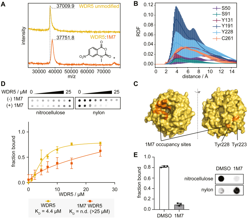
Figure 6 Treatment with chemical probes perturbs RNA binding of WDR5.A, MALDI-MS shows an increase in molecular weight of WDR5 treated with 1M7. Potential dimer formation of modified protein is highlighted with an asterisk. B, radial distribution plot of 1M7 against specific residues of WDR5, averaged across three simulation trials and reported with standard deviations. C, preferred 1M7 occupancy sites (orange) cluster on the structure of WDR5 (yellow). D, filter dot-blot assay of WDR5 and lncRNA UMLILO. WDR5 loses its binding ability to the target RNA when treated with 1M7. Errors from triplicate experiments shown in Figure S13. E, filter dot-blot assay of pre-formed UMLILO-WDR5 complex treated with DMSO or 1M7. Plot of median and standard deviation error bars from triplicate experiments shown in Figure S14.
Figure 6 Treatment with chemical probes perturbs RNA binding of WDR5.A, MALDI-MS shows an increase in molecular weight of WDR5 treated with 1M7. Potential dimer formation of modified protein is highlighted with an asterisk. B, radial distribution plot of 1M7 against specific residues of WDR5, averaged across three simulation trials and reported with standard deviations. C, preferred 1M7 occupancy sites (orange) cluster on the structure of WDR5 (yellow). D, filter dot-blot assay of WDR5 and lncRNA UMLILO. WDR5 loses its binding ability to the target RNA when treated with 1M7. Errors from triplicate experiments shown in Figure S13. E, filter dot-blot assay of pre-formed UMLILO-WDR5 complex treated with DMSO or 1M7. Plot of median and standard deviation error bars from triplicate experiments shown in Figure S14.CC BY-NC-ND · PubMed Central

Verification

Claims the paper makes

The claims and statistics of this paper haven’t been checked yet.

Run the check — free, no account needed. It reads the open-access text and checks the sources it cites.

A few free checks a day · papers already verified open instantly · results are public

Self-check

Percentages, sums and reported differences re-computed from the paper’s own numbers.

The self-check needs the paper’s full text.

Citations

Cites

Metadata

Authors
Klingler, D., Fallon, L., Cohn, D., Crawford, A., Marcus, L., & Jones, A. N.
Affiliations
New York University (US)
Corresponding author
Jones, Alisha N
Venue
Journal of Biological Chemistry
Publisher
openRxiv
Published
2024-03-21
Volume
301
Issue
8
Pages
110468-110468
ISSN
0021-9258 (ISSN-L)
OpenAlex
W4412392221
Cited by
2
References
1
Citations by year
2026: 2
FWCI
0.60
Type
journal article
Language
en
Open access
Locations
  • Journal of Biological Chemistry (publishedVersion, cc-by-nc-nd)
  • bioRxiv (Cold Spring Harbor Laboratory) (acceptedVersion)
  • PubMed Central (publishedVersion, cc-by-nc-nd)
  • PubMed (publishedVersion)
Funders
National Science Foundation (2243667)
Topics
  • Life Sciences › Biochemistry, Genetics and Molecular Biology › Molecular Biology › RNA and protein synthesis mechanisms
  • Life Sciences › Biochemistry, Genetics and Molecular Biology › Molecular Biology › DNA and Nucleic Acid Chemistry
  • Life Sciences › Biochemistry, Genetics and Molecular Biology › Molecular Biology › RNA Research and Splicing
Keywords
RNA-binding proteins, RNA-protein interactions, methylating reagents, acylating reagents, RNA structure, binding site identification
MeSH terms
Binding Sites, Humans, Protein Binding, RNA, RNA-Binding Proteins, Spectrometry, Mass, Matrix-Assisted Laser Desorption-Ionization, Molecular Dynamics Simulation

About these checks

Registry, retraction, Cites and statistics results are computed, not judged, with one exception: a printed reference that no index lists is read once by a model before it counts as a citation error. Claims are extracted by a model and kept only when their quote appears verbatim in the text; each cited source is then checked, with a model’s help, for whether it says what the claim relies on. Those verdicts can be wrong — read the claim’s quote and the cited source before relying on one.

Statistics and arithmetic are recomputed, not judged.

Confidence bands are shown only for fields calibrated on our annotated test set. None is calibrated yet, so no band is shown.

What counts as a claim ›How we measure confidence ›How we check this ›

Are you an author, or do you see a problem with this page? Request a review

Data sources

Bibliographic data: Semantic Scholar, OpenAlex, Crossref.

Contains information from the Semantic Scholar Academic Graph Datasets, made available under the ODC Attribution License (ODC-BY 1.0).

Semantic Scholar’s data is described in: Kinney, R., et al. (2023). The Semantic Scholar Open Data Platform. arXiv:2301.10140.