PreprintCited by 9Protein Structure and DynamicsChemical Synthesis and AnalysisAdvanced Physical and Chemical Molecular Interactions

The DOI matches its Crossref record; 1 of the 1 references it cites were found in a registry or an index.

An integrative characterisation of proline cis and trans conformers in a disordered peptide

Open access

Preprint — published as 10.1016/j.bpj.2024.09.028

Abstract

Intrinsically disordered proteins (IDPs) often contain proline residues, which undergo cis/trans isomerisation. While molecular dynamics (MD) simulations have the potential to fully characterise the proline cis and trans sub-ensembles, they are limited by the slow timescales of isomerisation and force field inaccuracies. Nuclear magnetic resonance (NMR) spectroscopy can report on ensemble-averaged observables for both the cis and trans proline states, but a full atomistic characterisation of these sub-ensembles is challenging. Given the importance of proline cis/trans isomerisation for influencing the conformational sampling of disordered proteins, we employed a combination of all-atom MD simulations with enhanced sampling (metadynamics), NMR, and small-angle X-ray scattering (SAXS) to characterise the two sub-ensembles of the ORF6 C-terminal region (ORF6 CTR ) from SARS-CoV-2 corresponding to the proline-57 (P57) cis and trans states. We performed MD simulations in three distinct force fields: AMBER03ws, AMBER99SB- disp , and CHARMM36m, which are all optimised for disordered proteins. Each simulation was run for an accumulated time of 180-220 µs until convergence was reached, as assessed by blocking analysis. A good agreement between the cis -P57 populations predicted from metadynamics simulations in AMBER03ws was observed with populations obtained from experimental NMR data. Moreover, we observed good agreement between the radius of gyration predicted from the metadynamics simulations in AMBER03ws and that measured using SAXS. Our findings suggest that both the cis -P57 and trans -P57 conformations of ORF6 CTR are extremely dynamic and that interdisciplinary approaches combining both multi-scale computations and experiments offer avenues to explore highly dynamic states that cannot be reliably characterised by either approach in isolation. SIGNIFICANCE: This study employs MD simulations (with metadynamics), NMR spectroscopy, and SAXS to elucidate the individual cis and trans proline conformations of ORF6 CTR from SARS-CoV-2. The good agreement on proline cis / trans populations observed in experiments (NMR) and those calculated from simulations in the AMBER03ws force field (with SAXS reweighting) showcases the efficiency of this interdisciplinary approach, which can be used to characterise highly dynamic disordered protein states, even for very slow processes. Furthermore, our study emphasises the importance of considering both computational and experimental methodologies to gain a more holistic understanding of highly dynamic proteins. The presented integrative approach sets a precedent for future studies aiming to explore complex and dynamic biological systems with slow transitions such as proline isomerisations.

Overview

The claims and statistics of this paper haven’t been checked yet.

Run the check — free, no account needed. It reads the open-access text and checks the sources it cites.

A few free checks a day · papers already verified open instantly · results are public

Integrity signals

Registry

Crossref · 10.1101/2024.05.14.594077

Cites

1 of 1 references verified0 citation errors · 0 not checkable

Statistics

Figures

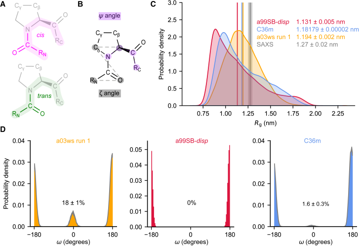
Figure 1 All three force fields predict NAc-ORF6CTR is disordered but only the a03ws and C36m force fields predict that NAc-ORF6CTR undergoes proline cis/trans isomerization. (A) cis-proline (pink) and trans-proline (green) conformations with the positions of Cγ and Cβ indicated. (B) Definition of the ζ (gray) and ψ (purple) dihedral angles used as CVs to enhance proline cis/trans isomerization. (C) Rg probability distributions were calculated using kernel density estimates to compare the a03ws run 1 (orange), a99SB-disp (red), and C36m (blue) ensembles. Ensemble-averaged Rg are shown for each system. The associated error represents the standard deviation between the first and second halves of the analyzed trajectories. The experimental SAXS data and error (standard deviation from the Guinier analysis) are shown in gray. (D) Probability distributions for the ω angle about the Q56-P57 peptide bond in the a03ws run 1 (orange), a99SB-disp (red), and C36m (blue) ensembles. The thickness of the gray distribution represents the uncertainty. The cis-P57 populations are displayed for each system and the error represents the standard deviation between the first and second halves of the analyzed trajectories.
Figure 1 All three force fields predict NAc-ORF6CTR is disordered but only the a03ws and C36m force fields predict that NAc-ORF6CTR undergoes proline cis/trans isomerization. (A) cis-proline (pink) and trans-proline (green) conformations with the positions of Cγ and Cβ indicated. (B) Definition of the ζ (gray) and ψ (purple) dihedral angles used as CVs to enhance proline cis/trans isomerization. (C) Rg probability distributions were calculated using kernel density estimates to compare the a03ws run 1 (orange), a99SB-disp (red), and C36m (blue) ensembles. Ensemble-averaged Rg are shown for each system. The associated error represents the standard deviation between the first and second halves of the analyzed trajectories. The experimental SAXS data and error (standard deviation from the Guinier analysis) are shown in gray. (D) Probability distributions for the ω angle about the Q56-P57 peptide bond in the a03ws run 1 (orange), a99SB-disp (red), and C36m (blue) ensembles. The thickness of the gray distribution represents the uncertainty. The cis-P57 populations are displayed for each system and the error represents the standard deviation between the first and second halves of the analyzed trajectories.CC BY · PubMed Central
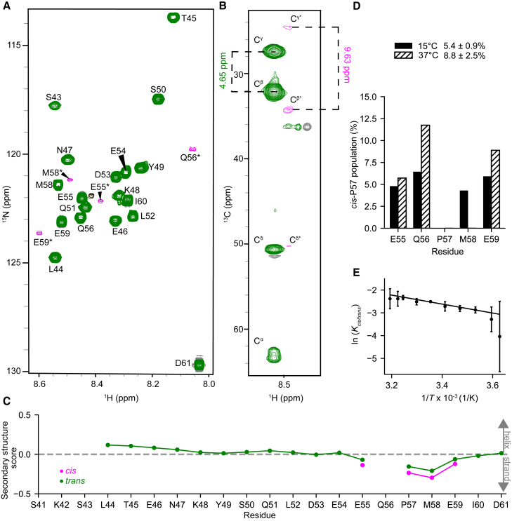
Figure 2 NMR verifies the disordered ORF6CTR undergoes proline cis/trans isomerization. (A) 2D 1H-15N HSQC spectrum of 300 μM 15N-labeled ORF6CTR measured at 15°C, pH 6.9, and 14.1 T. Residues colored in green originate from the trans-P57 conformational ensemble, while residues colored in pink and labeled with an asterisk (∗) report on the cis-P57 conformation. The black peak is unassigned. (B) 2D 1H-13C strip taken from the 3D CC(CO)NH spectrum of the 300 μM 13C,15N-labeled ORF6CTR at the 15N chemical shift of M58∗. (C) The ORF6CTR13Cα, 13Cβ, and 1Hα chemical shifts were used to calculate the secondary structure propensity scores for both the cis-P57 and trans-P57 subensembles. A positive value indicates α-helical propensity, a negative value indicates a β-strand propensity, whereas a value near zero indicates random coil. (D) The cis-P57 populations at 15°C (solid fill) and 37°C (dashed fill) are shown for each well-resolved cis-P57 and trans-P57 peak in the 2D 1H-15N HSQC spectrum. The integrated peak volume was used to calculate the mean cis-P57 population and standard deviation across residues E55, Q56, and E59. (E) The van ’t Hoff analysis of P57 cis/trans isomerization determined from the integrated peak volume. The mean natural logarithm of the equilibrium constant for proline cis/trans isomerization about the Q56-P57 peptide bond as a function of temperature was calculated using residues E55, Q56, and E59. Error bars represent the standard deviation across the three residues used in the analysis. The van ’t Hoff linear fit yielded a cis-P57 population of 10 ± 2% at 37°C.
Figure 2 NMR verifies the disordered ORF6CTR undergoes proline cis/trans isomerization. (A) 2D 1H-15N HSQC spectrum of 300 μM 15N-labeled ORF6CTR measured at 15°C, pH 6.9, and 14.1 T. Residues colored in green originate from the trans-P57 conformational ensemble, while residues colored in pink and labeled with an asterisk (∗) report on the cis-P57 conformation. The black peak is unassigned. (B) 2D 1H-13C strip taken from the 3D CC(CO)NH spectrum of the 300 μM 13C,15N-labeled ORF6CTR at the 15N chemical shift of M58∗. (C) The ORF6CTR13Cα, 13Cβ, and 1Hα chemical shifts were used to calculate the secondary structure propensity scores for both the cis-P57 and trans-P57 subensembles. A positive value indicates α-helical propensity, a negative value indicates a β-strand propensity, whereas a value near zero indicates random coil. (D) The cis-P57 populations at 15°C (solid fill) and 37°C (dashed fill) are shown for each well-resolved cis-P57 and trans-P57 peak in the 2D 1H-15N HSQC spectrum. The integrated peak volume was used to calculate the mean cis-P57 population and standard deviation across residues E55, Q56, and E59. (E) The van ’t Hoff analysis of P57 cis/trans isomerization determined from the integrated peak volume. The mean natural logarithm of the equilibrium constant for proline cis/trans isomerization about the Q56-P57 peptide bond as a function of temperature was calculated using residues E55, Q56, and E59. Error bars represent the standard deviation across the three residues used in the analysis. The van ’t Hoff linear fit yielded a cis-P57 population of 10 ± 2% at 37°C.CC BY · PubMed Central
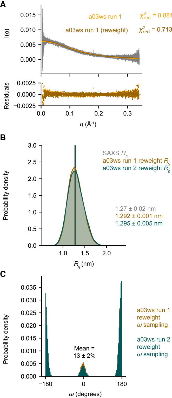
Figure 3 The a03ws force field most accurately predicts the disordered NAc-ORF6CTR ensemble and P57 cis/trans populations after SAXS BME reweighting. (A) The calculated SAXS intensities from the a03ws run 1 metadynamic simulation (orange) and the SAXS BME reweighted simulation (brown) compared with the experimental SAXS intensities (gray), and the error associated with each intensity (gray). (B) Rg probability distributions were calculated using kernel density estimates to compare the SAXS BME reweighted a03ws run 1 ensemble (brown) and the SAXS BME reweighted a03ws run 2 ensemble (teal). The associated error represents the standard deviation between the first and second halves of the analyzed trajectories. The experimental SAXS data and error (standard deviation from the Guinier analysis) are shown in gray. (C) Probability distributions for the ω angle about the Q56-P57 peptide bond in the SAXS BME reweighted a03ws run 1 ensemble (brown) and the SAXS BME reweighted a03ws run 2 ensemble (teal). The two reweighted independent a03ws ensembles were used to calculate the mean cis-P57 population and standard deviation. Additional plots for the SAXS BME reweighted a03ws run 2 and C36m ensembles can be found in the supporting material.
Figure 3 The a03ws force field most accurately predicts the disordered NAc-ORF6CTR ensemble and P57 cis/trans populations after SAXS BME reweighting. (A) The calculated SAXS intensities from the a03ws run 1 metadynamic simulation (orange) and the SAXS BME reweighted simulation (brown) compared with the experimental SAXS intensities (gray), and the error associated with each intensity (gray). (B) Rg probability distributions were calculated using kernel density estimates to compare the SAXS BME reweighted a03ws run 1 ensemble (brown) and the SAXS BME reweighted a03ws run 2 ensemble (teal). The associated error represents the standard deviation between the first and second halves of the analyzed trajectories. The experimental SAXS data and error (standard deviation from the Guinier analysis) are shown in gray. (C) Probability distributions for the ω angle about the Q56-P57 peptide bond in the SAXS BME reweighted a03ws run 1 ensemble (brown) and the SAXS BME reweighted a03ws run 2 ensemble (teal). The two reweighted independent a03ws ensembles were used to calculate the mean cis-P57 population and standard deviation. Additional plots for the SAXS BME reweighted a03ws run 2 and C36m ensembles can be found in the supporting material.CC BY · PubMed Central
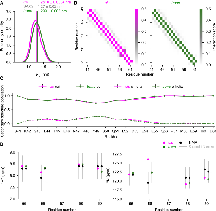
Figure 4 NAc-ORF6CTRcis-P57 and trans-P57 subensembles predicted by the a03ws run 1 SAXS BME reweighted metadynamic simulation have very similar profiles. (A) Rg probability distributions were calculated using kernel density estimates to compare the cis-P57 (pink) and trans-P57 (green) conformational subensembles. The associated error represents the standard deviation between the first and second halves of the analyzed trajectory. The experimental SAXS data and error (standard deviation from the Guinier analysis) are shown in gray. (B) Ensemble-averaged Cα minimum distance contact maps for the cis-P57 (pink) and trans-P57 (green) conformations. Color intensity represents the probability of a contact between residues, with white indicating no contact, and increasing color intensity suggesting higher contact probabilities. (C) Secondary structure populations for all residues in the cis-P57 (pink) and the trans-P57 (green) conformations based on SAXS BME statistical weights. Coil populations are represented by solid lines and α-helical populations by dashed lines. β-strand represents less than 0.5% of the population for each residue so was not included. Error bars (black) represent the standard deviation between the first and second halves of the analyzed trajectory. (D) Consistency of the 1HN and 15N cis-P57 and trans-P57 ORF6CTR experimental chemical shifts with the predicted chemical shifts from the reweighted NAc-ORF6CTR a03ws run 1 ensemble. The error in CamShift (silver) is shown. The standard deviation for predicted chemical shifts between the first and second halves of the analyzed trajectory were also plotted but are too small to see. See the supporting material for the a03ws run 2 and C36m analysis.
Figure 4 NAc-ORF6CTRcis-P57 and trans-P57 subensembles predicted by the a03ws run 1 SAXS BME reweighted metadynamic simulation have very similar profiles. (A) Rg probability distributions were calculated using kernel density estimates to compare the cis-P57 (pink) and trans-P57 (green) conformational subensembles. The associated error represents the standard deviation between the first and second halves of the analyzed trajectory. The experimental SAXS data and error (standard deviation from the Guinier analysis) are shown in gray. (B) Ensemble-averaged Cα minimum distance contact maps for the cis-P57 (pink) and trans-P57 (green) conformations. Color intensity represents the probability of a contact between residues, with white indicating no contact, and increasing color intensity suggesting higher contact probabilities. (C) Secondary structure populations for all residues in the cis-P57 (pink) and the trans-P57 (green) conformations based on SAXS BME statistical weights. Coil populations are represented by solid lines and α-helical populations by dashed lines. β-strand represents less than 0.5% of the population for each residue so was not included. Error bars (black) represent the standard deviation between the first and second halves of the analyzed trajectory. (D) Consistency of the 1HN and 15N cis-P57 and trans-P57 ORF6CTR experimental chemical shifts with the predicted chemical shifts from the reweighted NAc-ORF6CTR a03ws run 1 ensemble. The error in CamShift (silver) is shown. The standard deviation for predicted chemical shifts between the first and second halves of the analyzed trajectory were also plotted but are too small to see. See the supporting material for the a03ws run 2 and C36m analysis.CC BY · PubMed Central
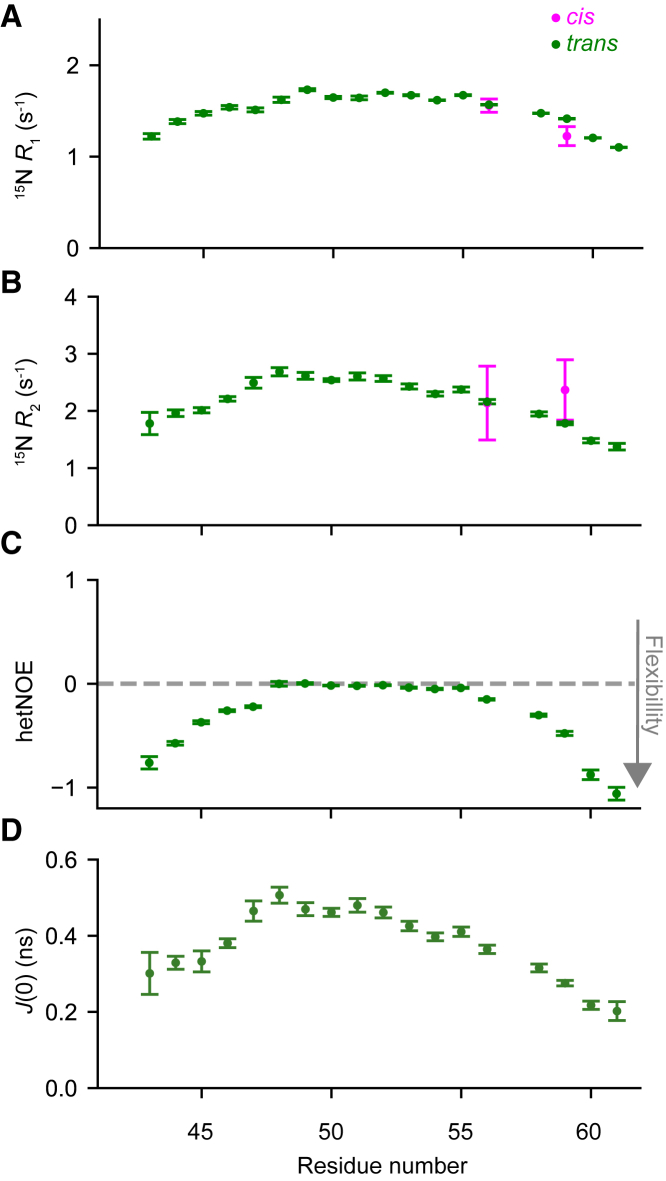
Figure 5 ORF6CTR is very dynamic in both the trans-P57 and cis-P57 conformations. (A) 15N longitudinal relaxation rates (R1), (B) 15N transverse relaxation rates (R2), (C) {1H}-15N steady-state hetNOEs, and (D) J(0) values of 300 μM 15N-labeled ORF6CTR at 15°C and pH 6.9. The backbone motions are shown for all peaks corresponding to the trans-P57 conformation (green), and only for the well-resolved peaks with a measurable intensity corresponding to the cis-P57 (pink) conformation. Error bars represent the fitting of the NMR data in (A) and (C). The error bars for (B) were calculated by error propagation. The error bars for (D) represent the standard deviations for each residue, which were calculated using Monte Carlo uncertainty propagation. All experiments were recorded at a static magnetic field strength of 14.1 T.
Figure 5 ORF6CTR is very dynamic in both the trans-P57 and cis-P57 conformations. (A) 15N longitudinal relaxation rates (R1), (B) 15N transverse relaxation rates (R2), (C) {1H}-15N steady-state hetNOEs, and (D) J(0) values of 300 μM 15N-labeled ORF6CTR at 15°C and pH 6.9. The backbone motions are shown for all peaks corresponding to the trans-P57 conformation (green), and only for the well-resolved peaks with a measurable intensity corresponding to the cis-P57 (pink) conformation. Error bars represent the fitting of the NMR data in (A) and (C). The error bars for (B) were calculated by error propagation. The error bars for (D) represent the standard deviations for each residue, which were calculated using Monte Carlo uncertainty propagation. All experiments were recorded at a static magnetic field strength of 14.1 T.CC BY · PubMed Central

Verification

Claims the paper makes

The claims and statistics of this paper haven’t been checked yet.

Run the check — free, no account needed. It reads the open-access text and checks the sources it cites.

A few free checks a day · papers already verified open instantly · results are public

Self-check

Percentages, sums and reported differences re-computed from the paper’s own numbers.

The self-check needs the paper’s full text.

Citations

Cites

Research works printed but not linked: 68

  1. 10.1016/j.tibs.2015.07.004 · DOI
  2. 10.1016/S0022-2836(02)00969-5 · DOI · Paper page
  3. 2014, Exceptionally abundant exceptions: Comprehensive characterization of intrinsic disorder in all domains of life, Cellular and Molecular Life Sciences, 72, 137
  4. 10.4161/idp.24360 · DOI
  5. 10.1021/bi00613a026 · DOI
  6. 10.1038/s41467-024-44859-2 · DOI
  7. 10.1021/jacs.6b10272 · DOI
  8. Zhang X , Vigers M , McCarty J , Rauch JN , Fredrickson GH , Wilson MZ , Shea JE , Han S , Kosik KS . 2020. The proline-rich domain promotes Tau liquid-liquid phase separation in cells. Journal of Cell Biology. 219. · DOI
  9. 10.1002/bip.1977.360160707 · DOI
  10. 10.1002/bip.1981.360201209 · DOI
  11. 10.1006/jmbi.1998.1770 · DOI
  12. 10.1002/cbic.201700548 · DOI
  13. 2021, An Unbound Proline-Rich Signaling Peptide Frequently Samples Cis Conformations in Gaussian Accelerated Molecular Dynamics Simulations, Front Mol Biosci, 8, 734169, 10.3389/fmolb.2021.734169 · DOI
  14. 10.1021/ct500569b · DOI
  15. 10.1073/pnas.1800690115 · DOI
  16. 10.1038/nmeth.4067 · DOI
  17. 10.1016/j.sbi.2016.12.004 · DOI
  18. 2020, Integrating Molecular Simulation and Experimental Data: A Bayesian/Maximum Entropy Reweighting Approach, Methods in molecular biology, 2112, 219, 10.1007/978-1-0716-0270-6_15 · DOI
  19. 2011, Metadynamics, Wiley Interdiscip Rev Comput Mol Sci, 1, 826, 10.1002/wcms.31 · DOI
  20. 10.1021/acs.jctc.5b00846 · DOI
  21. Heller GT , Aprile FA , Michaels TCT , Limbocker R , Perni M , Ruggeri FS , et al. 2020. Small-molecule sequestration of amyloid-β as a drug discovery strategy for Alzheimer’s disease. Sci Adv. 6. · DOI
  22. 10.1021/jp9046962 · DOI
  23. 2021, Proline isomerization effects in the amyloidogenic protein β 2 -microglobulin, Physical Chemistry Chemical Physics, 23, 356, 10.1039/D0CP04780E · DOI
  24. 10.1021/cr400688u · DOI
  25. 10.1016/j.jmr.2013.10.020 · DOI
  26. 10.1007/s12192-017-0791-z · DOI
  27. 2020, The Ambivalent Role of Proline Residues in an Intrinsically Disordered Protein: From Disorder Promoters to Compaction Facilitators, J Mol Biol, 432, 3093, 10.1016/j.jmb.2019.11.015 · DOI
  28. 10.1016/j.bpj.2022.12.013 · DOI
  29. 2022, Molecular Mechanism of SARS-CoVs Orf6 Targeting the Rae1–Nup98 Complex to Compete With mRNA Nuclear Export, Front Mol Biosci, 8, 1345
  30. 2022, Structural basis for Sarbecovirus ORF6 mediated blockage of nucleocytoplasmic transport, Nat Commun, 13, 4782, 10.1038/s41467-022-32489-5 · DOI
  31. 10.1073/pnas.2016650117 · DOI
  32. 2021, Refinement of α-Synuclein Ensembles Against SAXS Data: Comparison of Force Fields and Methods, Front Mol Biosci, 8, 654333, 10.3389/fmolb.2021.654333 · DOI
  33. 10.1021/ja072717t · DOI
  34. 10.1007/BF00197809 · DOI
  35. 10.1093/bioinformatics/btu830 · DOI
  36. 10.1110/ps.062465306 · DOI
  37. 10.1107/S0021889800012930 · DOI
  38. 10.1107/S1600576717011438 · DOI
  39. 2015, GROMACS: High performance molecular simulations through multi-level parallelism from laptops to supercomputers, SoftwareX, 1–2, 19
  40. 10.1038/s41592-019-0506-8 · DOI
  41. Abascal JLF , Vega C . 2005. A general purpose model for the condensed phases of water: TIP4P/2005. J Chem Phys. 123. · DOI
  42. 2015, Water dispersion interactions strongly influence simulated structural properties of disordered protein states, Journal of Physical Chemistry B, 119, 5113, 10.1021/jp508971m · DOI
  43. 10.1063/1.445869 · DOI
  44. 10.1021/ct5010406 · DOI
  45. 10.1063/1.2408420 · DOI
  46. 10.1063/1.328693 · DOI
  47. 10.1002/(SICI)1096-987X(199709)18:12 · DOI
  48. 10.1063/1.470117 · DOI
  49. 2019, Analyzing and Biasing Simulations with PLUMED, Methods in molecular biology, 2022, 529, 10.1007/978-1-4939-9608-7_21 · DOI
  50. 10.1016/j.bpj.2015.08.015 · DOI
  51. 10.1021/ja903772t · DOI
  52. 2017, Pepsi-SAXS: an adaptive method for rapid and accurate computation of small-angle X-ray scattering profiles, Acta Crystallogr D Struct Biol, 73, 449, 10.1107/S2059798317005745 · DOI
  53. 10.1021/acs.jctc.5b00736 · DOI
  54. 10.1021/acs.jctc.9b00251 · DOI
  55. 10.1016/j.jmb.2014.12.007 · DOI
  56. Wilson CJ , Choy WY , Karttunen M . 2022. AlphaFold2: A Role for Disordered Protein/Region Prediction? Int J Mol Sci. 23. · DOI · Paper page
  57. 10.1371/journal.pcbi.1005659 · DOI · Paper page
  58. 2023, Improved predictions of phase behaviour of intrinsically disordered proteins by tuning the interaction range, Open Research Europe, 2, 94, 10.12688/openreseurope.14967.2 · DOI
  59. 10.1038/s41586-023-07004-5 · DOI
  60. 2024, Direct prediction of intrinsically disordered protein conformational properties from sequences, Nat Methods, 21, 465, 10.1038/s41592-023-02159-5 · DOI
  61. 10.1023/A:1018334207887 · DOI
  62. 10.1006/jmrb.1993.1019 · DOI
  63. 10.1007/s10858-009-9395-y · DOI
  64. 10.1016/S1090-7807(03)00062-4 · DOI
  65. 2023, Not Only Expansion: Proline Content and Density Also Induce Disordered Protein Conformation Compaction, J Mol Biol, 435, 168196, 10.1016/j.jmb.2023.168196 · DOI
  66. 10.1021/bi00449a003 · DOI
  67. 10.1021/ja0204776 · DOI
  68. 2016, Identification of Dynamic Modes in an Intrinsically Disordered Protein Using Temperature-Dependent NMR Relaxation, J Am Chem Soc, 138, 6240, 10.1021/jacs.6b02424 · DOI

Metadata

Authors
Pettitt, A. J., Shukla, V. K., Figueiredo, A. M., Newton, L. S., McCarthy, S., Tabor, A. B., Heller, G. T., Lorenz, C. D., & Hansen, D. F.
Affiliations
  • King's College London (GB)
  • The Francis Crick Institute (GB)
  • Institute of Structural and Molecular Biology (GB)
Corresponding author
  • Lorenz, Christian D.
  • Hansen, D. Flemming
Venue
Biophysical Journal
Publisher
openRxiv
Published
2024-05-15
Volume
123
Issue
21
Pages
3798-3811
ISSN
0006-3495 (ISSN-L)
OpenAlex
W4402879499
Cited by
9
References
1
Citations by year
2026: 6 · 2025: 3
FWCI
1.22
Type
journal article
Language
en
Open access
Licence
cc-by
Locations
  • Biophysical Journal (publishedVersion, cc-by)
  • bioRxiv (Cold Spring Harbor Laboratory) (acceptedVersion, cc-by-nc-nd)
  • PubMed Central (publishedVersion, cc-by)
  • PubMed (publishedVersion)
  • HAL (submittedVersion, cc-by)
  • UCL Discovery (University College London) (submittedVersion, cc-by)
  • Figshare (submittedVersion, cc-by)
  • The Francis Crick Institute (cc-by)
Funders
  • Wellcome Trust (FC001029, 101569/Z/13/Z, MR/N013867/1, 109073/Z/15/Z)
  • Francis Crick Institute (FC001029)
  • Diamond Light Source
  • UK Research and Innovation (EP/X036782/1, MR/N013867/1)
  • Cancer Research UK (FC001029)
  • University College London
  • Partnership for Advanced Computing in Europe AISBL
  • Directorate for Biological Sciences
  • Medical Research Council (FC001029, MR/N013867/1, COVID-19)
  • Engineering and Physical Sciences Research Council (EP/X036782/1)
  • Biotechnology and Biological Sciences Research Council (BB/X009955/1, BB/T008709/1, BB/R000255/1, T008709)
  • Newnham College, University of Cambridge
Topics
  • Life Sciences › Biochemistry, Genetics and Molecular Biology › Molecular Biology › Protein Structure and Dynamics
  • Physical Sciences › Chemistry › Organic Chemistry › Chemical Synthesis and Analysis
  • Physical Sciences › Chemistry › Physical and Theoretical Chemistry › Advanced Physical and Chemical Molecular Interactions
Keywords
proline cis-trans isomerization, intrinsically disordered proteins, SARS-CoV-2, cis-trans conformers, metadynamics, force field comparison, CHARMM36m, radius of gyration, small-angle X-ray scattering, nuclear magnetic resonance, conformational dynamics
MeSH terms
SARS-CoV-2, Isomerism, Peptides, Proline, Protein Conformation, Viral Proteins, X-Ray Diffraction, Scattering, Small Angle, Molecular Dynamics Simulation, Intrinsically Disordered Proteins

About these checks

Registry, retraction, Cites and statistics results are computed, not judged, with one exception: a printed reference that no index lists is read once by a model before it counts as a citation error. Claims are extracted by a model and kept only when their quote appears verbatim in the text; each cited source is then checked, with a model’s help, for whether it says what the claim relies on. Those verdicts can be wrong — read the claim’s quote and the cited source before relying on one.

Statistics and arithmetic are recomputed, not judged.

Confidence bands are shown only for fields calibrated on our annotated test set. None is calibrated yet, so no band is shown.

What counts as a claim ›How we measure confidence ›How we check this ›

Are you an author, or do you see a problem with this page? Request a review