PreprintCited by 9CRISPR and Genetic EngineeringBiochemical and Structural CharacterizationBacterial Genetics and Biotechnology

The DOI matches its Crossref record; 1 of the 1 references it cites were found in a registry or an index.

Identification of cognate recombination directionality factors for large serine recombinases by virtual pulldown

Open access

Preprint — published as 10.1093/nar/gkaf691 · Paper page

Abstract

Integrases from the “large serine” family are simple, highly directional site-specific DNA recombinases that have great promise as synthetic biology and genome editing tools. Integrative recombination (mimicking phage or mobile element insertion) requires only integrase and two short (∼40 – 50) DNA sites. The reverse reaction, excisive recombination, does not occur until it is triggered by the presence of a second protein termed a Recombination Directionality Factor (RDF), which binds specifically to its cognate integrase. Identification of RDFs has been hampered due to their lack of sequence conservation and lack of synteny with the phage integrase gene. Here we use Alphafold2-multimer to identify putative RDFs for more than half of a test set of 98 large serine recombinases, and experimental methods to verify predicted RDFs for 4 of 5 integrases chosen as test cases. We find no universally conserved structural motifs among known and predicted RDFs, yet they are all predicted to bind a similar location on their cognate integrase, suggesting convergent evolution of function. Our methodology greatly expands the available genetic toolkit of cognate integrase – RDF pairs.

Overview

The claims and statistics of this paper haven’t been checked yet.

Run the check — free, no account needed. It reads the open-access text and checks the sources it cites.

A few free checks a day · papers already verified open instantly · results are public

Integrity signals

Registry

Crossref · 10.1101/2024.06.11.598349

Cites

1 of 1 references verified0 citation errors · 0 not checkable

Statistics

Figures

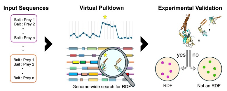
Graphical Abstract
Graphical Abstract CC BY-NC · PubMed Central
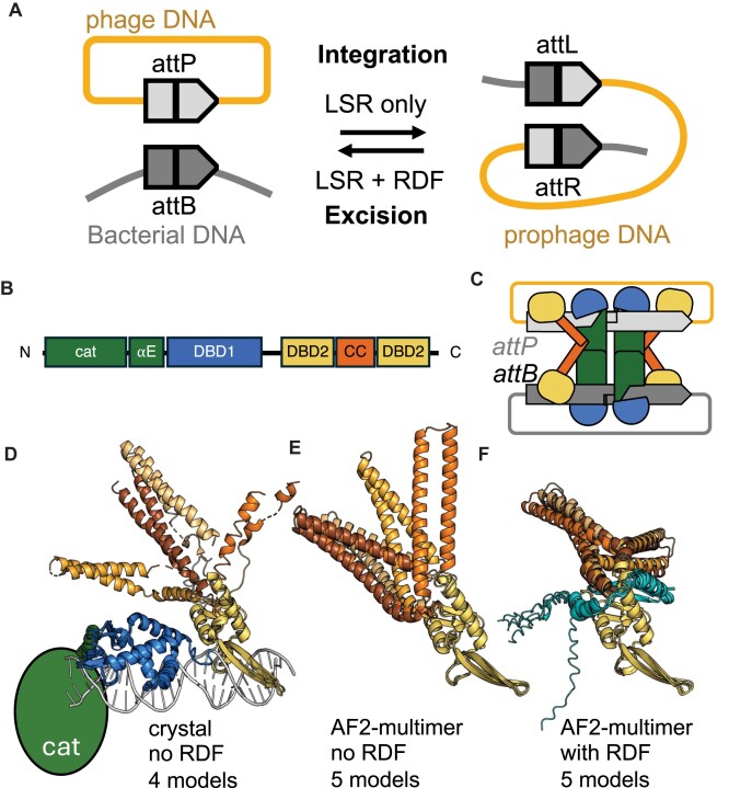
Figure 1. Recombination mechanism and domain structure. (A) Integrative recombination requires only the LSR and its cognate att sites but reverse reaction (excisive recombination) requires the presence of a second protein, the cognate RDF. (B) Linear cartoon of the domain structure of LSR domain structure. CAT: catalytic domain; αE: long helix connecting the CAT to the DNA-binding domains (DBDs); DBD1: DNA-binding domain 1 (also known as recombinase domain); DBD2: DNA-binding domain 2 (also known as Zn-binding domain); CC: coiled coil. (C) Cartoon of the proposed structure of the substrate complex for integrative recombination, as proposed by Van Duyne et al. [38]. (D) Structure of the DBDs of LI integrase bound to half an attP site [40]. The proteins from the four copies in the asymmetric unit are superimposed on one another, the second half of DBD2 as a guide. Domains are colored as in panel (C) except that varying shades of orange are used for the CCs to highlight their flexibility. (E) AlphaFold2-multimer modeling of LI integrase. DBD2 and the CC for five models are superimposed and colored as in panel (D). (F) AlphaFold2-multimer modeling of LI integrase in complex with its RDF [20]. The RDF (turquoise) partially restrains the flexibility of the CC when compared to the experimental or the AlphaFold2-multimer models in the absence of the RDF.
Figure 1. Recombination mechanism and domain structure. (A) Integrative recombination requires only the LSR and its cognate att sites but reverse reaction (excisive recombination) requires the presence of a second protein, the cognate RDF. (B) Linear cartoon of the domain structure of LSR domain structure. CAT: catalytic domain; αE: long helix connecting the CAT to the DNA-binding domains (DBDs); DBD1: DNA-binding domain 1 (also known as recombinase domain); DBD2: DNA-binding domain 2 (also known as Zn-binding domain); CC: coiled coil. (C) Cartoon of the proposed structure of the substrate complex for integrative recombination, as proposed by Van Duyne et al. [38]. (D) Structure of the DBDs of LI integrase bound to half an attP site [40]. The proteins from the four copies in the asymmetric unit are superimposed on one another, the second half of DBD2 as a guide. Domains are colored as in panel (C) except that varying shades of orange are used for the CCs to highlight their flexibility. (E) AlphaFold2-multimer modeling of LI integrase. DBD2 and the CC for five models are superimposed and colored as in panel (D). (F) AlphaFold2-multimer modeling of LI integrase in complex with its RDF [20]. The RDF (turquoise) partially restrains the flexibility of the CC when compared to the experimental or the AlphaFold2-multimer models in the absence of the RDF.CC BY-NC · PubMed Central
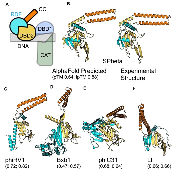
Figure 2. Predicted structures for known LSR–RDF pairs. (A) Cartoon similar to those in Fig. 1, but in the same relative orientation as the structural cartoons in panels (B–F). (B) AlphaFold-predicted versus experimental structure of SPbeta Int–RDF complex. (C–F) Predicted complexes of previously identified RDFs in complex with their cognate integrases. Only DBD2 and the CC inserted within it are shown for the LSR partner but the entire predicted structure for each RDF is shown.
Figure 2. Predicted structures for known LSR–RDF pairs. (A) Cartoon similar to those in Fig. 1, but in the same relative orientation as the structural cartoons in panels (B–F). (B) AlphaFold-predicted versus experimental structure of SPbeta Int–RDF complex. (C–F) Predicted complexes of previously identified RDFs in complex with their cognate integrases. Only DBD2 and the CC inserted within it are shown for the LSR partner but the entire predicted structure for each RDF is shown.CC BY-NC · PubMed Central
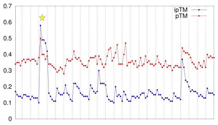
Figure 3. A scatter plot of the virtual pulldown for the phage-like element A118. Five models were predicted for the complex of each element-encoded protein with the A118 integrase (dotted vertical lines separate sets of models). The plot shows two measures of confidence for each—pTM (red) and ipTM (blue) scores—which were calculated using AlphaFold2-multimer v3 model in ColabFold v.1.5.2 [30, 45, 46]. The highest-ranking model for the known RDF is highlighted with a yellow star. See Supplementary Fig. S3 scatter plots for the other LSR–RDF pairs shown in Fig. 2 and for the PAE plots for the each of these known LSR–RDF pairs.
Figure 3. A scatter plot of the virtual pulldown for the phage-like element A118. Five models were predicted for the complex of each element-encoded protein with the A118 integrase (dotted vertical lines separate sets of models). The plot shows two measures of confidence for each—pTM (red) and ipTM (blue) scores—which were calculated using AlphaFold2-multimer v3 model in ColabFold v.1.5.2 [30, 45, 46]. The highest-ranking model for the known RDF is highlighted with a yellow star. See Supplementary Fig. S3 scatter plots for the other LSR–RDF pairs shown in Fig. 2 and for the PAE plots for the each of these known LSR–RDF pairs.CC BY-NC · PubMed Central
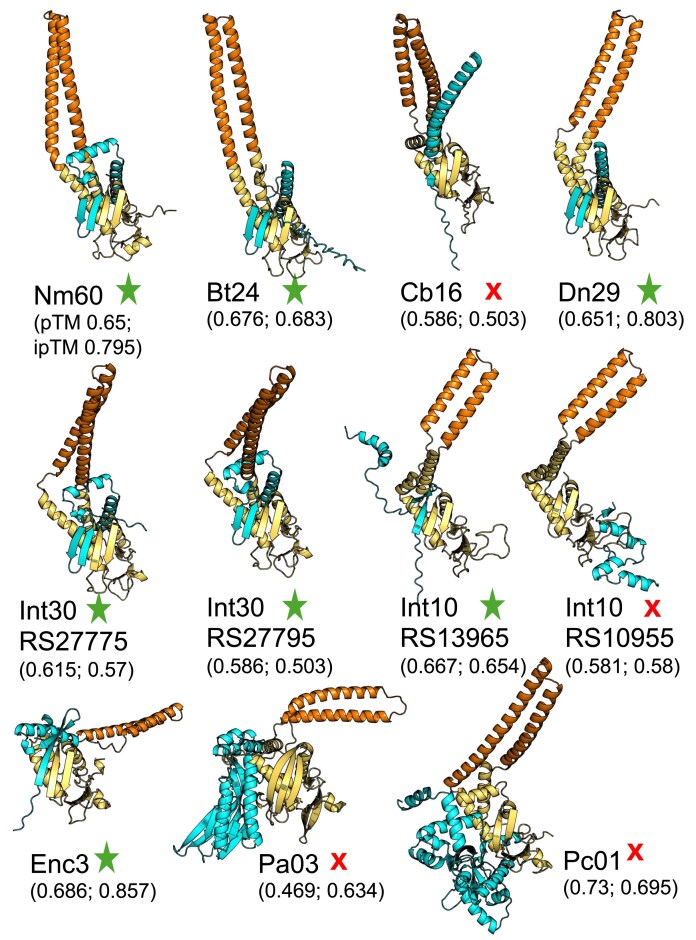
Figure 4. Predicted DBD2–RDF complex structures for the 11 LSR–RDF pairs tested experimentally. DBD2 of the LSR is shown in yellow, the CC in orange, and the RDF in cyan. If more than one potential RDF was predicted, a partial locus tag is added to disambiguate. Functional RDFs are marked with stars and false positives with X’s. PAE plots for each pair are shown in Supplementary Fig. S5. The confidence scores are given beneath each model; closer to 1 denotes higher confidence.
Figure 4. Predicted DBD2–RDF complex structures for the 11 LSR–RDF pairs tested experimentally. DBD2 of the LSR is shown in yellow, the CC in orange, and the RDF in cyan. If more than one potential RDF was predicted, a partial locus tag is added to disambiguate. Functional RDFs are marked with stars and false positives with X’s. PAE plots for each pair are shown in Supplementary Fig. S5. The confidence scores are given beneath each model; closer to 1 denotes higher confidence.CC BY-NC · PubMed Central
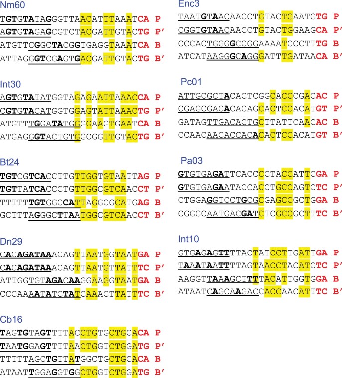
Figure 5. Alignment of LSR half-site sequences. The central dinucleotides are shown in bold and red font. DBD1 (also called RD) is expected to bind the innermost 12 bases, and DBD2 (also called ZD) is expected to bind the outer underlined segment. Bases that are conserved in three or four positions are highlighted in yellow in the DBD1 motif and in bold in the DBD2 motif. This convention follows [38].
Figure 5. Alignment of LSR half-site sequences. The central dinucleotides are shown in bold and red font. DBD1 (also called RD) is expected to bind the innermost 12 bases, and DBD2 (also called ZD) is expected to bind the outer underlined segment. Bases that are conserved in three or four positions are highlighted in yellow in the DBD1 motif and in bold in the DBD2 motif. This convention follows [38].CC BY-NC · PubMed Central
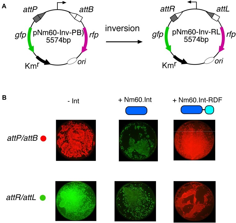
Figure 6. In vivo recombination reactions of Nm60 integrase and its fusion with the RDF. (A) Schematic illustration of the in vivo recombination (inversion) assay. In the plasmid pNm60-invPB, the promoter, flanked by attP and attB sites, constitutively drives the expression of an rfp gene (pink arrow). To prevent transcriptional read-through to the gfp gene (green arrow), a terminator sequence is inserted upstream of the promoter. LSR-catalyzed recombination (inversion) of attP × attB to give attR × attL products (plasmid pNm60-invRL) flips the orientation of the promoter to allow the expression of GFP, and block RFP production. (B) Recombination activities of Nm60 integrase and its fusion to the RDF on attP × attB (pNm60-invPB) and attR × attL (pNm60-invRL) substrates. The integrase is depicted as a long oval (blue) and the RDF as a short oval (cyan).
Figure 6. In vivo recombination reactions of Nm60 integrase and its fusion with the RDF. (A) Schematic illustration of the in vivo recombination (inversion) assay. In the plasmid pNm60-invPB, the promoter, flanked by attP and attB sites, constitutively drives the expression of an rfp gene (pink arrow). To prevent transcriptional read-through to the gfp gene (green arrow), a terminator sequence is inserted upstream of the promoter. LSR-catalyzed recombination (inversion) of attP × attB to give attR × attL products (plasmid pNm60-invRL) flips the orientation of the promoter to allow the expression of GFP, and block RFP production. (B) Recombination activities of Nm60 integrase and its fusion to the RDF on attP × attB (pNm60-invPB) and attR × attL (pNm60-invRL) substrates. The integrase is depicted as a long oval (blue) and the RDF as a short oval (cyan).CC BY-NC · PubMed Central
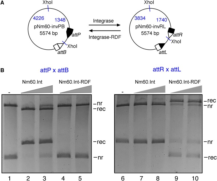
Figure 7. In vitro recombination reactions of Nm60 integrase and its fusion with the RDF. (A) Schematic illustration of the in vitro recombination (inversion) assay. LSR-catalyzed recombination (inversion) of attP and attB sites in pNm60-invPB gives rise to attR and attL sites in the product plasmid (pNm60-invRL), and vice versa. (B) Reactions were carried out for 2 h as described in the “Materials and methods” section. Integrase and integrase–RDF fusions were used at two final concentrations of 50 and 100 nM. Reaction products were digested with the restriction endonuclease, XhoI, prior to 1.2% agarose gel electrophoresis. As illustrated in panel (A), digestion of the plasmids with XhoI gives different restriction patterns for recombined and non-recombined DNAs. The bands on the gel are labeled nr (non-recombinant, i.e. substrate) and rec (recombination product).
Figure 7. In vitro recombination reactions of Nm60 integrase and its fusion with the RDF. (A) Schematic illustration of the in vitro recombination (inversion) assay. LSR-catalyzed recombination (inversion) of attP and attB sites in pNm60-invPB gives rise to attR and attL sites in the product plasmid (pNm60-invRL), and vice versa. (B) Reactions were carried out for 2 h as described in the “Materials and methods” section. Integrase and integrase–RDF fusions were used at two final concentrations of 50 and 100 nM. Reaction products were digested with the restriction endonuclease, XhoI, prior to 1.2% agarose gel electrophoresis. As illustrated in panel (A), digestion of the plasmids with XhoI gives different restriction patterns for recombined and non-recombined DNAs. The bands on the gel are labeled nr (non-recombinant, i.e. substrate) and rec (recombination product).CC BY-NC · PubMed Central

Verification

Claims the paper makes

The claims and statistics of this paper haven’t been checked yet.

Run the check — free, no account needed. It reads the open-access text and checks the sources it cites.

A few free checks a day · papers already verified open instantly · results are public

Self-check

Percentages, sums and reported differences re-computed from the paper’s own numbers.

The self-check needs the paper’s full text.

Citations

Cites

Show all 1 references

Research works printed but not linked: 51

  1. 10.1042/BJ20151112 · DOI
  2. 10.1093/nar/gkt1101 · DOI
  3. 10.1007/978-1-4939-7169-5_19 · DOI
  4. 10.1016/j.ymben.2019.01.005 · DOI
  5. 10.1126/science.aad8559 · DOI
  6. 10.1126/science.1232758 · DOI
  7. 2020, Genetic switches designed for eukaryotic cells and controlled by serine integrases, Commun Biol, 3, 255, 10.1038/s42003-020-0971-8 · DOI
  8. 10.1038/s41587-021-01133-w · DOI · Paper page
  9. 10.1101/2021.11.01.466786 · DOI
  10. 2023, Systematic discovery of recombinases for efficient integration of large DNA sequences into the human genome, Nat Biotechnol, 41, 488, 10.1038/s41587-022-01494-w · DOI
  11. 10.1128/mBio.01467-18 · DOI
  12. Smith, M.C.M . (2015) Phage-encoded Serine Integrases and Other Large Serine Recombinases. Microbiol Spectr, 3. · DOI
  13. 10.1046/j.1365-2958.1997.3081671.x · DOI
  14. 10.1128/JB.181.23.7291-7297.1999 · DOI
  15. 10.1046/j.1365-2958.2002.03130.x · DOI
  16. 10.1111/j.1365-2958.2011.07696.x · DOI
  17. 10.1371/journal.pone.0080434 · DOI
  18. 10.1371/journal.pbio.0040186 · DOI
  19. 2017, Control of Recombination Directionality by the Listeria Phage A118 Protein Gp44 and the Coiled-Coil Motif of Its Serine Integrase, J Bacteriol, 199, e00019
  20. 2017, A novel non prophage(-like) gene-intervening element within gerE that is reconstituted during sporulation in Bacillus cereus ATCC10987, Sci Rep, 7, 11426, 10.1038/s41598-017-11796-8 · DOI
  21. 10.1371/journal.pgen.1006312 · DOI
  22. 2021, Extreme C-terminal element of SprA serine integrase is a potential component of the ‘molecular toggle switch’ which controls the recombination and its directionality, Mol Microbiol, 115, 1110, 10.1111/mmi.14654 · DOI
  23. 10.1093/nar/gkx1233 · DOI
  24. 2020, Compatibility of Site-Specific Recombination Units between Mobile Genetic Elements, iScience, 23, 100805, 10.1016/j.isci.2019.100805 · DOI
  25. 10.1038/s41467-018-04786-5 · DOI
  26. 10.1101/2021.10.04.463034 · DOI · Paper page
  27. 2023, AlphaPulldown-a python package for protein-protein interaction screens using AlphaFold-Multimer, Bioinformatics, 39, btac749, 10.1093/bioinformatics/btac749 · DOI · Paper page
  28. 2015, The λ Integrase Site-specific Recombination Pathway, Microbiol Spectr, 3, MDNA3-0051
  29. 10.1016/j.mib.2017.04.006 · DOI
  30. 10.1038/nmeth.3147 · DOI · Paper page
  31. 2016, Rapid Optimization of Engineered Metabolic Pathways with Serine Integrase Recombinational Assembly (SIRA), Methods Enzymol, 575, 285, 10.1016/bs.mie.2016.02.009 · DOI
  32. 2023, High fidelity one-pot DNA assembly using orthogonal serine integrases, Biotechnol J, 18, e2200411, 10.1002/biot.202200411 · DOI
  33. 10.1146/annurev.biochem.73.011303.073908 · DOI
  34. 10.1093/nar/gkt580 · DOI
  35. 2017, Coiled-coil interactions mediate serine integrase directionality, Nucleic Acids Res, 45, 7339, 10.1093/nar/gkx474 · DOI
  36. 10.1016/j.str.2008.04.018 · DOI
  37. 2019, PHAST, PHASTER and PHASTEST: Tools for finding prophage in bacterial genomes, Brief Bioinform, 20, 1560, 10.1093/bib/bbx121 · DOI
  38. 10.1126/science.ade2574 · DOI · Paper page
  39. 10.1101/2021.08.15.456425 · DOI · Paper page
  40. 10.1021/bi5002689 · DOI
  41. 2019, Control of ϕC31 integrase-mediated site-specific recombination by protein trans-splicing, Nucleic Acids Res, 47, 11452, 10.1093/nar/gkz936 · DOI
  42. 10.1101/2024.04.03.587898 · DOI
  43. 2019, A single-input binary counting module based on serine integrase site-specific recombination, Nucleic Acids Res, 47, 4896, 10.1093/nar/gkz245 · DOI
  44. 2017, Control of serine integrase recombination directionality by fusion with the directionality factor, Nucleic Acids Res, 45, 8635, 10.1093/nar/gkx567 · DOI
  45. 10.3109/10409238.2013.831807 · DOI
  46. 1988, Resolution of ColE1 dimers requires a DNA sequence implicated in the three-dimensional organization of the cer site, EMBO J, 7, 851, 10.1002/j.1460-2075.1988.tb02884.x · DOI
  47. 10.1016/0092-8674(95)90308-9 · DOI
  48. 10.1111/mmi.12253 · DOI
  49. 2023, Signal Amplification for Cell-Free Biosensors, an Analog-to-Digital Converter, ACS Synth Biol, 12, 2819, 10.1021/acssynbio.3c00227 · DOI
  50. 2023, An integrase toolbox to record gene-expression during plant development, Nat Commun, 14, 1844, 10.1038/s41467-023-37607-5 · DOI
  51. 10.1073/pnas.1202344109 · DOI

Metadata

Authors
Shin, H., Holland, A., Alsaleh, A., Retiz, A. D., Pigli, Y. Z., Taiwo-Aiyerin, O. T., Reyes, T. P., Bello, A. J., Olorunniji, F. J., & Rice, P. A.
Affiliations
  • University of Chicago (US)
  • Liverpool John Moores University (GB)
Corresponding author
  • Olorunniji, Femi J.
  • Rice, Phoebe A.
Venue
Nucleic Acids Research
Publisher
openRxiv
Published
2024-06-11
Volume
53
Issue
14
ISSN
0305-1048 (ISSN-L)
OpenAlex
W4412649839
Cited by
9
References
1
Citations by year
2026: 5 · 2025: 3
FWCI
2.37
Type
journal article
Language
en
Open access
Licence
cc-by-nc
Locations
  • Nucleic Acids Research (publishedVersion, cc-by-nc)
  • bioRxiv (Cold Spring Harbor Laboratory) (acceptedVersion, cc-by-nc-nd)
  • PubMed Central (publishedVersion, cc-by-nc)
  • PubMed (publishedVersion)
  • Liverpool John Moores University (acceptedVersion)
Funders
  • National Science Foundation
  • UK Research and Innovation (UKRI/BBSRC BB/X012085/1, NSF/BIO 2223480)
  • Biotechnology and Biological Sciences Research Council (BB/X012085/1)
Topics
  • Life Sciences › Biochemistry, Genetics and Molecular Biology › Molecular Biology › CRISPR and Genetic Engineering
  • Life Sciences › Biochemistry, Genetics and Molecular Biology › Molecular Biology › Biochemical and Structural Characterization
  • Life Sciences › Biochemistry, Genetics and Molecular Biology › Genetics › Bacterial Genetics and Biotechnology
Keywords
site-specific recombination, integrative recombination, protein-protein interaction prediction, AlphaFold2-Multimer, convergent evolution, genome editing tools, synthetic biology
MeSH terms
DNA Nucleotidyltransferases, Recombination, Genetic, Serine, Integrases

About these checks

Registry, retraction, Cites and statistics results are computed, not judged, with one exception: a printed reference that no index lists is read once by a model before it counts as a citation error. Claims are extracted by a model and kept only when their quote appears verbatim in the text; each cited source is then checked, with a model’s help, for whether it says what the claim relies on. Those verdicts can be wrong — read the claim’s quote and the cited source before relying on one.

Statistics and arithmetic are recomputed, not judged.

Confidence bands are shown only for fields calibrated on our annotated test set. None is calibrated yet, so no band is shown.

What counts as a claim ›How we measure confidence ›How we check this ›

Are you an author, or do you see a problem with this page? Request a review

How to cite

Cite CoChat for the checks on this page, and the paper for its own findings.

This page
CoChat. (2026). Citation check: Identification of cognate recombination directionality factors for large serine recombinases by virtual pulldown. https://app.cochat.ai/papers/10.1101/2024.06.11.598349
The paper
Shin, H., Holland, A., Alsaleh, A., Retiz, A. D., Pigli, Y. Z., Taiwo-Aiyerin, O. T., Reyes, T. P., Bello, A. J., Olorunniji, F. J., & Rice, P. A. (2024). Identification of cognate recombination directionality factors for large serine recombinases by virtual pulldown. Nucleic Acids Research. https://doi.org/10.1101/2024.06.11.598349