

The DOI matches its Crossref record; 1 of the 1 references it cites were found in a registry or an index.
Structural characterization of pyruvic oxime dioxygenase, a key enzyme of heterotrophic nitrification
Open accessPreprint — published as 10.1128/jb.00342-24 · Paper page
Abstract
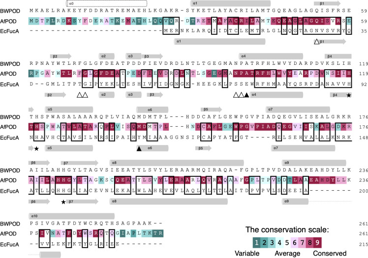
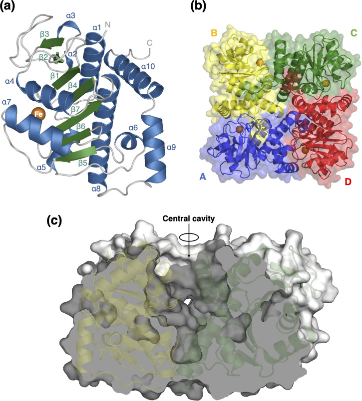
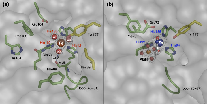
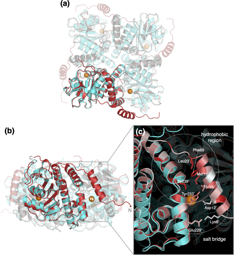
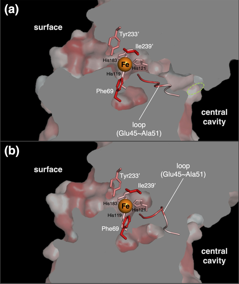
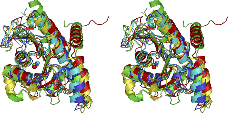
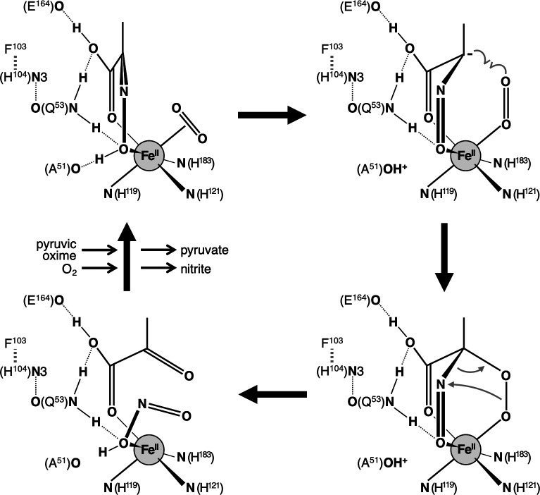
Nitrification by heterotrophic microorganisms is an important part of the nitrogen cycle in the environment. The enzyme responsible for the core function of heterotrophic nitrification is pyruvic oxime dioxygenase (POD). POD is a non-heme Fe(II)-dependent enzyme that catalyzes the dioxygenation of pyruvic oxime to produce pyruvate and nitrite. To analyze the catalytic mechanism of POD, the crystal structure of POD from Alcaligenes faecalis (AfPOD) was determined at 1.76 Å resolution. The enzyme is a homo-tetramer and the subunit structure is homologous to those of class II aldolases, in particular a zinc-dependent L-fuculose-1-phosphate aldolase. The active site of the subunit is located at the bottom of a cleft formed with an adjacent subunit. The iron ion at the active site is coordinated by three histidines and three water molecules in an octahedral geometry. The putative oxygen tunnel was connected between the active site and the central cavity of the tetramer. The N-terminal region of AfPOD, which is essential for catalytic activity, is disordered in the crystal. Structure prediction with AlphaFold2 combined with mutational experiments suggested that the disordered N-terminal region adopts an α-helix conformation and participates in the formation of the active site. The catalytic mechanism of the dioxygenase reaction by POD is discussed on the basis of the molecular docking model. IMPORTANCE: Our knowledge of nitrification has increased considerably in recent decades with the discovery of new nitrifying microorganisms and the characterization of their biochemical processes. Some heterotrophic bacteria and fungi are known to show nitrification activities, but the molecular mechanisms had been poorly understood. Here, we performed a structural characterization of POD, a key enzyme in heterotrophic nitrification that produces nitrite from ammonia using pyruvic oxime as an intermediate. Structural and enzymatic analyses revealed that POD is a unique dioxygenase with features such as an aldolase backbone, an N-terminal α-helix, and an oxygen tunnel. Our results provide insights not only into the molecular mechanisms but also into the design of specific inhibitors of heterotrophic nitrification.
Overview
The claims and statistics of this paper haven’t been checked yet.
Run the check — free, no account needed. It reads the open-access text and checks the sources it cites.
Integrity signals
Registry
Cites
Statistics
Figures
Verification
Claims the paper makes
The claims and statistics of this paper haven’t been checked yet.
Run the check — free, no account needed. It reads the open-access text and checks the sources it cites.
Self-check
Percentages, sums and reported differences re-computed from the paper’s own numbers.
The self-check needs the paper’s full text.
Citations
Cites
Highly accurate protein structure prediction with AlphaFold
Jumper, J., Evans, R., Pritzel, A., Green, T., Figurnov, M., Ronneberger, O., Tunyasuvunakool, K., Bates, R., Žídek, A., Potapenko, A., et al. (2021) · Nature · doi:10.1038/s41586-021-03819-2 · Cited by 43,764
Research works printed but not linked: 46
- 2020, The chemistry, biology, and modulation of ammonium nitrification in soil, Angew Chem Int Ed Engl, 59, 2182, 10.1002/anie.201903014 · DOI
- 2020, Technologies for biological removal and recovery of nitrogen from wastewater, Biotechnol Adv, 43, 107570, 10.1016/j.biotechadv.2020.107570 · DOI
- Stein LY . 2011. Heterotrophic nitrification and nitrifier denitrification, p 95–114. In Ward BB , Arp DJ , Klotz MG (ed), Nitrification. ASM Press, Washington, DC. · DOI
- 1998, A Most Probable Number method (MPN) for the estimation of cell numbers of heterotrophic nitrifying bacteria in soil, Plant Soil, 199, 123, 10.1023/A:1004243810473 · DOI
- 10.1099/00221287-5-2-360 · DOI
- 1982, Nitrogen redox metabolism of a heterotrophic, nitrifying-denitrifying Alcaligenes sp. from soil, Appl Environ Microbiol, 44, 923, 10.1128/aem.44.4.923-928.1982 · DOI
- 1984, Heterotrophic nitrification among denitrifiers, Appl Environ Microbiol, 47, 620, 10.1128/aem.47.4.620-623.1984 · DOI
- 10.1111/j.1574-6968.1996.tb08187.x · DOI
- 10.1038/srep39991 · DOI
- 2021, Gene expression analysis of Alcaligenes faecalis during induction of heterotrophic nitrification, Sci Rep, 11, 23105, 10.1038/s41598-021-02579-3 · DOI
- 2023, Phylogenetic diversity, distribution, and gene structure of the pyruvic oxime dioxygenase involved in heterotrophic nitrification, Antonie Van Leeuwenhoek, 116, 1037, 10.1007/s10482-023-01862-9 · DOI
- 10.1016/0003-2697(87)90587-2 · DOI
- 2021, A fully automated crystallization apparatus for small protein quantities, Acta Crystallogr F, 77, 29, 10.1107/S2053230X20015514 · DOI
- 2018, Anaerobic crystallization of proteins, Biophys Rev, 10, 183, 10.1007/s12551-017-0345-8 · DOI
- 10.1107/S0907444909047337 · DOI · Paper page
- 10.1038/ncomms3777 · DOI
- 2016, On the influence of crystal size and wavelength on native SAD phasing, Acta Crystallogr D, 7, 728
- 2011, Data processing and analysis with the autoPROC toolbox, Acta Crystallogr D, 6, 293
- 10.1107/S0021889897006766 · DOI
- 10.1107/S0907444904019158 · DOI · Paper page
- 10.1107/S0907444911001314 · DOI
- Bricogne G , Blanc E , Brandl M , Flensburg C , Keller P , Paciorek W , Roversi P , Sharff A , Smart O , Vonrhein C , Womack T . 2022. BUSTER version 2.10.4. Cambridge, United Kingdom: Global Phasing Ltd.
- 2022, Search and sequence analysis tools services from EMBL-EBI in 2022, Nucleic Acids Res, 50, W276, 10.1093/nar/gkac240 · DOI
- 10.1093/molbev/msy096 · DOI
- 10.1093/nar/gkw408 · DOI · Paper page
- 10.1093/nar/gkac387 · DOI
- 10.1038/S41592-022-01488-1 · DOI · Paper page
- 10.1016/S0076-6879(57)03489-8 · DOI
- 10.1042/bj0510278 · DOI
- 2021, GNINA 1.0: molecular docking with deep learning, J Cheminform, 13, 43, 10.1186/s13321-021-00522-2 · DOI
- 10.1006/jmbi.1996.0332 · DOI
- 10.1016/j.jmb.2007.05.022 · DOI
- 2022, AlphaFold2: A role for disordered protein/region prediction?, Int J Mol Sci, 23, 4591, 10.3390/ijms23094591 · DOI · Paper page
- 2015, Coupling oxygen consumption with hydrocarbon oxidation in bacterial multicomponent monooxygenases, Acc Chem Res, 48, 2632, 10.1021/acs.accounts.5b00312 · DOI
- 10.1111/tpj.12711 · DOI
- 10.1038/s41467-018-05589-4 · DOI
- 10.1021/bi0112513 · DOI
- 10.1107/S0907444902004614 · DOI
- 10.1021/bi701439j · DOI
- 2009, Why do cysteine dioxygenase enzymes contain a 3-His ligand motif rather than a 2His/1Asp motif like most nonheme dioxygenases?, J Phys Chem A, 113, 1835, 10.1021/jp809700f · DOI
- 10.1006/jmbi.1996.0060 · DOI
- 10.1107/S0907444996009146 · DOI
- 10.1021/bi9927686 · DOI
- 2000, Structures of l-fuculose-1-phosphate aldolase mutants outlining motions during catalysis, J Mol Biol, 303, 531, 10.1006/jmbi.2000.4153 · DOI
- 10.1038/srep39991 · DOI
- Sambrook J , Russell DW . 2001. Molecular cloning: a laboratory manual, 3rd edn. Cold Spring Harbor Laboratory Press, New York.
Metadata
- Authors
- Tsujino, S., Yamada, Y., Senda, M., Nakamura, A., Senda, T., & Fujiwara, T.
- Affiliations
- Shizuoka University (JP)
- Tohoku University (JP)
- High Energy Accelerator Research Organization (JP)
- Corresponding author
- Tsujino, Shuhei
- Fujiwara, Taketomo
- Venue
- Journal of Bacteriology
- Publisher
- openRxiv
- Published
- 2024-10-08
- Volume
- 207
- Issue
- 2
- Pages
- e0034224-e0034224
- ISSN
- 0021-9193 (ISSN-L)
- PMC
- PMC11841055
- OpenAlex
- W4406154292
- Cited by
- 0
- References
- 1
- FWCI
- 0.00
- Type
- journal article
- Language
- en
- Open access
- diamond
- doi.org
- Licence
- Locations
- Journal of Bacteriology (publishedVersion, cc-by)
- bioRxiv (Cold Spring Harbor Laboratory) (acceptedVersion, cc-by-nc-nd)
- PubMed Central (publishedVersion, cc-by)
- PubMed (publishedVersion)
- Institutional Repositories DataBase (IRDB) (acceptedVersion, cc-by)
- DOAJ (DOAJ: Directory of Open Access Journals) (submittedVersion)
- Institutional Repositories DataBase (IRDB) (submittedVersion, cc-by-nc-nd)
- Funders
- Japan Agency for Medical Research and Development (JP23ama121001)
- Ministry of Education, Culture, Sports, Science and Technology (21K12220, 17K00517, 21K12220, 24K15258, 24K15258, 17K00517)
- Shizuoka University
- High Energy Accelerator Research Organization
- Amano Institute of Technology
- Japan Society for the Promotion of Science (21K12220, 24K15258, 17K00517)
- Topics
- Life Sciences › Biochemistry, Genetics and Molecular Biology › Structural Biology › Enzyme Structure and Function
- Physical Sciences › Chemistry › Inorganic Chemistry › Metal-Catalyzed Oxygenation Mechanisms
- Life Sciences › Biochemistry, Genetics and Molecular Biology › Biochemistry › Metalloenzymes and iron-sulfur proteins
- Keywords
- heterotrophic nitrification, Alcaligenes faecalis, crystal structure, N-terminal disordered region, active site, catalytic mechanism, nitrite production, nitrification inhibitors, nitrogen cycling
- MeSH terms
- Bacterial Proteins, Models, Molecular, Protein Conformation, Crystallography, X-Ray, Catalytic Domain, Alcaligenes faecalis, Dioxygenases, Heterotrophic Processes, Nitrification
About these checks
Registry, retraction, Cites and statistics results are computed, not judged, with one exception: a printed reference that no index lists is read once by a model before it counts as a citation error. Claims are extracted by a model and kept only when their quote appears verbatim in the text; each cited source is then checked, with a model’s help, for whether it says what the claim relies on. Those verdicts can be wrong — read the claim’s quote and the cited source before relying on one.
Statistics and arithmetic are recomputed, not judged.
Confidence bands are shown only for fields calibrated on our annotated test set. None is calibrated yet, so no band is shown.
What counts as a claim ›How we measure confidence ›How we check this ›
Are you an author, or do you see a problem with this page? Request a review
