PreprintCited by 7Machine Learning in BioinformaticsProtein Structure and DynamicsCell Image Analysis Techniques

The DOI matches its Crossref record; its reference list is not available.

Deep generative model for protein subcellular localization prediction

Open access
Yuan, Guo-Hua, Li, Jinzhe, Yang, Zejun, Chen, Yao-Qi, Yuan, Zhonghang, Chen, Tao, Ouyang, Wanli, Dong, Nanqing, Yang, Li·2024·Briefings in Bioinformatics

Preprint — published as 10.1093/bib/bbaf152

Abstract

Protein sequence determines not only its structure but also its subcellular localization. Although a series of artificial intelligence models have been reported to predict protein subcellular localization, most of them provide only textual outputs. Here, we present deepGPS, a deep g enerative model for p rotein s ubcellular localization prediction. After trained with both protein primary sequences and protein subcellular localization fluorescence images, deepGPS shows the ability to predict cytoplasmic and nuclear localizations by reporting both textual labels and generative images as outputs. In addition, deepGPS shows potential to be further extended for other types of subcellular localization prediction, even with limited input data volumes for training. Finally, an openGPS website ( https://bits.fudan.edu.cn/opengps ) is constructed to provide a public and convenient platform for protein subcellular localization prediction with the scientific community.

Overview

The claims and statistics of this paper haven’t been checked yet.

Run the check — free, no account needed. It reads the open-access text and checks the sources it cites.

A few free checks a day · papers already verified open instantly · results are public

Integrity signals

Registry

Crossref · 10.1101/2024.10.29.620765

Cites

Statistics

Figures

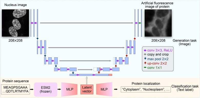
Figure 1 An overview design of deepGPS. A schematic diagram illustrating the architecture of deepGPS with a nucleus image and a protein sequence as inputs. DeepGPS enables the prediction of protein subcellular localization with generating a text label and an artificial fluorescence image as outputs.
Figure 1 An overview design of deepGPS. A schematic diagram illustrating the architecture of deepGPS with a nucleus image and a protein sequence as inputs. DeepGPS enables the prediction of protein subcellular localization with generating a text label and an artificial fluorescence image as outputs.CC BY-NC · PubMed Central
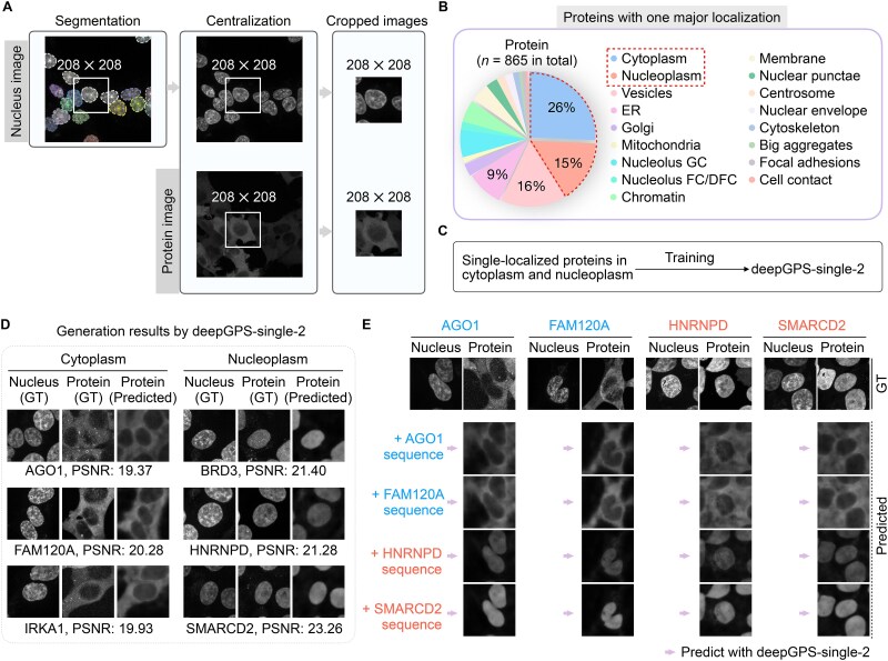
Figure 2 Construction and evaluation for deepGPS-single-2. (a) A specific example of image processing, illustrating the workflow from image segmentation to image cropping. (b) Distribution of proteins with only one major localization in the OpenCell database. (c) Strategy for training deepGPS-single-2. (d) Six examples generated by deepGPS-single-2. GT, ground truth; PSNR, peak signal-to-noise ratio. (e) A cross-assay using the ground-truth nucleus image as a nuclear fiducial marker and inputting different protein sequences to deepGPS-single-2. AGO1 and FAM120A are cytoplasmic proteins shown in blue, while HNRNPD and SMARCD2 are nuclear proteins shown in red. Of note, protein images generated by deepGPS in panels (d and e) were all from the test set, which were not used for model training.
Figure 2 Construction and evaluation for deepGPS-single-2. (a) A specific example of image processing, illustrating the workflow from image segmentation to image cropping. (b) Distribution of proteins with only one major localization in the OpenCell database. (c) Strategy for training deepGPS-single-2. (d) Six examples generated by deepGPS-single-2. GT, ground truth; PSNR, peak signal-to-noise ratio. (e) A cross-assay using the ground-truth nucleus image as a nuclear fiducial marker and inputting different protein sequences to deepGPS-single-2. AGO1 and FAM120A are cytoplasmic proteins shown in blue, while HNRNPD and SMARCD2 are nuclear proteins shown in red. Of note, protein images generated by deepGPS in panels (d and e) were all from the test set, which were not used for model training.CC BY-NC · PubMed Central
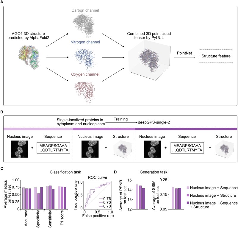
Figure 3 Performance comparison of deepGPS-single-2 variants using different input formats. (a) Schematic diagram illustrating the conversion of a protein structure predicted by AlphaFold2 into a point cloud tensor with carbon, nitrogen, and oxygen channels using PyUUL. (b) Strategies for training three variants of deepGPS-single-2 with different inputs of “nucleus image + protein sequence”, “nucleus image + protein structure”, and “nucleus image + protein sequence + protein structure”. (c) General performance of deepGPS-single-2 variants on the classification task including accuracy, specificity, sensitivity, and F1 score in left and ROC curve in right. (d) General performance of deepGPS-single-2 variants on the generation task.
Figure 3 Performance comparison of deepGPS-single-2 variants using different input formats. (a) Schematic diagram illustrating the conversion of a protein structure predicted by AlphaFold2 into a point cloud tensor with carbon, nitrogen, and oxygen channels using PyUUL. (b) Strategies for training three variants of deepGPS-single-2 with different inputs of “nucleus image + protein sequence”, “nucleus image + protein structure”, and “nucleus image + protein sequence + protein structure”. (c) General performance of deepGPS-single-2 variants on the classification task including accuracy, specificity, sensitivity, and F1 score in left and ROC curve in right. (d) General performance of deepGPS-single-2 variants on the generation task.CC BY-NC · PubMed Central
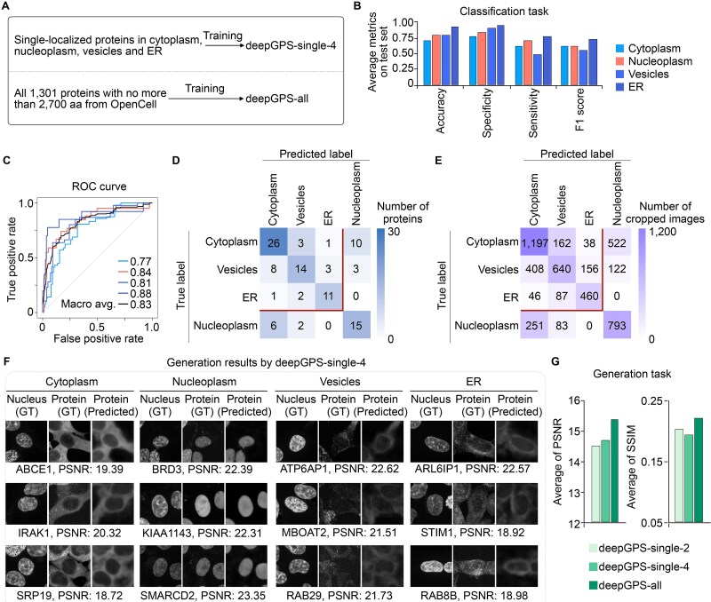
Figure 4 Extended deepGPS models for predicting other subcellular localization types. (a) Strategies for training deepGPS-single-4 and deepGPS-all. (b and c) General performance of deepGPS-single-4 on the classification task including accuracy, specificity, sensitivity, and F1 score in panel b and ROC curve in panel c. (d and e) Confusion matrix of proteins (d) and cropped images (e) for the classification task achieved by deepGPS-single-4. (f) Twelve examples generated by deepGPS-single-4. GT, ground truth; PSNR, peak signal-to-noise ratio. (g) General performance of deepGPS-single-2, deepGPS-single-4, and deepGPS-all on the generation task.
Figure 4 Extended deepGPS models for predicting other subcellular localization types. (a) Strategies for training deepGPS-single-4 and deepGPS-all. (b and c) General performance of deepGPS-single-4 on the classification task including accuracy, specificity, sensitivity, and F1 score in panel b and ROC curve in panel c. (d and e) Confusion matrix of proteins (d) and cropped images (e) for the classification task achieved by deepGPS-single-4. (f) Twelve examples generated by deepGPS-single-4. GT, ground truth; PSNR, peak signal-to-noise ratio. (g) General performance of deepGPS-single-2, deepGPS-single-4, and deepGPS-all on the generation task.CC BY-NC · PubMed Central
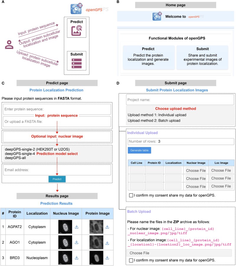
Figure 5 A shared platform for protein subcellular localization prediction provided by openGPS. (a) Schematic diagram illustrating the user interaction with the openGPS website, including two functional modules of “Predict” and “Submit”. (b) “Home” page of the openGPS website. (c) “Predict” and “Results” pages of the openGPS website. After entering protein sequences and selecting the prediction model, users can submit the prediction process to receive tabulated results, which are available for download. (d) “Submit” page of the openGPS website. Users can submit true images of protein localization with “Individual Upload” or “Batch Upload” modes.
Figure 5 A shared platform for protein subcellular localization prediction provided by openGPS. (a) Schematic diagram illustrating the user interaction with the openGPS website, including two functional modules of “Predict” and “Submit”. (b) “Home” page of the openGPS website. (c) “Predict” and “Results” pages of the openGPS website. After entering protein sequences and selecting the prediction model, users can submit the prediction process to receive tabulated results, which are available for download. (d) “Submit” page of the openGPS website. Users can submit true images of protein localization with “Individual Upload” or “Batch Upload” modes.CC BY-NC · PubMed Central

Verification

Claims the paper makes

The claims and statistics of this paper haven’t been checked yet.

Run the check — free, no account needed. It reads the open-access text and checks the sources it cites.

A few free checks a day · papers already verified open instantly · results are public

Self-check

Percentages, sums and reported differences re-computed from the paper’s own numbers.

The self-check needs the paper’s full text.

Citations

No citation lists for this paper

No citation lists for this paper yet — checked again daily.

References 32 · Cited by 7

Research works printed but not linked: 23

  1. 10.1126/science.1169375 · DOI
  2. 10.1016/j.cell.2016.03.014 · DOI · Paper page
  3. 10.1038/s41580-020-0250-z · DOI · Paper page
  4. 10.1016/j.cell.2022.06.035 · DOI
  5. 10.1002/ana.410220514 · DOI
  6. 10.1038/sj.onc.1210970 · DOI
  7. 10.1016/j.molcel.2024.05.011 · DOI
  8. 10.1093/NAR/GKAC1052 · DOI · Paper page
  9. 10.1093/bioinformatics/17.8.721 · DOI
  10. 10.1186/1471-2105-10-274 · DOI
  11. 10.1093/bioinformatics/btx548 · DOI
  12. 10.1126/science.abi6983 · DOI · Paper page
  13. 10.1038/s41592-022-01541-z · DOI
  14. 10.1126/science.ade2574 · DOI · Paper page
  15. Lin, P. , Yan, Y. & Huang, S.Y . DeepHomo2.0: improved protein-protein contact prediction of homodimers by transformer-enhanced deep learning. Brief Bioinform 24 (2023). · DOI
  16. Weigert, M. & Schmidt, U. Nuclei instance segmentation and classification in histopathology images with StarDist. arXiv:2203.02284 (2022). · DOI
  17. Orlando, G. et al. PyUUL provides an interface between biological structures and deep learning algorithms. Nat Commun 13, 961 (2022). · DOI
  18. Qi, C.R. , Su, H. , Mo, K. & Guibas, L.J. PointNet: Deep Learning on Point Sets for 3D Classification and Segmentation. arXiv:1612.00593 (2016). · arXiv
  19. 10.1038/s41586-020-2077-3 · DOI · Paper page
  20. 10.1038/s41580-018-0094-y · DOI · Paper page
  21. 10.1038/s41592-018-0216-7 · DOI
  22. Luo, Z. et al. Interpretable feature extraction and dimensionality reduction in ESM2 for protein localization prediction. Brief Bioinform 25 (2024). · DOI
  23. Ronneberger, O. , Fischer, P. & Brox, T . U-Net: Convolutional Networks for Biomedical Image Segmentation. arXiv:1505.04597 (2015). · DOI · Paper page

Metadata

Authors
Yuan, G.-H., Li, J., Yang, Z., Chen, Y.-Q., Yuan, Z., Chen, T., Ouyang, W., Dong, N., & Yang, L.
Affiliations
  • Fudan University (CN)
  • Shanghai Artificial Intelligence Laboratory (CN)
Corresponding author
  • Dong, Nanqing
  • Yang, Li
Venue
Briefings in Bioinformatics
Publisher
openRxiv
Published
2024-11-03
Volume
26
Issue
2
ISSN
1467-5463 (ISSN-L)
OpenAlex
W4409346328
Cited by
7
References
32
Citations by year
2026: 5 · 2025: 2
FWCI
2.09
Type
journal article
Language
en
Open access
Locations
  • Briefings in Bioinformatics (publishedVersion, cc-by-nc)
  • bioRxiv (Cold Spring Harbor Laboratory) (acceptedVersion, cc-by-nc-nd)
  • PubMed Central (publishedVersion, cc-by-nc)
  • PubMed (publishedVersion)
Funders
  • National Natural Science Foundation of China (32430018)
  • Science and Technology Commission of Shanghai Municipality (23JS1400300, 23DX1900102)
  • National Key Research and Development Program of China (2021YFA1300503, 2024YFC3405902)
Topics
  • Life Sciences › Biochemistry, Genetics and Molecular Biology › Molecular Biology › Machine Learning in Bioinformatics
  • Life Sciences › Biochemistry, Genetics and Molecular Biology › Molecular Biology › Protein Structure and Dynamics
  • Life Sciences › Biochemistry, Genetics and Molecular Biology › Biophysics › Cell Image Analysis Techniques
Keywords
protein localization, deep generative models, fluorescence microscopy images, cytoplasmic localization, nuclear localization, protein primary sequence
MeSH terms
Deep Learning, Cell Nucleus, Cytoplasm, Humans, Proteins, Subcellular Fractions, Computational Biology, Protein Transport

About these checks

Registry, retraction, Cites and statistics results are computed, not judged, with one exception: a printed reference that no index lists is read once by a model before it counts as a citation error. Claims are extracted by a model and kept only when their quote appears verbatim in the text; each cited source is then checked, with a model’s help, for whether it says what the claim relies on. Those verdicts can be wrong — read the claim’s quote and the cited source before relying on one.

Statistics and arithmetic are recomputed, not judged.

Confidence bands are shown only for fields calibrated on our annotated test set. None is calibrated yet, so no band is shown.

What counts as a claim ›How we measure confidence ›How we check this ›

Are you an author, or do you see a problem with this page? Request a review

How to cite

Cite CoChat for the checks on this page, and the paper for its own findings.

This page
CoChat. (2026). Citation check: Deep generative model for protein subcellular localization prediction. https://app.cochat.ai/papers/10.1101/2024.10.29.620765
The paper
Yuan, G.-H., Li, J., Yang, Z., Chen, Y.-Q., Yuan, Z., Chen, T., Ouyang, W., Dong, N., & Yang, L. (2024). Deep generative model for protein subcellular localization prediction. Briefings in Bioinformatics. https://doi.org/10.1101/2024.10.29.620765