Cited by 20

The DOI matches its Crossref record.

The structure of bradyzoite-specific enolase from Toxoplasma gondii reveals insights into its dual cytoplasmic and nuclear functions

Ruan, Jiapeng, Mouveaux, Thomas, Light, Samuel H., Minasov, George, Anderson, Wayne F., Tomavo, Stanislas, Ngô, Huân M.·2015·Acta Crystallographica Section D Biological Crystallography

Abstract

In addition to catalyzing a central step in glycolysis, enolase assumes a remarkably diverse set of secondary functions in different organisms, including transcription regulation as documented for the oncogene c-Myc promoter-binding protein 1. The apicomplexan parasite Toxoplasma gondii differentially expresses two nuclear-localized, plant-like enolases: enolase 1 (TgENO1) in the latent bradyzoite cyst stage and enolase 2 (TgENO2) in the rapidly replicative tachyzoite stage. A 2.75 Å resolution crystal structure of bradyzoite enolase 1, the second structure to be reported of a bradyzoite-specific protein in Toxoplasma, captures an open conformational state and reveals that distinctive plant-like insertions are located on surface loops. The enolase 1 structure reveals that a unique residue, Glu164, in catalytic loop 2 may account for the lower activity of this cyst-stage isozyme. Recombinant TgENO1 specifically binds to a TTTTCT DNA motif present in the cyst matrix antigen 1 (TgMAG1) gene promoter as demonstrated by gel retardation. Furthermore, direct physical interactions of both nuclear TgENO1 and TgENO2 with the TgMAG1 gene promoter are demonstrated in vivo using chromatin immunoprecipitation (ChIP) assays. Structural and biochemical studies reveal that T. gondii enolase functions are multifaceted, including the coordination of gene regulation in parasitic stage development. Enolase 1 provides a potential lead in the design of drugs against Toxoplasma brain cysts.

Overview

The claims and statistics of this paper haven’t been checked yet.

Run the check — free, no account needed. It reads the open-access text and checks the sources it cites.

A few free checks a day · papers already verified open instantly · results are public

Integrity signals

Registry

Crossref · 10.1107/s1399004714026479

Cites

Statistics

Figures

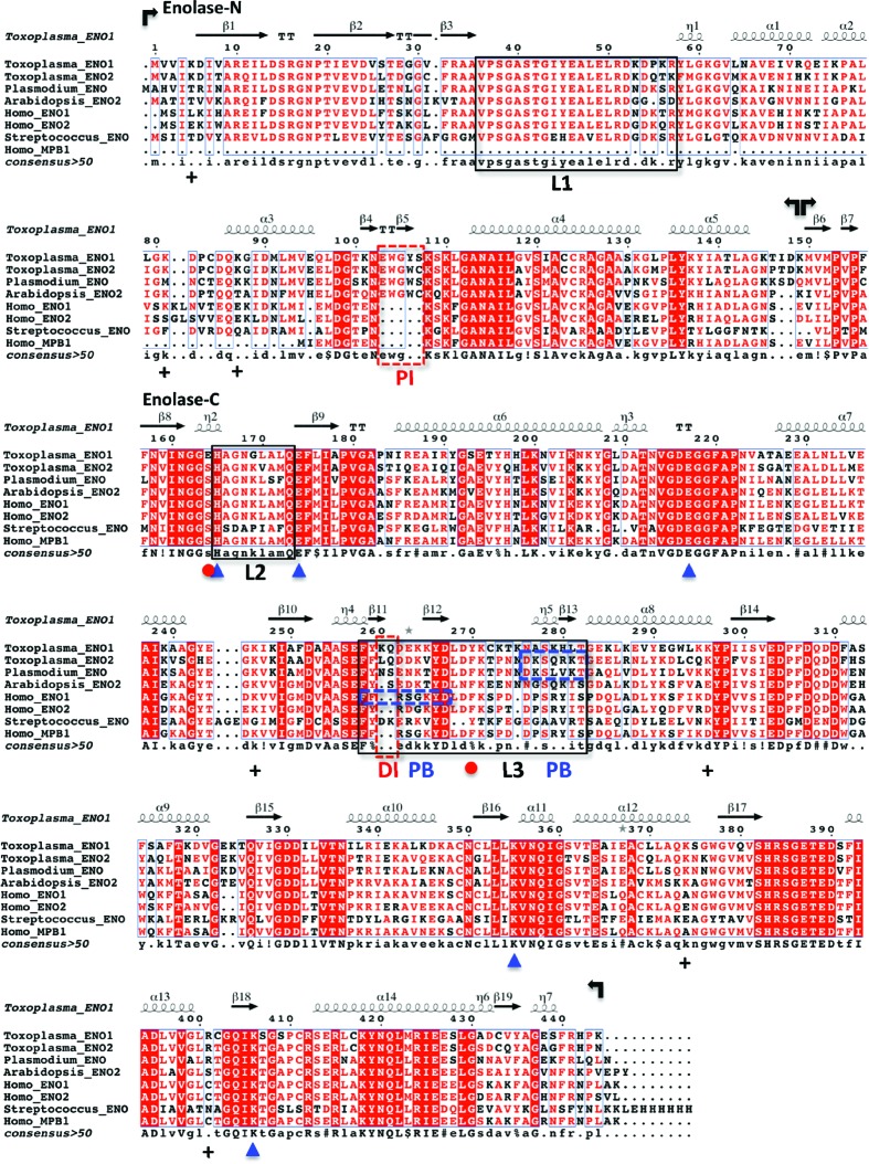
Figure 1 Secondary-structural alignment of enolases. Bacterial (Streptococcus suis), protozoan parasite (Toxoplasma gondii and Plasmodium falciparum), plant (Arabidopsis thaliana) and human enolases are aligned for comparison. The enolases are all nuclear-localized, with the exceptions of human ENO2 and Streptococcus ENO. Human MPB1 is an alternatively spliced form of ENO1 and is truncated at the N-terminal domain. The five conserved active-site residues (His165, Glu174, Glu217, Lys355 and Lys406) are marked by blue arrowheads. The atypical TgENO1 residue replacement E164S at the N-­terminus of loop 2 and the interacting residue Tyr270 in loop 3 are indicated by red dots. Conserved positively charged residues on the surface of transcription regulatory enolases are labelled with plus signs (+). Boxed regions are plant-like insertions (red dashes; DI, dipeptide insertion; PI, pentapeptide insertion), catalytic mobile loops (black; L, loop) and plasminogen-binding motifs (blue dashes; PB, plasminogen binding).
Figure 1 Secondary-structural alignment of enolases. Bacterial (Streptococcus suis), protozoan parasite (Toxoplasma gondii and Plasmodium falciparum), plant (Arabidopsis thaliana) and human enolases are aligned for comparison. The enolases are all nuclear-localized, with the exceptions of human ENO2 and Streptococcus ENO. Human MPB1 is an alternatively spliced form of ENO1 and is truncated at the N-terminal domain. The five conserved active-site residues (His165, Glu174, Glu217, Lys355 and Lys406) are marked by blue arrowheads. The atypical TgENO1 residue replacement E164S at the N-­terminus of loop 2 and the interacting residue Tyr270 in loop 3 are indicated by red dots. Conserved positively charged residues on the surface of transcription regulatory enolases are labelled with plus signs (+). Boxed regions are plant-like insertions (red dashes; DI, dipeptide insertion; PI, pentapeptide insertion), catalytic mobile loops (black; L, loop) and plasminogen-binding motifs (blue dashes; PB, plasminogen binding).CC BY · PubMed Central
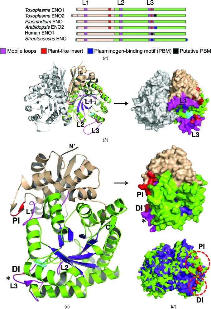
Figure 2 Structure of bradyzoite TgENO1. (a) The domain topology of TgENO1 is compared with selected enolases. (b) TgENO1 dimer. (c) TgENO1 monomer. The N-terminal domain (in wheat), C-terminal domain (in green), canonical TIM barrel (TIM), three mobile loops (L1–L3; magenta) and plant-like insertions (PI and DI; red) are shown. The Plasmodium-derived plasminogen-binding motif (asterisk) is located on surface loop 3 in TgENO2 but not in TgENO1. (d) Plant-like insertions (circled) are labelled red in TgENO1 (green) and superposed with human ENO1 (purple; PDB entry 2psn; J. K. Hyo, J. K. Seung, J. C. Sang & J. Suk-Kyeong, unpublished work) which does not contain the plant-like insertion.
Figure 2 Structure of bradyzoite TgENO1. (a) The domain topology of TgENO1 is compared with selected enolases. (b) TgENO1 dimer. (c) TgENO1 monomer. The N-terminal domain (in wheat), C-terminal domain (in green), canonical TIM barrel (TIM), three mobile loops (L1–L3; magenta) and plant-like insertions (PI and DI; red) are shown. The Plasmodium-derived plasminogen-binding motif (asterisk) is located on surface loop 3 in TgENO2 but not in TgENO1. (d) Plant-like insertions (circled) are labelled red in TgENO1 (green) and superposed with human ENO1 (purple; PDB entry 2psn; J. K. Hyo, J. K. Seung, J. C. Sang & J. Suk-Kyeong, unpublished work) which does not contain the plant-like insertion.CC BY · PubMed Central
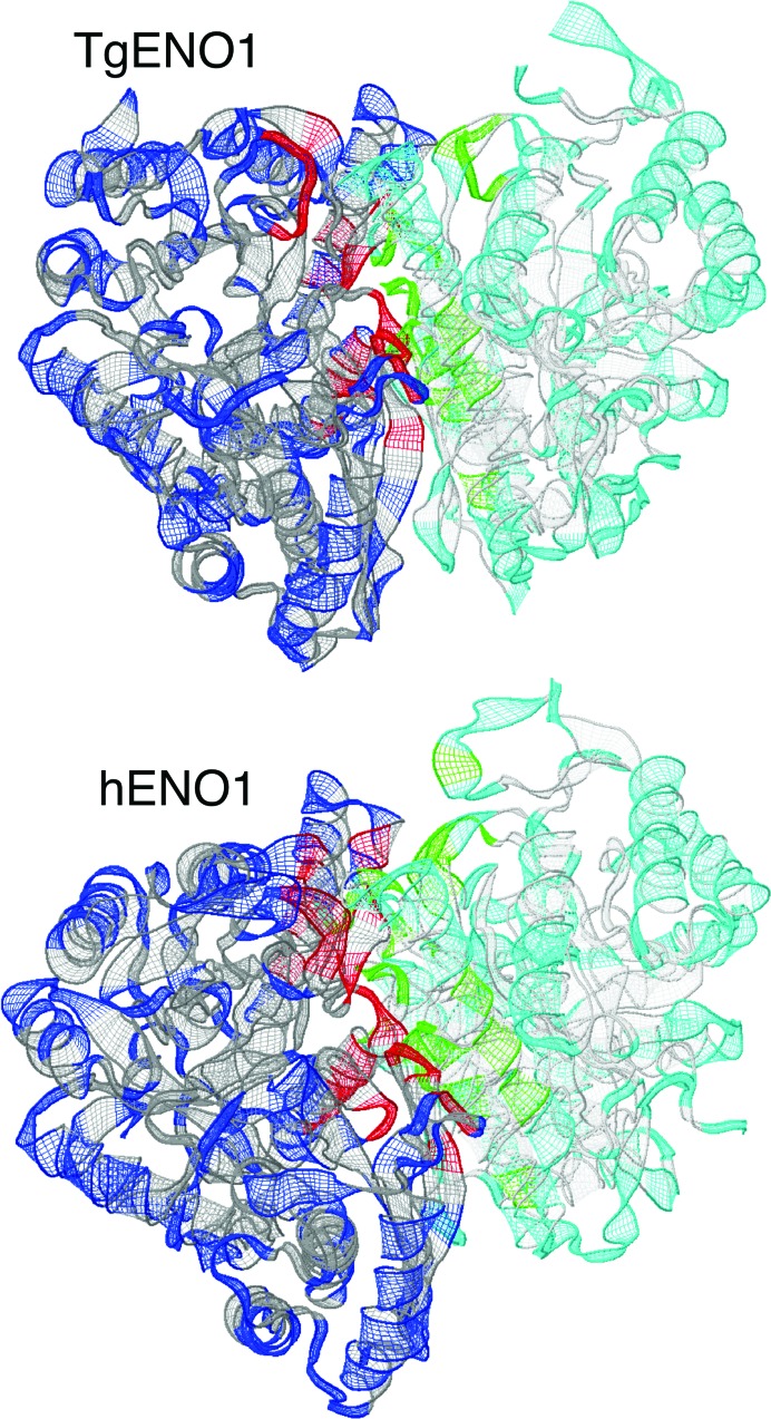
Figure 3 Enolase dimer interface. The subunit interfaces in Toxoplasma and human ENO1 dimers are illustrated in red and green. The Toxoplasma ENO1 dimer is formed by 33 hydrogen bonds, yielding an average surface area of 1891.7 Å2, as determined by PISA and 26 hydrogen bonds from LigPlot+ (Supplementary Fig. S1a), whereas the human ENO1 dimer interface contains 43 (PISA) and 31 (LigPlot+) hydrogen bonds (Supplementary Fig. S1b) with an average surface area of 2003.2 Å2.
Figure 3 Enolase dimer interface. The subunit interfaces in Toxoplasma and human ENO1 dimers are illustrated in red and green. The Toxoplasma ENO1 dimer is formed by 33 hydrogen bonds, yielding an average surface area of 1891.7 Å2, as determined by PISA and 26 hydrogen bonds from LigPlot+ (Supplementary Fig. S1a), whereas the human ENO1 dimer interface contains 43 (PISA) and 31 (LigPlot+) hydrogen bonds (Supplementary Fig. S1b) with an average surface area of 2003.2 Å2.CC BY · PubMed Central
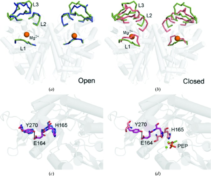
Figure 4 The TgENO1 active site. (a) Superposition of the TgENO1 dimer (green) with the ‘open-loop’ ScENO dimer (blue; PDB entry 1ebh; Wedekind et al., 1995 ▶). The r.m.s.d. is 0.56 Å over 777 Cα atoms. (b) Superposition of the TgENO1 dimer with the ‘closed-loop’ hENO1 dimer (orange; PDB entry 3b97; Kang et al., 2008 ▶). The r.m.s.d. is 0.68 Å over 645 Cα atoms. (c) A different perspective of the superposition in (a) highlights the hydrogen bond (dashed line) between the unique TgENO1 residue Glu164 in loop 2 and residue Tyr270 in loop 3. (d) Superposition of TgENO1 on the closed-loop PEP-bound complex (PDB entry 3ucd; Qin et al., 2012 ▶) illustrates that L2 closure necessitates breakage of the Glu164–Tyr270 hydrogen bond and should allow His165 to hydrogen-bond to a PEP O atom.
Figure 4 The TgENO1 active site. (a) Superposition of the TgENO1 dimer (green) with the ‘open-loop’ ScENO dimer (blue; PDB entry 1ebh; Wedekind et al., 1995 ▶). The r.m.s.d. is 0.56 Å over 777 Cα atoms. (b) Superposition of the TgENO1 dimer with the ‘closed-loop’ hENO1 dimer (orange; PDB entry 3b97; Kang et al., 2008 ▶). The r.m.s.d. is 0.68 Å over 645 Cα atoms. (c) A different perspective of the superposition in (a) highlights the hydrogen bond (dashed line) between the unique TgENO1 residue Glu164 in loop 2 and residue Tyr270 in loop 3. (d) Superposition of TgENO1 on the closed-loop PEP-bound complex (PDB entry 3ucd; Qin et al., 2012 ▶) illustrates that L2 closure necessitates breakage of the Glu164–Tyr270 hydrogen bond and should allow His165 to hydrogen-bond to a PEP O atom.CC BY · PubMed Central
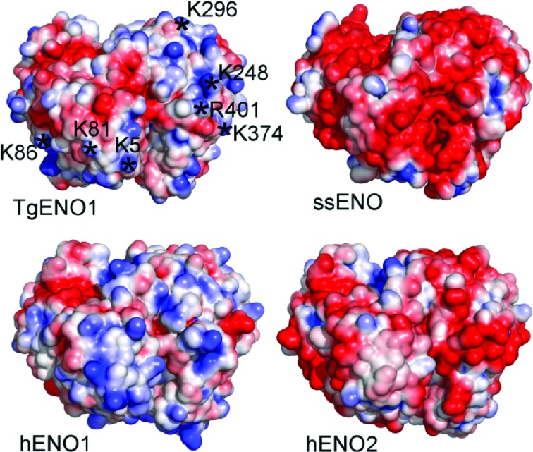
Figure 5 Surface electrostatic potentials of enolases. The cytosolic ssENO and hENO2 and the transcription factors TgENO1 and hENO1 are shown in surface representation and coloured by charge. Blue represents positive charge and red negative charge. Asterisks denote identical and/or conserved positively charged residues in nuclear enolases (see Fig. 1 ▶). Electrostatics were calculated and visualized using the APBS plugin in PyMOL. ssENO, S. suis, PDB entry 4ewj (Lu et al., 2012 ▶). hENO2, Homo sapiens, PDB entry 1te6 (Chai et al., 2004 ▶).
Figure 5 Surface electrostatic potentials of enolases. The cytosolic ssENO and hENO2 and the transcription factors TgENO1 and hENO1 are shown in surface representation and coloured by charge. Blue represents positive charge and red negative charge. Asterisks denote identical and/or conserved positively charged residues in nuclear enolases (see Fig. 1 ▶). Electrostatics were calculated and visualized using the APBS plugin in PyMOL. ssENO, S. suis, PDB entry 4ewj (Lu et al., 2012 ▶). hENO2, Homo sapiens, PDB entry 1te6 (Chai et al., 2004 ▶).CC BY · PubMed Central
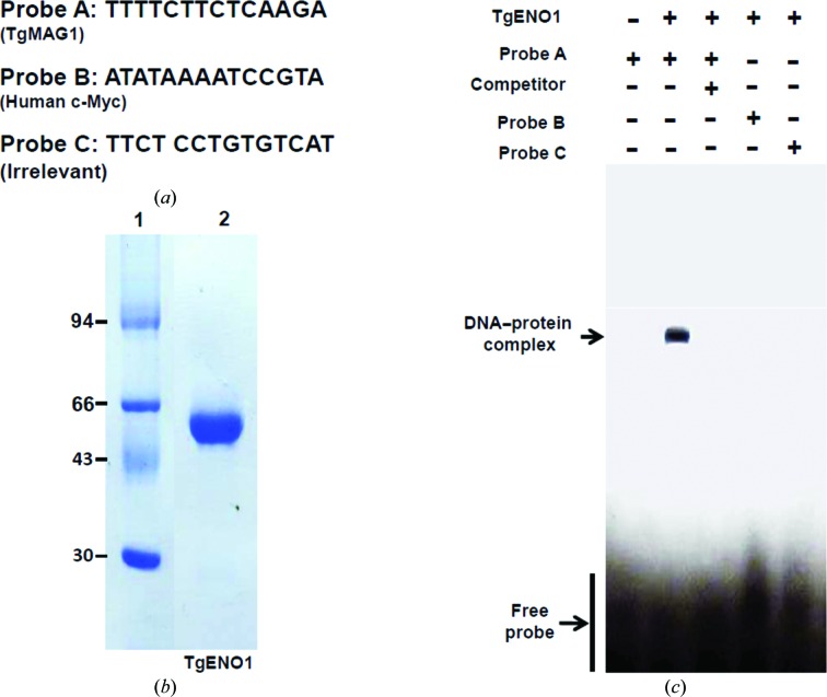
Figure 6 In vitro interaction of TgENO1 with the TgMAG1 promoter sequence. (a) Oligonucleotide probes were designed for the TTTTCT motif in the bradyzoite TgMAG1 promoter, the TATA box in the human c-­Myc promoter and a negative random sequence. (b) SDS–PAGE of purified recombinant TgENO1. Lane 1 contains molecular-weight marker (labelled in kDa). (c) Electrophoretic mobility shift assay of biotinylated TgENO1 incubated with probes and detected for retardation mobility of the DNA–protein complex. The TgENO1–TgMAG1 complex (second lane) was competed with an unlabelled probe prior to incubation with labelled TGMAG1 probe (third lane).
Figure 6 In vitro interaction of TgENO1 with the TgMAG1 promoter sequence. (a) Oligonucleotide probes were designed for the TTTTCT motif in the bradyzoite TgMAG1 promoter, the TATA box in the human c-­Myc promoter and a negative random sequence. (b) SDS–PAGE of purified recombinant TgENO1. Lane 1 contains molecular-weight marker (labelled in kDa). (c) Electrophoretic mobility shift assay of biotinylated TgENO1 incubated with probes and detected for retardation mobility of the DNA–protein complex. The TgENO1–TgMAG1 complex (second lane) was competed with an unlabelled probe prior to incubation with labelled TGMAG1 probe (third lane).CC BY · PubMed Central
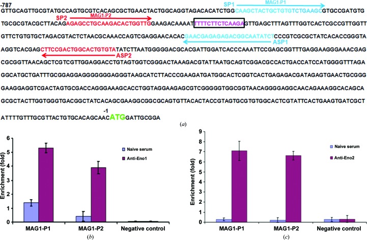
Figure 7 In vitro interaction of TgENO1 and TgENO2 with the bradyzoite TgMAG1 promoter as shown by chromatin immunoprecipitation (ChIP) analyses. (a) Upstream and downstream primers of the TTTTCT motif in TgMAG1 for PCR of the ChIP products. (b) Polyclonal antibodies to bradyzoite-specific TgENO1 precipitated ChIP fragments from the TgMAG1 promoter. (c) Polyclonal antibodies to tachyzoite-specific TgENO2 also pulled down ChIP fragments from TgMAG1 promoter.
Figure 7 In vitro interaction of TgENO1 and TgENO2 with the bradyzoite TgMAG1 promoter as shown by chromatin immunoprecipitation (ChIP) analyses. (a) Upstream and downstream primers of the TTTTCT motif in TgMAG1 for PCR of the ChIP products. (b) Polyclonal antibodies to bradyzoite-specific TgENO1 precipitated ChIP fragments from the TgMAG1 promoter. (c) Polyclonal antibodies to tachyzoite-specific TgENO2 also pulled down ChIP fragments from TgMAG1 promoter.CC BY · PubMed Central

Verification

Claims the paper makes

The claims and statistics of this paper haven’t been checked yet.

Run the check — free, no account needed. It reads the open-access text and checks the sources it cites.

A few free checks a day · papers already verified open instantly · results are public

Claims other papers make about this one

Each is checked against this paper’s own text: does it say what the citing paper says it does?

Self-check

Percentages, sums and reported differences re-computed from the paper’s own numbers.

The self-check needs the paper’s full text.

Citations

Cites

No references could be resolved yet.

Research works printed but not linked: 40

  1. Bergmann, 2005, Thromb. Haemost., 94, 304, 10.1160/TH05-05-0369 · DOI
  2. Brandina, 2006, Biochim. Biophys. Acta, 1757, 1217, 10.1016/j.bbabio.2006.07.001 · DOI
  3. Broennimann, 2006, J. Synchrotron Rad., 13, 120, 10.1107/S0909049505038665 · DOI
  4. Chai, 2004, J. Mol. Biol., 341, 1015, 10.1016/j.jmb.2004.05.068 · DOI
  5. Chandran, 2006, J. Mol. Biol., 358, 8, 10.1016/j.jmb.2006.02.012 · DOI
  6. Coppin, 2003, Biochimie, 85, 353, 10.1016/S0300-9084(03)00076-2 · DOI
  7. Dzierszinski, 2001, J. Mol. Biol., 309, 1017, 10.1006/jmbi.2001.4730 · DOI
  8. Dzierszinski, 2004, Eukaryot. Cell, 3, 992, 10.1128/EC.3.4.992-1003.2004 · DOI
  9. Dzierszinski, 1999, J. Biol. Chem., 274, 24888, 10.1074/jbc.274.35.24888 · DOI
  10. Emsley, 2004, Acta Cryst. D, 60, 2126, 10.1107/S0907444904019158 · DOI · Paper page
  11. Ferguson, 2002, Int. J. Parasitol., 32, 1399, 10.1016/S0020-7519(02)00129-7 · DOI
  12. Flegr, 2013, Trends Parasitol., 29, 156, 10.1016/j.pt.2013.01.007 · DOI
  13. Gajria, 2008, Nucleic Acids Res., 36, D553, 10.1093/nar/gkm981 · DOI
  14. Gerlt, 2012, J. Biol. Chem., 287, 29, 10.1074/jbc.R111.240945 · DOI
  15. Ghosh, 2011, Proc. Natl Acad. Sci. USA, 108, 17153, 10.1073/pnas.1103657108 · DOI
  16. Harper, 2004, Gene, 340, 227, 10.1016/j.gene.2004.06.048 · DOI
  17. Hsu, 2009, Mol. Biol. Cell, 20, 5127, 10.1091/mbc.E09-05-0386 · DOI
  18. Kannan, 2012, Schizophr. Bull., 38, 1155, 10.1093/schbul/sbs079 · DOI
  19. Kang, 2008, Acta Cryst. D, 64, 651, 10.1107/S0907444908008561 · DOI
  20. Kibe, 2005, Nucleic Acids Res., 33, 1722, 10.1093/nar/gki314 · DOI
  21. Laskowski, 2011, J. Chem. Inf. Model., 51, 2778, 10.1021/ci200227u · DOI
  22. Lebioda, 1989, J. Biol. Chem., 264, 3685, 10.1016/S0021-9258(19)84906-2 · DOI
  23. Lee, 2002, EMBO J., 21, 2692, 10.1093/emboj/21.11.2692 · DOI
  24. Lu, 2012, Protein Cell, 3, 769, 10.1007/s13238-012-2040-7 · DOI
  25. McCoy, 2005, Acta Cryst. D, 61, 458, 10.1107/S0907444905001617 · DOI
  26. Mouveaux, 2014, PLoS One, 9, e105820, 10.1371/journal.pone.0105820 · DOI
  27. Murshudov, 2011, Acta Cryst. D, 67, 355, 10.1107/S0907444911001314 · DOI
  28. Nadassy, 1999, Biochemistry, 38, 1999, 10.1021/bi982362d · DOI
  29. Nurmohamed, 2010, Acta Cryst. D, 66, 1036, 10.1107/S0907444910030015 · DOI
  30. Olguin-Lamas, 2011, PLoS Pathog., 7, e1001328, 10.1371/journal.ppat.1001328 · DOI
  31. Pal-Bhowmick, 2009, Malar. J., 8, 179, 10.1186/1475-2875-8-179 · DOI
  32. Pal-Bhowmick, 2007, Malar. J., 6, 45, 10.1186/1475-2875-6-45 · DOI
  33. Qin, 2012, J. Inorg. Biochem., 111, 187, 10.1016/j.jinorgbio.2012.02.011 · DOI
  34. Sedoris, K. C., Thomas, S. D. & Miller, D. M. (2010). BMC Cancer, 10, 157. · DOI
  35. Tomavo, 2001, Int. J. Parasitol., 31, 1023, 10.1016/S0020-7519(01)00193-X · DOI
  36. Tovy, 2010, PLoS Pathog., 6, e1000775, 10.1371/journal.ppat.1000775 · DOI
  37. Vora, 2009, Arch. Biochem. Biophys., 485, 128, 10.1016/j.abb.2009.02.012 · DOI
  38. Wedekind, 1995, Biochemistry, 34, 4325, 10.1021/bi00013a022 · DOI
  39. Xu, 2008, J. Mol. Biol., 381, 487, 10.1016/j.jmb.2008.06.002 · DOI
  40. Zhang, 1997, Biochemistry, 36, 12526, 10.1021/bi9712450 · DOI

Cited by

Sorted by citations

Metadata

Authors
Ruan, J., Mouveaux, T., Light, S. H., Minasov, G., Anderson, W. F., Tomavo, S., & Ngô, H. M.
Venue
Acta Crystallographica Section D Biological Crystallography
Published
2015-02-26
Volume
71
Issue
3
Pages
417-426
ISSN
1399-0047
Cited by
20
References
0

About these checks

Registry, retraction, Cites and statistics results are computed, not judged, with one exception: a printed reference that no index lists is read once by a model before it counts as a citation error. Claims are extracted by a model and kept only when their quote appears verbatim in the text; each cited source is then checked, with a model’s help, for whether it says what the claim relies on. Those verdicts can be wrong — read the claim’s quote and the cited source before relying on one.

Statistics and arithmetic are recomputed, not judged.

Confidence bands are shown only for fields calibrated on our annotated test set. None is calibrated yet, so no band is shown.

What counts as a claim ›How we measure confidence ›How we check this ›

Are you an author, or do you see a problem with this page? Request a review