

The DOI matches its Crossref record; 74 of the 80 references it cites were found in a registry or an index.
Results of the 2020 fastMRI Challenge for Machine Learning MR Image Reconstruction
Open accessAbstract
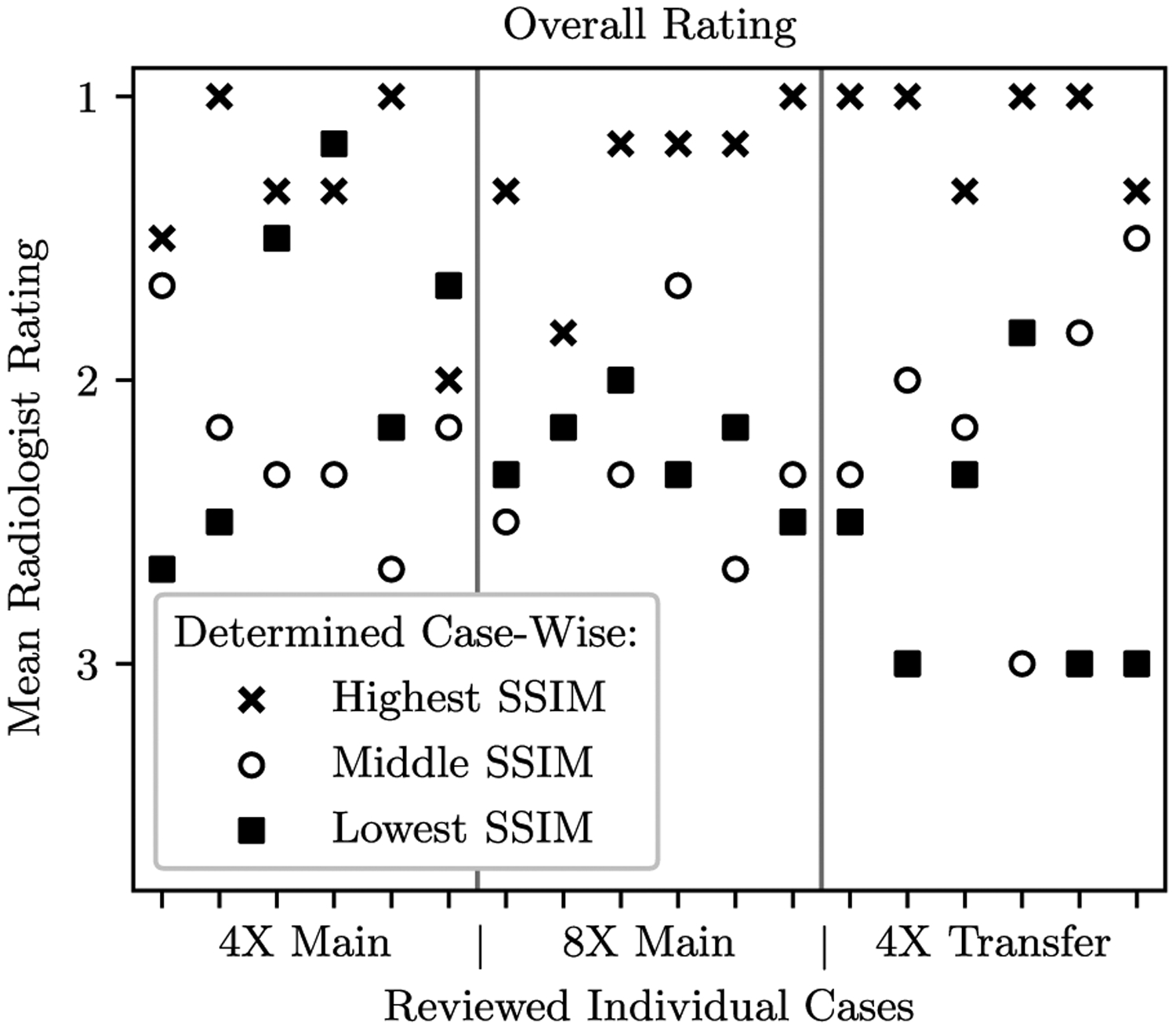
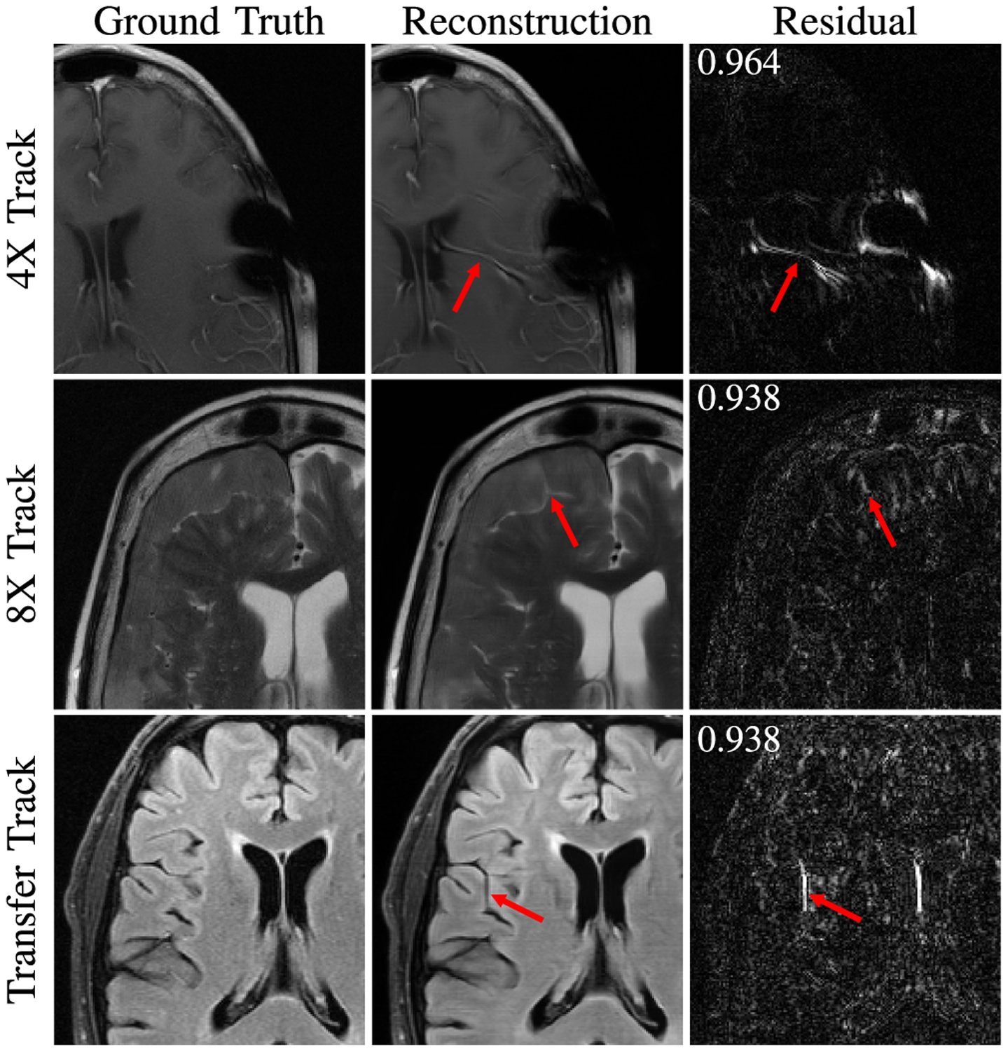
Accelerating MRI scans is one of the principal outstanding problems in the MRI research community. Towards this goal, we hosted the second fastMRI competition targeted towards reconstructing MR images with subsampled k-space data. We provided participants with data from 7,299 clinical brain scans (de-identified via a HIPAA-compliant procedure by NYU Langone Health), holding back the fully-sampled data from 894 of these scans for challenge evaluation purposes. In contrast to the 2019 challenge, we focused our radiologist evaluations on pathological assessment in brain images. We also debuted a new Transfer track that required participants to submit models evaluated on MRI scanners from outside the training set. We received 19 submissions from eight different groups. Results showed one team scoring best in both SSIM scores and qualitative radiologist evaluations. We also performed analysis on alternative metrics to mitigate the effects of background noise and collected feedback from the participants to inform future challenges. Lastly, we identify common failure modes across the submissions, highlighting areas of need for future research in the MRI reconstruction community.
Overview
The claims and statistics of this paper haven’t been checked yet.
Run the check — free, no account needed. It reads the open-access text and checks the sources it cites.
Integrity signals
Registry
Cites
Statistics
Figures
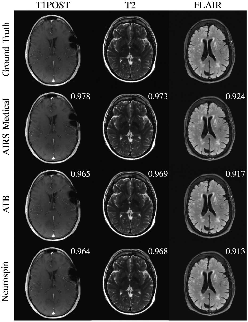
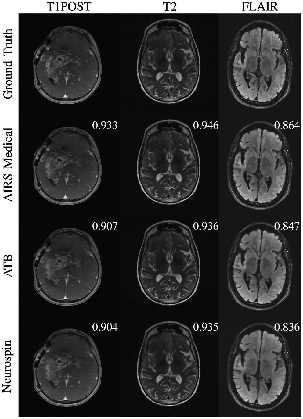
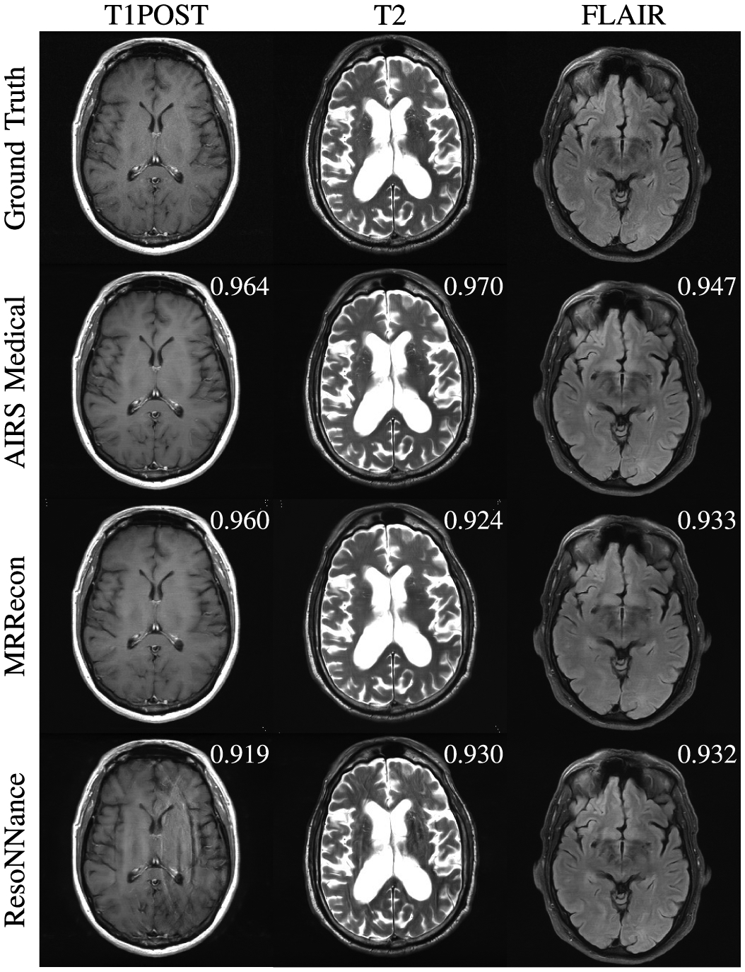
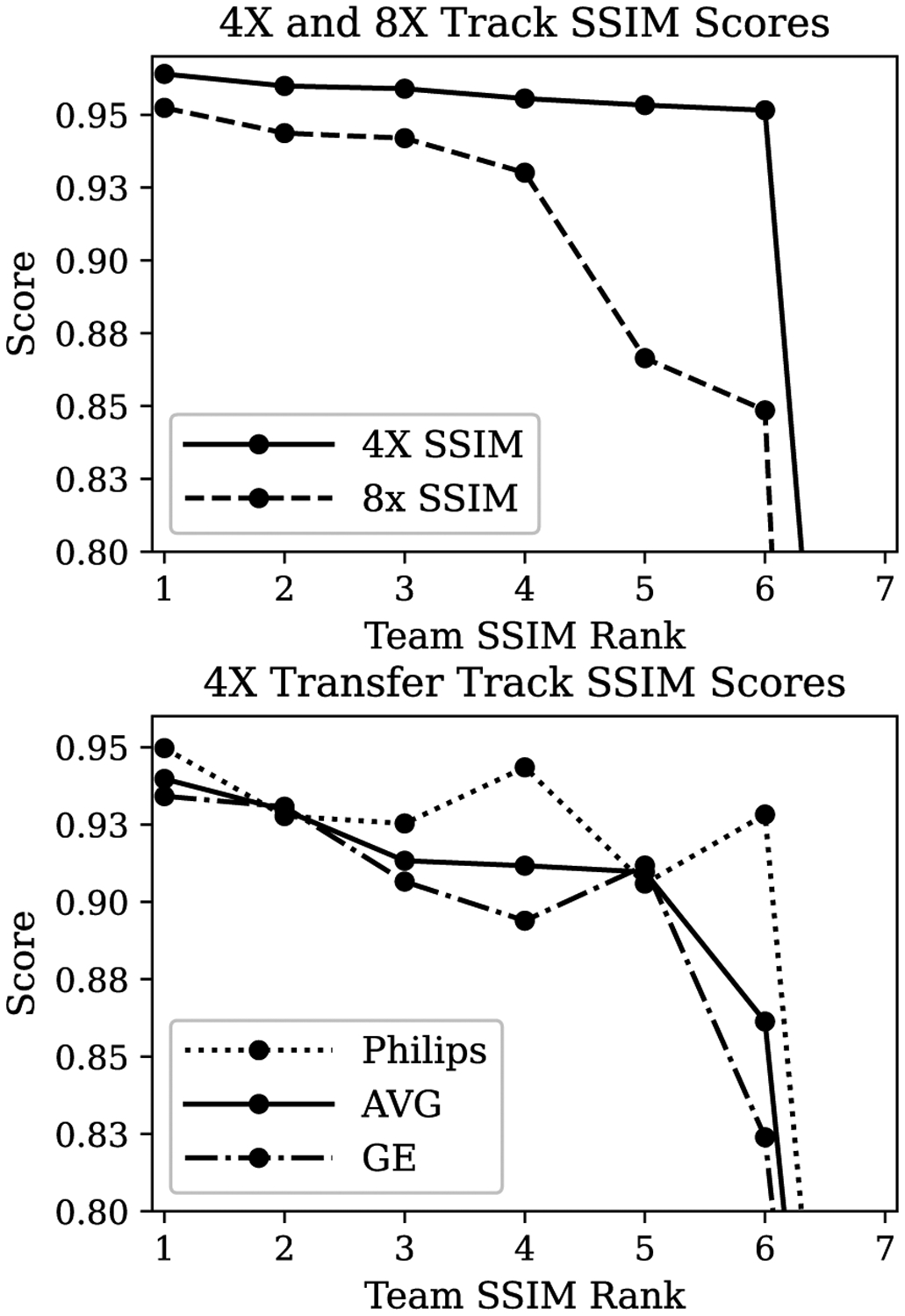
Verification
Claims the paper makes
The claims and statistics of this paper haven’t been checked yet.
Run the check — free, no account needed. It reads the open-access text and checks the sources it cites.
Claims other papers make about this one
Each is checked against this paper’s own text: does it say what the citing paper says it does?
In the citing paper
“The fastMRI [117,118] 15 is a large-scale dataset for medical image reconstruction using machine learning approaches.”
From Federated learning for medical image analysis: A survey (2024) · cites this paper as [118]
Self-check
Percentages, sums and reported differences re-computed from the paper’s own numbers.
The self-check needs the paper’s full text.
Citations
Cites
Deep Residual Learning for Image Recognition
He, K., Zhang, X., Ren, S., & Sun, J. (2016) · 2016 IEEE Conference on Computer Vision and Pattern Recognition (CVPR) · doi:10.1109/cvpr.2016.90 · Cited by 235,474
Very Deep Convolutional Networks for Large-Scale Image Recognition
Simonyan, K., & Zisserman, A. (2014) · arXiv · doi:10.48550/arxiv.1409.1556 · Cited by 114,148
ImageNet classification with deep convolutional neural networks
Krizhevsky, A., Sutskever, I., & Hinton, G. E. (2017) · Communications of the ACM · doi:10.1145/3065386 · Cited by 112,155
U-Net: Convolutional Networks for Biomedical Image Segmentation
Ronneberger, O., Fischer, P., & Brox, T. (2015) · Lecture notes in computer science · doi:10.1007/978-3-319-24574-4_28 · Cited by 99,404
Adam: A Method for Stochastic Optimization
Kingma, D. P., & Ba, J. (2014) · arXiv · doi:10.48550/arxiv.1412.6980 · Cited by 98,946
ImageNet: A large-scale hierarchical image database
Deng, J., Dong, W., Socher, R., Li, L.-J., Li, K., & Fei-Fei, L. (2009) · 2009 IEEE Conference on Computer Vision and Pattern Recognition · doi:10.1109/cvpr.2009.5206848 · Cited by 65,333
Gradient-based learning applied to document recognition
Lecun, Y., Bottou, L., Bengio, Y., & Haffner, P. (1998) · Proceedings of the IEEE · doi:10.1109/5.726791 · Cited by 58,805
Image quality assessment: from error visibility to structural similarity
Wang, Z., Bovik, A. C., Sheikh, H. R., & Simoncelli, E. P. (2004) · IEEE Transactions on Image Processing · doi:10.1109/tip.2003.819861 · Cited by 57,892
Densely Connected Convolutional Networks
Huang, G., Liu, Z., Van Der Maaten, L., & Weinberger, K. Q. (2017) · 2017 IEEE Conference on Computer Vision and Pattern Recognition (CVPR) · doi:10.1109/cvpr.2017.243 · Cited by 48,566
Rethinking the Inception Architecture for Computer Vision
Szegedy, C., Vanhoucke, V., Ioffe, S., Shlens, J., & Wojna, Z. (2016) · 2016 IEEE Conference on Computer Vision and Pattern Recognition (CVPR) · doi:10.1109/cvpr.2016.308 · Cited by 32,138
Research works printed but not linked: 2
- kotelnikov, 1933, On the carrying capacity of the ‘ether’ and wire in telecommunications, Proc 1st All-Union Conf Questions Commun
- uecker, 2015, Berkeley advanced reconstruction toolbox, Proc Int Soc Magn Res Med, 2486
Cited by
Sorted by citations
Items 101–200 of 312
Duan, J., Liu, Y., & Wang, J. (2023) · IEEE Transactions on Computational Imaging · doi:10.1109/tci.2023.3252260 · Cited by 10
Improved characterization of lenticulostriate arteries using compressed sensing time-of-flight at 7T
Zhang, Z., Kong, Q., Zhang, Y., Zhu, W., Wei, N., Xu, Y., Suo, Y., Meng, X., Liebig, P. A., Zhang, Z., et al. (2023) · European Radiology · doi:10.1007/s00330-023-09629-6 · Cited by 10
IR-FRestormer: Iterative Refinement with Fourier-Based Restormer for Accelerated MRI Reconstruction
Darestani, M. Z., Nath, V., Li, W., He, Y., Roth, H. R., Xu, Z., Xu, D., Heckel, R., & Zhao, C. H. (2024) · IEEE Winter Conference on Applications of Computer Vision · doi:10.1109/wacv57701.2024.00748 · Cited by 10
The beating heart: artificial intelligence for cardiovascular application in the clinic
Villegas-Martínez, M., de Naide, V. D. V., Muthurangu, V., & Bustin, A. (2024) · Magnetic Resonance Materials in Physics Biology and Medicine · doi:10.1007/s10334-024-01180-9 · Cited by 10
Optimal MRI undersampling patterns for ultimate benefit of medical vision tasks
Razumov, A. A., Rogov, O. Y., & Dylov, D. V. (2023) · Magnetic Resonance Imaging · doi:10.1016/j.mri.2023.06.020 · Cited by 9
Pooja, K., Ramzi, Z., Radhakrishna, C. G., & Ciuciu, P. (2022) · IEEE International Symposium on Biomedical Imaging (ISBI) · doi:10.1109/isbi52829.2022.9761583 · Cited by 9
Self-citationRyu, K., Alkan, C., & Vasanawala, S. S. (2022) · Magnetic Resonance in Medicine · doi:10.1002/mrm.29261 · Cited by 9
MRI reconstruction with enhanced self-similarity using graph convolutional network
Ma, Q., Lai, Z., Wang, Z., Qiu, Y., Qu, B., Zhang, H., & Qu, X. (2024) · BMC Medical Imaging · doi:10.1186/s12880-024-01297-2 · Cited by 9
An End-to-End Recurrent Neural Network for Radial MR Image Reconstruction
Oh, C., Chung, J., & Han, Y. (2022) · Sensors · doi:10.3390/s22197277 · Cited by 8
Learning-Assisted Fast Determination of Regularization Parameter in Constrained Image Reconstruction
Guan, Y., Li, Y., Ke, Z., Peng, X., Liu, R., Li, Y., Du, Y. P., & Liang, Z. (2024) · IEEE Transactions on Biomedical Engineering · doi:10.1109/tbme.2024.3367762 · Cited by 8
Nagaraj, U. D., Dillman, J. R., Tkach, J. A., Greer, J. S., & Leach, J. L. (2024) · Pediatric Radiology · doi:10.1007/s00247-024-05968-8 · Cited by 8
Agreement of image quality metrics with radiological evaluation in the presence of motion artifacts
Marchetto, E., Eichhorn, H., Gallichan, D., Schnabel, J. A., & Ganz, M. (2025) · Magnetic Resonance Materials in Physics Biology and Medicine · doi:10.1007/s10334-025-01266-y · Cited by 8
Instabilities in Conventional Multi-Coil MRI Reconstruction with Small Adversarial Perturbations
Zhang, C., Jia, J., Yaman, B., Moeller, S., Liu, S., Hong, M., & Akçakaya, M. (2021) · Asilomar Conference on Signals, Systems and Computers (ACSSC) · doi:10.1109/ieeeconf53345.2021.9723363 · Cited by 8
Self-citationHemidi, Z. A.-H., Vogt, N., Quillien, L., Weihsbach, C., Heinrich, M. P., & Oster, J. (2024) · Lecture notes in computer science · doi:10.1007/978-3-031-52448-6_44 · Cited by 7
Duan, J., & Ren, X. (2024) · IET Signal Processing · doi:10.1049/2024/7006156 · Cited by 7
Enhancing High-Resolution Image Compression Through Local-Global Joint Attention Mechanism
Jiang, Z., Liu, X., Li, A., & Wang, G. (2024) · IEEE Signal Processing Letters · doi:10.1109/lsp.2024.3383963 · Cited by 7
Do, H. P., Lockard, C. A., Berkeley, D., Tymkiw, B., Dulude, N., Tashman, S., Gold, G. E., Gross, J. S., Kelly, E., & Ho, C. P. (2024) · Skeletal Radiology · doi:10.1007/s00256-024-04679-3 · Cited by 7
Jeon, Y. H., Park, C., Lee, K. H., Choi, K. S., Lee, J. Y., Hwang, I., Yoo, R., Yun, T. J., Choi, S. H., Kim, J., et al. (2025) · Neuroradiology · doi:10.1007/s00234-025-03564-7 · Cited by 7
Xia, M., Bayerlein, R., Chemli, Y., Liu, X., Ouyang, J., Lin, M., Fakhri, G. E., Badawi, R. D., Li, Q., & Liu, C. (2025) · Journal of Nuclear Medicine · doi:10.2967/jnumed.125.270653 · Cited by 9
Beljaards, L., Pezzotti, N., Rao, C., Doneva, M., van Osch, M. J. P., & Staring, M. (2024) · Medical Physics · doi:10.1002/mp.16918 · Cited by 7
Synthesizing Complex-Valued Multicoil MRI Data from Magnitude-Only Images
Deveshwar, N., Rajagopal, A., Sahin, S. I., Shimron, E., & Larson, P. E. Z. (2023) · Bioengineering · doi:10.3390/bioengineering10030358 · Cited by 7
Promoting fast MR imaging pipeline by full-stack AI
Wang, Z., Li, B., Yu, H., Zhang, Z., Ran, M., Xia, W., Yang, Z., Lu, J., Chen, H., Zhou, J., et al. (2023) · iScience · doi:10.1016/j.isci.2023.108608 · Cited by 8
MIP-Enhanced Uncertainty-Aware Network for Fast 7T Time-of-Flight MRA Reconstruction
Sun, K., Duan, C., Lou, X., & Shen, D. G. (2025) · IEEE Transactions on Medical Imaging · doi:10.1109/tmi.2025.3528402 · Cited by 6
To Be or Not to Be Stable, That Is the Question: Understanding Neural Networks for Inverse Problems
Evangelista, D., Piccolomini, E. L., Morotti, E., & Nagy, J. G. (2025) · SIAM Journal on Scientific Computing · doi:10.1137/23m1586872 · Cited by 6
AeSPa : Attention-guided Self-supervised Parallel imaging for MRI Reconstruction
Joo, J., Kim, H., Won, H., Lee, D., Eo, T., & Hwang, D. (2025) · IEEE Computer Society Conference on Computer Vision and Pattern Recognition/Proceedings - IEEE Computer Society Conference on Computer Vision and Pattern Recognition/Proceedings · doi:10.1109/cvpr52734.2025.00492 · Cited by 6
Self-citationZhang, J., Zuo, L., Dewey, B. E., Remedios, S. W., Liu, Y., Hays, S. P., Pham, D. L., Mowry, E. M., Newsome, S. D., Calabresi, P. A., et al. (2026) · Medical Image Analysis · doi:10.1016/j.media.2026.103954 · Cited by 6
Data‐efficient Bayesian learning for radial dynamic MR reconstruction
Brahma, S., Kolbitsch, C., Martin, J., Schaeffter, T., & Kofler, A. (2023) · Medical Physics · doi:10.1002/mp.16543 · Cited by 6
Bugler, H., Berto, R., de Souza, R. M., & Harris, A. D. (2024) · Magnetic Resonance Imaging · doi:10.1016/j.mri.2024.05.008 · Cited by 6
Nagaraj, U. D., Dillman, J. R., Tkach, J. A., Greer, J. S., & Leach, J. (2024) · Neuroradiology · doi:10.1007/s00234-024-03417-9 · Cited by 6
Deep learning-based Intraoperative MRI reconstruction
Ottesen, J. A., Storås, T. H., Vatnehol, S. A. S., Løvland, G., Vik-Mo, E. O., Schellhorn, T., Skogen, K., Larsson, C., Bjørnerud, A., Groote, I. R., et al. (2025) · European Radiology Experimental · doi:10.1186/s41747-024-00548-9 · Cited by 6
MRI Reconstruction with Regularized 3D Diffusion Model (R3DM)
Bangun, A., Cao, Z., Quercia, A., Scharr, H., & Pfaehler, E. A. G. (2025) · IEEE Winter Conference on Applications of Computer Vision · doi:10.1109/wacv61041.2025.00078 · Cited by 8
Ryu, K., Alkan, C., Choi, C., Jang, I., & Vasanawala, S. S. (2021) · IEEE International Conference on Computer Vision Workshops · doi:10.1109/iccvw54120.2021.00446 · Cited by 6
Deep Network Series for Large-Scale High-Dynamic Range Imaging
Aghabiglou, A., Terris, M., Jackson, A., & Wiaux, Y. (2023) · IEEE International Conference on Acoustics Speech and Signal Processing · doi:10.1109/icassp49357.2023.10094843 · Cited by 6
Validation and Generalizability of Self-Supervised Image Reconstruction Methods for Undersampled MRI
Yu, T. V., Hilbert, T., Piredda, G. F., Joseph, A. A., Bonanno, G., Zenkhri, S., Omoumi, P., Cuadra, M. B., Canales-Rodríguez, E. J., Kober, T., et al. (2022) · The Journal of Machine Learning for Biomedical Imaging · doi:10.59275/j.melba.2022-6g33 · Cited by 6
Computationally Efficient 3D MRI Reconstruction with Adaptive MLP
Chen, E. Z., Zhang, C., Chen, X., Liu, Y., Chen, T., & Sun, S. (2023) · Lecture notes in computer science · doi:10.1007/978-3-031-43999-5_19 · Cited by 6
Self-citationResults of the 2023 ISBI challenge to reduce GABA-edited MRS acquisition time
Berto, R., Bugler, H., Dias, G., Oliveira, M. C., Ueda, L. H., Dertkigil, S. S. J., Costa, P. D. P., Rittner, L., Merkofer, J. P., van de Sande, D., et al. (2024) · Magnetic Resonance Materials in Physics Biology and Medicine · doi:10.1007/s10334-024-01156-9 · Cited by 6
Spectro-ViT: A vision transformer model for GABA-edited MEGA-PRESS reconstruction using spectrograms
Dias, G., Berto, R. P., Oliveira, M. C., Ueda, L. H., Dertkigil, S. S. J., Costa, P. D. P., Shamaei, A., Bugler, H., de Souza, R. M., Harris, A. D., et al. (2024) · Magnetic Resonance Imaging · doi:10.1016/j.mri.2024.110219 · Cited by 8
Self-supervised learning for MRI reconstruction: a review and new perspective
Li, X., Huang, J., Sun, G., & Yang, Z. (2025) · Magnetic Resonance Materials in Physics Biology and Medicine · doi:10.1007/s10334-025-01274-y · Cited by 5
Applications of Artificial Intelligence in MR Image Acquisition and Reconstruction
Kang, J., & Nam, Y. (2022) · Journal of the Korean Society of Radiology · doi:10.3348/jksr.2022.0156 · Cited by 5
Artificial Intelligence for Image Reconstruction in Children: Counterpoint—Proceed With Caution
Chung, T., & Muthupillai, R. (2024) · American Journal of Roentgenology · doi:10.2214/ajr.23.30746 · Cited by 5
Liu, D., & Fill, H. (2025) · Sustainability · doi:10.3390/su17135778 · Cited by 7
Invertible Sharpening Network for MRI Reconstruction Enhancement
Dong, S., Chen, E. Z., Zhao, L., Chen, X., Liu, Y., Chen, T., & Sun, S. (2022) · Lecture notes in computer science · doi:10.1007/978-3-031-16446-0_55 · Cited by 5
Azour, L., Hu, Y., Ko, J. P., Chen, B., Knöll, F., Alpert, J. B., Brusca-Augello, G. T., Mason, D. M., Wickstrom, M. L., Kwon, Y. J., et al. (2023) · Journal of Computer Assisted Tomography · doi:10.1097/rct.0000000000001405 · Cited by 5
Self-citationGülle, M., Demirel, Ö. B., Dowdle, L. T., Moeller, S., Yacoub, E. S., Uğurbil, K., & Akçakaya, M. (2023) · IEEE International Workshop on Computational Advances in Multi-Sensor Adaptive Processing · doi:10.1109/camsap58249.2023.10403495 · Cited by 5
Zijlstra, F., & While, P. T. (2024) · Magnetic Resonance Materials in Physics Biology and Medicine · doi:10.1007/s10334-024-01193-4 · Cited by 6
Low-Power and Fast-Swing-Restoration GDI-Based Magnitude Comparator for Digital Images Processing
Rafiee, M., Shiri, N., Sadeghi, A., Darabi, A., & Abiri, E. (2022) · Circuits Systems and Signal Processing · doi:10.1007/s00034-022-01997-6 · Cited by 5
Martinini, F., Mangia, M., Marchioni, A., Rovatti, R., & Setti, G. (2022) · IEEE Journal of Selected Topics in Signal Processing · doi:10.1109/jstsp.2022.3171082 · Cited by 5
Optimal MRI Undersampling Patterns for Pathology Localization
Razumov, A. A., Rogov, O. Y., & Dylov, D. V. (2022) · Lecture notes in computer science · doi:10.1007/978-3-031-16446-0_73 · Cited by 5
Unsupervised Deep Learning Methods for Biological Image ReconstructionPreprint
Akçakaya, M., Yaman, B., Chung, H., & Ye, J. C. (2021) · arXiv (Cornell University) · Cited by 5
Liu, C., Xu, K., Shen, L. L., Huguet, G., Wang, Z., Tong, A., Bzdok, D., Stewart, J., Wang, J. C., Priore, L. V. D., et al. (2024) · doi:10.36227/techrxiv.172297920.01199828/v1 · Cited by 6
Karkalousos, D., Išgum, I., Marquering, H. A., & Caan, M. W. A. (2024) · Computer Methods and Programs in Biomedicine · doi:10.1016/j.cmpb.2024.108377 · Cited by 4
Self-citationLiebrand, L. C., Karkalousos, D., Poirion, É., Emmer, B. J., Roosendaal, S. D., Marquering, H. A., Majoie, C. B. L. M., Savatovsky, J., & Caan, M. W. A. (2024) · Magnetic Resonance Materials in Physics Biology and Medicine · doi:10.1007/s10334-024-01200-8 · Cited by 5
Self-citationPixCUE: Joint Uncertainty Estimation and Image Reconstruction in MRI using Deep Pixel Classification
Ekanayake, M. M., Pawar, K., Chen, Z., Egan, G. F., & Chen, Z. (2024) · Journal of Imaging Informatics in Medicine · doi:10.1007/s10278-024-01250-3 · Cited by 4
Li, Z., Sun, A., Wei, H., Chen, W., Liu, C., Sun, H., Du, C., & Li, R. (2025) · Medical Image Analysis · doi:10.1016/j.media.2025.103769 · Cited by 4
Few‐shot learning for highly accelerated 3D time‐of‐flight MRA reconstruction
Li, H., Chiew, M., Dragonu, I., Jezzard, P., & Okell, T. W. (2025) · Magnetic Resonance in Medicine · doi:10.1002/mrm.70072 · Cited by 4
Deep learning in fetal, infant, and toddler neuroimaging research
Chin, J., Wyburd, M. K., Ayzenberg, V., Bayet, L., Bilgiç, B., Chen, E. M., Chen, Y., Dineen, Á. T., Fujita, S., Liu, J., et al. (2026) · Developmental Cognitive Neuroscience · doi:10.1016/j.dcn.2026.101680 · Cited by 5
Self-citationHigh-Quality 0.5mm Isotropic fMRI: Random Matrix Theory Meets Physics-Driven Deep Learning
Demirel, Ö. B., Moeller, S., Vizioli, L., Yaman, B., Dowdle, L. T., Yacoub, E. S., Uǧurbil, K., & Akçakaya, M. (2023) · International IEEE/EMBS Conference on Neural Engineering · doi:10.1109/ner52421.2023.10123799 · Cited by 4
Fujita, N., Yokosawa, S., Shirai, T., & Terada, Y. (2023) · Magnetic Resonance in Medical Sciences · doi:10.2463/mrms.mp.2023-0031 · Cited by 5
DeDA: Deep Directed Accumulator
Zhang, H., Wang, R., Hu, R., Zhang, J., & Li, J. (2023) · Lecture notes in computer science · doi:10.1007/978-3-031-43895-0_72 · Cited by 4
Reinforcement Learning and Transformer for Fast Magnetic Resonance Imaging Scan
Liu, Y., Pang, Y., Jin, R., Hou, Y., & Li, X. (2024) · IEEE Transactions on Emerging Topics in Computational Intelligence · doi:10.1109/tetci.2024.3358180 · Cited by 4
Lemaire, R., Raboutet, C., Leleu, T., Jaudet, C., Dessoude, L., Missohou, F., Poirier, Y., Deslandes, P.-Y., Lechervy, A., Lacroix, J., et al. (2024) · Cancer/Radiothérapie · doi:10.1016/j.canrad.2023.11.004 · Cited by 4
OCUCFormer: An Over-Complete Under-Complete Transformer Network for accelerated MRI reconstruction
Fahim, M. A., Ramanarayanan, S., Rahul, G. S., Gayathri, M. N., Sarkar, A., Ram, K., & Sivaprakasam, M. (2024) · Image and Vision Computing · doi:10.1016/j.imavis.2024.105228 · Cited by 4
Robust Plug-and-Play Methods for Highly Accelerated Non-Cartesian MRI Reconstruction
Comby, P.-A., Lapostolle, B., Terris, M., & Ciuciu, P. (2025) · IEEE International Symposium on Biomedical Imaging (ISBI) · doi:10.1109/isbi60581.2025.10980851 · Cited by 4
Self-citationZhang, Y., Gui, H., Yang, N., & Hu, Y. (2025) · Magnetic Resonance Imaging · doi:10.1016/j.mri.2025.110337 · Cited by 4
Ekanayake, M. M., Chen, Z., Egan, G. F., Harandi, M. T., & Chen, Z. (2025) · IEEE Winter Conference on Applications of Computer Vision · doi:10.1109/wacv61041.2025.00022 · Cited by 4
Deep learning‐assisted model‐based off‐resonance correction for non‐Cartesian SWI
Daval‐Frérot, G., Massire, A., Mailhé, B., Nadar, M. S., Bapst, B., Luciani, A., Vignaud, A., & Ciuciu, P. (2023) · Magnetic Resonance in Medicine · doi:10.1002/mrm.29738 · Cited by 4
Self-citationBayesian reconstruction of magnetic resonance images using Gaussian processes
Xu, Y., Farris, C. W., Anderson, S. W., Zhang, X., & Brown, K. A. (2023) · Scientific Reports · doi:10.1038/s41598-023-39533-4 · Cited by 4
Can Un-trained Neural Networks Compete with Trained Neural Networks at Image Reconstruction?Preprint
Darestani, M. Z., & Heckel, R. (2020) · arXiv (Cornell University) · Cited by 4
Zhao, R., Zhang, Y., Yaman, B., Lungren, M. P., & Hansen, M. S. (2021) · arXiv (Cornell University) · doi:10.48550/arxiv.2109.11524 · Cited by 4
An end‐to‐end deep learning method for reconstructing SMS‐PI accelerated musculoskeletal MRI
Mostapha, M., Koerzdoerfer, G., Raithel, E., Janardhanan, N., Nadar, M. S., Leonhardt, Y., Vosshenrich, J., Bruno, M. T., & Fritz, J. M. (2025) · Medical Physics · doi:10.1002/mp.70178 · Cited by 3
Self-citationPark, J., Han, K., Oh, J. S., Chae, H. D., Kim, A., Park, S. Y., Yoo, H. J., & Lee, Y. H. (2026) · European Journal of Radiology · doi:10.1016/j.ejrad.2026.112695 · Cited by 3
van Lohuizen, Q., Fransen, S. J., Yiasemis, G., Twilt, J. J., Roest, C., Arita, Y., Borstlap, J., Fütterer, J. J., de Rooij, M., Rouw, D. B., et al. (2026) · European Radiology · doi:10.1007/s00330-026-12479-7 · Cited by 3
Self-citationBayesian Optimization of Sampling Densities in MRI
Gossard, A., de Gournay, F., & Weiss, P. O. (2023) · The Journal of Machine Learning for Biomedical Imaging · doi:10.59275/j.melba.2023-8172 · Cited by 3
AVS-Net: Attention-based Variable Splitting Network for P-MRI Acceleration
Zhang, Y., Li, J., Wang, Z., Duan, J., & Li, J. (2023) · IEEE International Conference on Bioinformatics and Biomedicine (BIBM) · doi:10.1109/bibm58861.2023.10385266 · Cited by 3
Li, Y., Chan, R. H., Shen, L., Zhuang, X., Wu, R., Huang, Y., & Liu, J. (2024) · SIAM Journal on Imaging Sciences · doi:10.1137/23m1571150 · Cited by 3
Effect of MR head coil geometry on deep‐learning‐based MR image reconstruction
Dubljevic, N., Moore, S., Lauzon, M. L., de Souza, R. M., & Frayne, R. (2024) · Magnetic Resonance in Medicine · doi:10.1002/mrm.30130 · Cited by 3
ISRSL0: compressed sensing MRI with image smoothness regularized-smoothed $$\:{\ell}_{0}$$ norm
Hassan, E., & Ghaffari, A. (2024) · Scientific Reports · doi:10.1038/s41598-024-74074-4 · Cited by 3
White, O. A., Shur, J. D., Castagnoli, F., Charles‐Edwards, G., Whitcher, B. J., Collins, D. J., Cashmore, M., Hall, M. G., Thomas, S. A., Thompson, A., et al. (2025) · Magnetic Resonance Materials in Physics Biology and Medicine · doi:10.1007/s10334-025-01253-3 · Cited by 6
Rhee, C., Hwang, J., Choi, J. W., Cho, Y. J., Lee, S., Cheon, J., & Choi, Y. H. (2025) · Pediatric Radiology · doi:10.1007/s00247-025-06413-0 · Cited by 3
Palla, A., Ramanarayanan, S., Ram, K., & Sivaprakasam, M. (2023) · Annual International Conference of the IEEE Engineering in Medicine and Biology Society (EMBC) · doi:10.1109/embc40787.2023.10341123 · Cited by 3
NoSENSE: Learned Unrolled Cardiac MRI Reconstruction Without Explicit Sensitivity Maps
Zimmermann, F. F., & Kofler, A. (2024) · Lecture notes in computer science · doi:10.1007/978-3-031-52448-6_43 · Cited by 3
DeepVibes: Correcting Microvibrations in Satellite Imaging With Pushbroom Cameras
Nguyen, M. H., de Vieilleville, F., & Weiss, P. O. (2024) · IEEE Transactions on Geoscience and Remote Sensing · doi:10.1109/tgrs.2024.3415372 · Cited by 3
Recurrent inference machine for medical image registration
Zhang, Y., Zhao, Y., Xue, H., Kellman, P., Klein, S., & Tao, Q. (2025) · Medical Image Analysis · doi:10.1016/j.media.2025.103748 · Cited by 4
Genzel, M., Macdonald, J., & März, M. (2021) · arXiv (Cornell University) · doi:10.48550/arxiv.2106.00280 · Cited by 3
Evaluating Machine Learning-Based MRI Reconstruction Using Digital Image Quality Phantoms
Tan, F. Z., Delfino, J. G., & Zeng, R. (2024) · Bioengineering · doi:10.3390/bioengineering11060614 · Cited by 2
Diffusion probabilistic generative models for accelerated, in‐NICU permanent magnet neonatal MRI
Arefeen, Y., Levac, B., Patel, B., Ho, C. Y., & Tamir, J. I. (2025) · Magnetic Resonance in Medicine · doi:10.1002/mrm.30585 · Cited by 2
Kolukısa, B., Çelebi, F., Ersu, N., Yücel, K. S., & Canger, E. M. (2025) · Concurrency and Computation Practice and Experience · doi:10.1002/cpe.70289 · Cited by 2
PixelINR: Scan-specific self-supervised MRI reconstruction based on implicit neural representations
Yang, S., Ou, Y., & Okutomi, M. (2025) · Biomedical Signal Processing and Control · doi:10.1016/j.bspc.2025.108838 · Cited by 2
MR imaging in the low-field: Leveraging the power of machine learning
Kofler, A., Si, D., Schote, D., Botnar, R. M., Kolbitsch, C., & Prieto, C. (2025) · Advances in magnetic resonance technology and applications · doi:10.1016/b978-0-443-14109-6.00005-5 · Cited by 2
Universal Behavior of the Image Resolution for Different Scanning Trajectories
Mukhatov, A., Le, T., Duc, T., & Pham, T. T. (2023) · Applied System Innovation · doi:10.3390/asi6060103 · Cited by 2
Chan, C., Wang, J., Nadeem, T., & Haldar, J. P. (2023) · Asilomar Conference on Signals, Systems and Computers (ACSSC) · doi:10.1109/ieeeconf59524.2023.10476746 · Cited by 2
Cycle-Consistent Self-Supervised Learning for Improved Highly-Accelerated MRI Reconstruction
Zhang, C., Demirel, Ö. B., & Akçakaya, M. (2024) · IEEE International Symposium on Biomedical Imaging (ISBI) · doi:10.1109/isbi56570.2024.10635895 · Cited by 2
Self-citationAccelerated cardiac cine with spatio‐coil regularized deep learning reconstruction
Demirel, Ö. B., Ghanbari, F., Morales, M. A., Pierce, P., Johnson, S., Rodriguez, J., Street, J., & Nezafat, R. (2024) · Magnetic Resonance in Medicine · doi:10.1002/mrm.30337 · Cited by 2
Simultaneous Image Quality Improvement and Artefacts Correction in Accelerated MRI
Kanli, G., Perlo, D., Boudissa, S., Jiřík, R., & Keunen, O. (2024) · Lecture notes in computer science · doi:10.1007/978-3-031-73284-3_23 · Cited by 2
Enhanced Partial Fourier MRI With Zero-Shot Deep Untrained Priors
Kang, S. H., Kim, J., Cho, J., Cooley, C. Z., Heydari, A., Bilgiç, B., & Kim, T. H. (2024) · IEEE Access · doi:10.1109/access.2024.3508761 · Cited by 2
van Lohuizen, Q., Fransen, S. J., Yiasemis, G., Roest, C., Simonis, F. F. J., Kwee, T. C., Yakar, D., & Huisman, H. J. (2025) · European Journal of Radiology Artificial Intelligence · doi:10.1016/j.ejrai.2025.100017 · Cited by 2
Chen, Z., Xing, T., Zhou, Q., Hu, H., & Li, X. (2022) · IEEE International Conference on Bioinformatics and Biomedicine (BIBM) · doi:10.1109/bibm55620.2022.9995612 · Cited by 2
Taneja, V., Chen, P., Yao, Y., & Liu, S. (2022) · IEEE International Conference on Acoustics Speech and Signal Processing · doi:10.1109/icassp43922.2022.9746433 · Cited by 3
Kleineisel, J., Lauer, K., Borzı̀, A., Bley, T. A., Köstler, H., & Wech, T. (2023) · Zeitschrift für Medizinische Physik · doi:10.1016/j.zemedi.2023.08.007 · Cited by 2
Noise2Recon: A Semi-Supervised Framework for Joint MRI Reconstruction and Denoising.Preprint
Desai, A., Ozturkler, B., Sandino, C. M., Vasanawala, S. S., Hargreaves, B. A., Ré, C., Pauly, J. M., & Chaudhari, A. S. (2021) · arXiv (Cornell University) · Cited by 2
Metadata
- Authors
- Muckley, M. J., Riemenschneider, B., Radmanesh, A., Kim, S., Jeong, G., Ko, J., Jun, Y., Shin, H., Hwang, D., Mostapha, M., Arberet, S., Nickel, D., Ramzi, Z., Ciuciu, P., Starck, J.-L., Teuwen, J., Karkalousos, D., Zhang, C., Sriram, A., Huang, Z., Yakubova, N., Lui, Y. W., & Knoll, F.
- Affiliations
- Meta (United States) (US)
- NYU Langone Health (US)
- Yonsei University (KR)
- Siemens Healthcare (United States) (US)
- Institut national de recherche en sciences et technologies du numérique (FR)
- Commissariat à l'Énergie Atomique et aux Énergies Alternatives (FR)
- Radboud University Nijmegen (NL)
- Amsterdam University Medical Centers (NL)
- ORCID
- Muckley, Matthew J.
- Riemenschneider, Bruno
- Radmanesh, Alireza
- Kim, Sunwoo
- Jeong, Geunu
- Jun, Yohan
- Shin, Hyungseob
- Hwang, Dosik
- Mostapha, Mahmoud
- Arberet, Simon
- Nickel, Dominik
- Ramzi, Zaccharie
- Ciuciu, Philippe
- Starck, Jean-Luc
- Teuwen, Jonas
- Karkalousos, Dimitrios
- Zhang, Chaoping
- Sriram, Anuroop
- Lui, Yvonne W.
- Knoll, Florian
- Venue
- IEEE Transactions on Medical Imaging
- Published
- 2021-09
- Volume
- 40
- Issue
- 9
- Pages
- 2306-2317
- ISSN
- 0278-0062 (ISSN-L)
- 1558-254X
- PMC
- PMC8428775
- OpenAlex
- W3157723514
- Cited by
- 313
- References
- 80
- Citations by year
- 2026: 40 · 2025: 76 · 2024: 76 · 2023: 56 · 2022: 48 · 2021: 15
- FWCI
- 25.73
- Type
- journal article
- Language
- en
- Open access
- hybrid
- doi.org
- Locations
- IEEE Transactions on Medical Imaging (publishedVersion, cc-by)
- PubMed (publishedVersion)
- Pure Amsterdam UMC (publishedVersion, cc-by)
- Europe PMC (PubMed Central) (acceptedVersion, cc-by)
- PubMed Central (acceptedVersion, cc-by)
- Data Archiving and Networked Services (DANS) (submittedVersion, other-oa)
- HAL (submittedVersion, other-oa)
- arXiv (Cornell University)
- Funders
- NIH (R01EB024532, P41EB017183, R21EB027241)
- NIH funding was awarded to NYU
- Topics
- Health Sciences › Medicine › Radiology, Nuclear Medicine and Imaging › Advanced MRI Techniques and Applications
- Health Sciences › Medicine › Radiology, Nuclear Medicine and Imaging › Medical Imaging Techniques and Applications
- Physical Sciences › Computer Science › Computer Vision and Pattern Recognition › Image and Signal Denoising Methods
- Keywords
- MRI reconstruction, accelerated MRI, brain MRI, radiologist evaluation, transfer learning, failure modes, background noise
- MeSH terms
- Machine Learning, Brain, Humans, Image Processing, Computer-Assisted, Magnetic Resonance Imaging, Neuroimaging
About these checks
Registry, retraction, Cites and statistics results are computed, not judged, with one exception: a printed reference that no index lists is read once by a model before it counts as a citation error. Claims are extracted by a model and kept only when their quote appears verbatim in the text; each cited source is then checked, with a model’s help, for whether it says what the claim relies on. Those verdicts can be wrong — read the claim’s quote and the cited source before relying on one.
Statistics and arithmetic are recomputed, not judged.
Confidence bands are shown only for fields calibrated on our annotated test set. None is calibrated yet, so no band is shown.
What counts as a claim ›How we measure confidence ›How we check this ›
Are you an author, or do you see a problem with this page? Request a review
