

The DOI matches its Crossref record; the references it cites are not checked yet.
Influence of feeding and UVB exposition on the absorption mechanisms of calcium in the gastrointestinal tract of veiled chameleons (Chamaeleo calyptratus)
Open accessAbstract
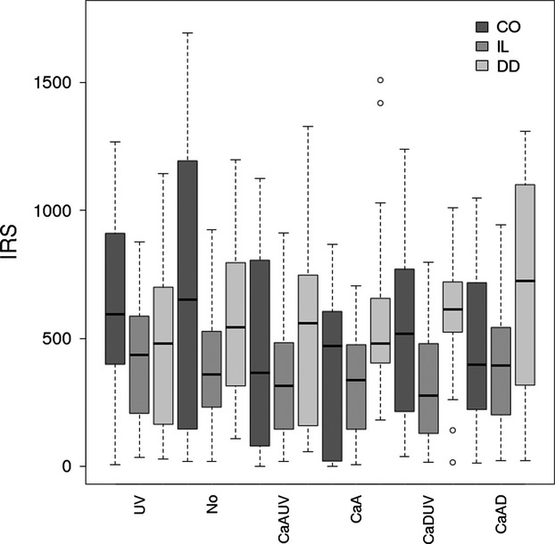
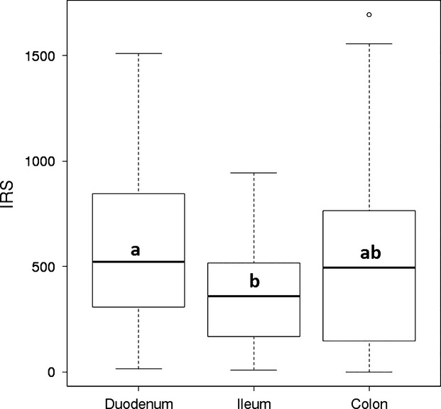
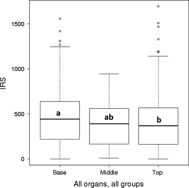
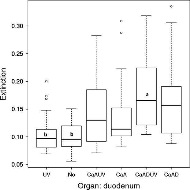
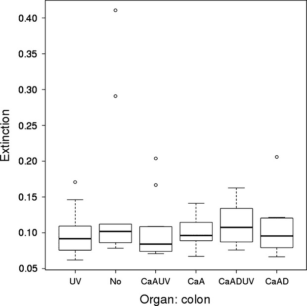
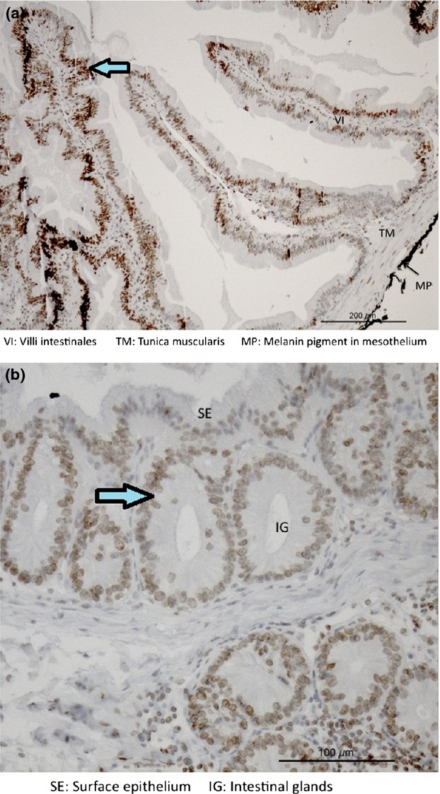
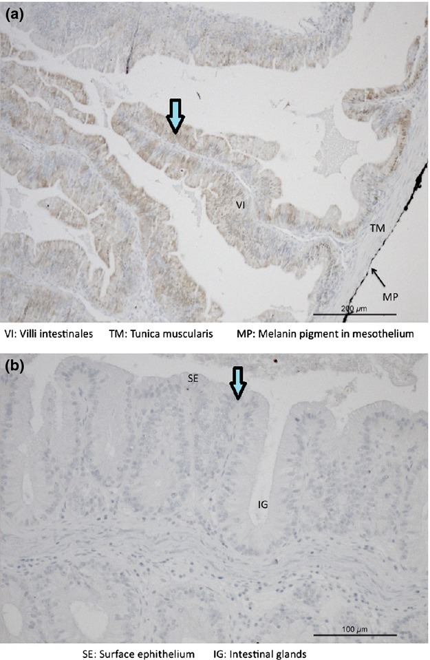
Summary: The purpose of this study was to investigate the influence of feeding and UVB exposition on the occurrence and distribution patterns of vitamin D receptors (VDR) and calbindin D28k (Cb‐D28k) in the gastrointestinal tract of veiled chameleons. Thus, 56 veiled chameleon hatchlings were divided into six treatment groups: UV (with UVB exposure); No (no supplements, no UVB exposure); CaAUV (with calcium (Ca), vitamin A supplementation, UVB exposure); CaA (with Ca, vitamin A supplementation); CaADUV (with Ca, vitamin A, vitamin D supplementation, UVB exposure); and CaAD (with Ca, vitamin A, vitamin D supplementation). Animals were reared under the suspected conditions for 6 months on locust‐based diets. Tissue samples of stomach, duodenum, ileum and colon were taken, and semi‐quantitative immunohistochemical methods (IHC) were performed to detect Cb‐D28k and VDR. VDR immunoreactions were higher in the luminal epithelium of the duodenum than in that of the ileum. VDR immunoreactions in the luminal epithelium were higher at the base of the villi of the duodenum as compared to the tip. Cb‐D28k immunoreactions were mainly observed in the luminal epithelium of the duodenum. The two groups treated with all dietary supplements (CaADUV, CaAD) exhibited a higher Cb‐D28k immunoreaction as those with no supplements and UVB exposure only. No immunoreaction for both proteins could be detected in the stomach. This study suggests that the duodenum plays an important role in the active transcellular absorption of Ca in veiled chameleons as shown by the immunohistochemical detection of VDR and Cb‐D28k. Expression of Cb‐D28k, in particular, appears to be regulated by dietary supplementation of vitamin D and vitamin A. VDRs, however, tended to be upregulated when animals were not supplemented with Ca, vitamin D and vitamin A. This may be due to the decreased Ca concentrations which caused vitamin D activation in the skin without any supplementation, but UVB exposure.
Overview
The claims and statistics of this paper haven’t been checked yet.
Run the check — free, no account needed. It reads the open-access text and checks the sources it cites.
Integrity signals
Registry
Cites
Statistics
Figures
Verification
Claims the paper makes
The claims and statistics of this paper haven’t been checked yet.
Run the check — free, no account needed. It reads the open-access text and checks the sources it cites.
Self-check
Percentages, sums and reported differences re-computed from the paper’s own numbers.
The self-check needs the paper’s full text.
Citations
The citation lists haven’t been fetched yet
References 39 · Cited by 13
Research works printed but not linked: 44
- Abate A. L., 2003, Chameleons and Vitamin A, Journal of Herpetological Medicine and Surgery, 13, 23, 10.5818/1529-9651.13.2.23 · DOI
- 10.1093/ps/77.4.585 · DOI
- Barker D., 1997, Preliminary observations on nutrient composition differences between adult and pinhead crickets, Acheta domestica, Bulletin of the Association of Reptilian Amphibian Veterinarians, 7, 10, 10.5818/1076-3139.7.1.10 · DOI
- 10.1126/science.92.2387.291 · DOI
- 10.1007/BF02571309 · DOI
- Bronner F., 2003, The role of passive transport in calcium absorption [letter], Journal of Nutrition, 133, 1426, 10.1093/jn/133.5.1426 · DOI
- 10.1643/0045-8511(2000)2000[0245:POAAFU]2.0.CO;2 · DOI
- 10.1016/j.biocon.2004.03.002 · DOI
- 10.1016/0306-4522(90)90091-H · DOI
- Coke R., 1998, Old‐World chameleons: captive care and breeding, Bulletin of the Association of Reptilian Amphibian Veterinarians, 8, 4, 10.5818/1076-3139.8.2.4 · DOI
- De Vries de Heekelingen T. 2008:Immunhistochemical detection of the distribution of Vitamin D receptors in the intestine of rabbits. Vet.‐Med. Dissertation Veterinär‐Anatomisches Institut Vetsuisse‐Fakultät Universität Zürich.
- Dierenfeld E. S., 1995, Vitamin A and insectivore nutrition, Verhandlungsbericht des Erkrankung der Zootiere, 37, 245
- 10.1002/(SICI)1098-2361(1996)15:3<279::AID-ZOO7>3.0.CO;2-8 · DOI
- 10.1016/0006-291X(90)92339-2 · DOI
- 10.1073/pnas.82.24.8780 · DOI
- 10.1002/jbmr.5650050404 · DOI
- 10.1016/0169-328X(91)90124-G · DOI
- 10.1139/o72-106 · DOI
- 10.3945/jn.110.120998 · DOI · Paper page
- Hoenderop J. G. J., 1999, Hormone‐stimulated Ca transport in rabbit kidney: multiple sites of inhibition by exogenous ATP, American Journal of Physiology, 277, F899
- 10.1152/physrev.00003.2004 · DOI
- 10.1073/pnas.92.8.3124 · DOI
- 10.3181/00379727-125-32011 · DOI
- 10.1016/j.domaniend.2006.05.008 · DOI
- 10.1111/j.1439-0396.2007.00782.x · DOI
- 10.1111/j.1748-1090.1976.tb00130.x · DOI
- 10.1111/j.1748-1090.2005.tb00006.x · DOI
- 10.1002/jcb.240580306 · DOI
- Olson J. A., 1984, Handbook of Vitamins: Nutritional Biochemical, and Clinical Aspects, 1
- 10.1016/0016-6480(87)90125-0 · DOI
- Pennino M., 1991, Retinol, α‐tocopherol, and proximate nutrient composition of invertebrates used as feed, The International Zoo Yearbook, 30, 143, 10.1111/j.1748-1090.1991.tb03477.x · DOI
- 10.1073/pnas.76.11.5485 · DOI
- R Core Team, 2013, R: A Language and Environment for Statistical Computing
- Robbins C. T., 1993, Wildlife Feeding and Nutrition, 352
- Sabatini J. A., 1998, Effects of internal or external supplementation on the nutrient content of crickets, The Vivarium, 9, 23
- 10.1113/jphysiol.1996.sp021340 · DOI
- Schroeter‐Vogt C. 2011:Immunohistochemical detection of Calbindin D9k in the different parts of the intestine of the rabbit. Vet.‐Med. Dissertation Veterinär‐Anatomisches Institut Vetsuisse‐Fakultät Universität Zürich.
- 10.2527/jas.2009-2682 · DOI
- Stanford M. D., 2005, Calcium metabolism in grey parrots: the effects of husbandry, The Royal College of Veterinary Surgeons, 2, 99
- Taylor A. N., 1968, A vitamin D‐dependent calcium binding protein in canine intestinal mucosa, Federation Proceedings, 27, 675
- Theofan G., 1986, Regulation of calbindin‐D28K gene expression by 1,25‐dihydroxyvitamin D3 is correlated to receptor occupancy, The Journal of Biological Chemistry, 261, 16943, 10.1016/S0021-9258(19)75981-X · DOI
- Varghese S., 1988, Analysis of rat vitamin D–dependent calbindin‐D28K gene expression, The Journal of Biological Chemistry, 263, 9776, 10.1016/S0021-9258(19)81585-5 · DOI
- 10.1210/mend-3-3-495 · DOI
- 10.1074/jbc.275.6.3963 · DOI
Metadata
- Authors
- Haxhiu, D., Hoby, S., Wenker, C., Boos, A., Kowalewski, M. P., Lewis, F., & Liesegang, A.
- Affiliations
- University of Zurich (CH)
- Zoo Basel Basel Switzerland
- Corresponding author
- Liesegang, A.
- Venue
- Journal of Animal Physiology and Animal Nutrition
- Publisher
- Wiley
- Published
- 2014-05-22
- Volume
- 98
- Issue
- 6
- Pages
- 1021-1030
- ISSN
- 0931-2439 (ISSN-L)
- 1439-0396
- PMC
- PMC4280901
- OpenAlex
- W2066020080
- Cited by
- 13
- References
- 39
- Citations by year
- 2026: 2 · 2023: 1 · 2020: 2 · 2019: 1 · 2018: 1 · 2017: 1 · 2015: 4 · 2014: 1
- FWCI
- 1.48
- Type
- journal article
- Language
- en
- Open access
- hybrid
- doi.org
- Licence
- Locations
- Journal of Animal Physiology and Animal Nutrition (publishedVersion, cc-by-nc-nd)
- PubMed Central (publishedVersion, cc-by-nc-nd)
- PubMed (publishedVersion)
- Zurich Open Repository and Archive (University of Zurich) (submittedVersion, cc-by-nc-nd)
- Topics
- Health Sciences › Medicine › Endocrinology, Diabetes and Metabolism › Vitamin D Research Studies
- Life Sciences › Agricultural and Biological Sciences › Ecology, Evolution, Behavior and Systematics › Amphibian and Reptile Biology
- Life Sciences › Agricultural and Biological Sciences › Animal Science and Zoology › Animal Nutrition and Physiology
- Keywords
- veiled chameleon, Chamaeleo calyptratus, calcium absorption, vitamin D receptor, calbindin-D28k, UVB exposure, dietary supplementation, vitamin A, vitamin D, duodenum, gastrointestinal tract, reptile nutrition, transcellular calcium transport
- MeSH terms
- Animals, Animal Feed, Animal Nutritional Physiological Phenomena, Calcium, Diet, Intestinal Mucosa, Lizards, Ultraviolet Rays, Vitamin A, Receptors, Calcitriol, Gastrointestinal Tract, Calbindin 1
About these checks
Registry, retraction, Cites and statistics results are computed, not judged, with one exception: a printed reference that no index lists is read once by a model before it counts as a citation error. Claims are extracted by a model and kept only when their quote appears verbatim in the text; each cited source is then checked, with a model’s help, for whether it says what the claim relies on. Those verdicts can be wrong — read the claim’s quote and the cited source before relying on one.
Statistics and arithmetic are recomputed, not judged.
Confidence bands are shown only for fields calibrated on our annotated test set. None is calibrated yet, so no band is shown.
What counts as a claim ›How we measure confidence ›How we check this ›
Are you an author, or do you see a problem with this page? Request a review
How to cite
Cite CoChat for the checks on this page, and the paper for its own findings.
- This page
- CoChat. (2026). Citation check: Influence of feeding and UVB exposition on the absorption mechanisms of calcium in the gastrointestinal tract of veiled chameleons (Chamaeleo calyptratus). https://app.cochat.ai/papers/10.1111/jpn.12206
- The paper
- Haxhiu, D., Hoby, S., Wenker, C., Boos, A., Kowalewski, M. P., Lewis, F., & Liesegang, A. (2014). Influence of feeding and UVB exposition on the absorption mechanisms of calcium in the gastrointestinal tract of veiled chameleons (Chamaeleo calyptratus). Journal of Animal Physiology and Animal Nutrition. https://doi.org/10.1111/jpn.12206
