Cited by 8Viral-associated cancers and disordersHerpesvirus Infections and TreatmentsCytomegalovirus and herpesvirus research

The DOI matches its Crossref record; 1 of the 1 references it cites were found in a registry or an index.

Kaposi’s Sarcoma-Associated Herpesvirus ORF67.5 Functions as a Component of the Terminase Complex

Open access

Published version of a preprint

Abstract

Although the formation and function of the alpha- and betaherpesvirus terminase complexes are well understood, the Kaposi’s sarcoma-associated herpesvirus (KSHV) terminase complex is still largely uncharacterized. This complex presumably contains KSHV open reading frame 7 (ORF7), ORF29, and ORF67.5.

Overview

The claims and statistics of this paper haven’t been checked yet.

Run the check — free, no account needed. It reads the open-access text and checks the sources it cites.

A few free checks a day · papers already verified open instantly · results are public

Integrity signals

Registry

Crossref · 10.1128/jvi.00475-23

Cites

1 of 1 references verified0 citation errors · 0 not checkable

Statistics

Figures

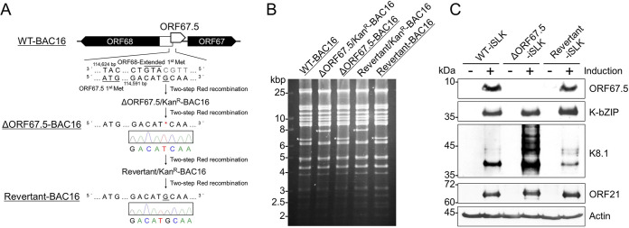
FIG 1 Construction of ΔORF67.5-BAC16 and Revertant-BAC16. (A) Diagram showing the location of KSHV ORF67.5 (nucleotides 114382 to 114624; accession number: GQ994935) and its neighboring ORFs on the KSHV-BAC (WT-BAC16). The adjacent DNA sequence of the mutation sites in ΔORF67.5-BAC16 and Revertant-BAC16 clones was confirmed by Sanger sequencing. (B) Agarose gel electrophoresis image of EcoRV-digested recombinant BAC16 clones. The asterisks indicate the insertion or deletion of a kanamycin resistance cassette in each BAC clone. (C) Western blot showing the disappearance of ORF67.5 expression in lytic-induced (+) or uninduced (−) ΔORF67.5-BAC16-harboring iSLK cells (ΔORF67.5-iSLK). WT-iSLK, ΔORF67.5-iSLK, and Revertant-iSLK were treated (or untreated) with Dox and SB for 72 h to induce lytic replication. An antibody against β-actin (actin) was used as a loading control. The lytic gene products, K-bZIP (DE), K8.1 (L), and ORF21 (L) were expressed in all cell lines, unlike ORF67.5, which was absent in ΔORF67.5-iSLK.
FIG 1 Construction of ΔORF67.5-BAC16 and Revertant-BAC16. (A) Diagram showing the location of KSHV ORF67.5 (nucleotides 114382 to 114624; accession number: GQ994935) and its neighboring ORFs on the KSHV-BAC (WT-BAC16). The adjacent DNA sequence of the mutation sites in ΔORF67.5-BAC16 and Revertant-BAC16 clones was confirmed by Sanger sequencing. (B) Agarose gel electrophoresis image of EcoRV-digested recombinant BAC16 clones. The asterisks indicate the insertion or deletion of a kanamycin resistance cassette in each BAC clone. (C) Western blot showing the disappearance of ORF67.5 expression in lytic-induced (+) or uninduced (−) ΔORF67.5-BAC16-harboring iSLK cells (ΔORF67.5-iSLK). WT-iSLK, ΔORF67.5-iSLK, and Revertant-iSLK were treated (or untreated) with Dox and SB for 72 h to induce lytic replication. An antibody against β-actin (actin) was used as a loading control. The lytic gene products, K-bZIP (DE), K8.1 (L), and ORF21 (L) were expressed in all cell lines, unlike ORF67.5, which was absent in ΔORF67.5-iSLK.CC BY · PubMed Central
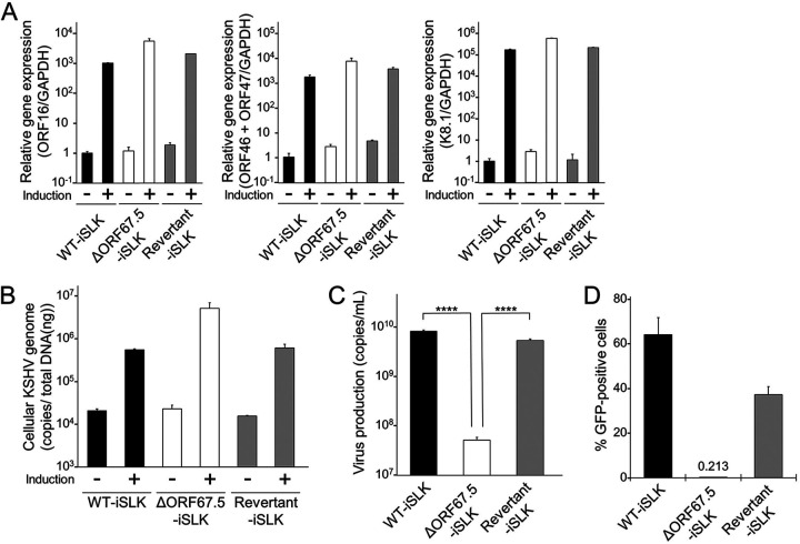
FIG 2 Characterization of ΔORF67.5 KSHV. (A) mRNA expression analysis of the following KSHV lytic genes: IE, ORF16; DE, ORF46 and ORF47; and L, K8.1. WT-iSLK, ΔORF67.5-iSLK, and Revertant-iSLK were treated with Dox and SB for 72 h to induce the lytic phase (+) or were uninduced (−). Total RNA was purified from harvested iSLK cells and subjected to RT-qPCR. The lytic gene mRNA levels were normalized to GAPDH mRNA levels. The values obtained from uninduced WT-iSLK were defined as 1.0. (B) Quantification of intracellular KSHV genomic DNA. WT-iSLK, ΔORF67.5-iSLK, and Revertant-iSLK were treated with Dox and SB to induce the lytic phase (+) or were uninduced (−). At 72 h postinduction, viral DNA was purified from the harvested cells. The intracellular viral genome copy number was then measured by qPCR and normalized to the amount of total DNA. (C) Quantification of extracellular encapsidated KSHV genomes. Each iSLK cell line was treated with Dox and SB for 72 h to induce the lytic phase, and the culture supernatants were harvested. Next, encapsidated KSHV genomic DNA from the culture supernatants was measured by qPCR; ****, P < 0.001. (D) Measurement of infectious virus. Each iSLK cell line was cultured with medium containing Dox and SB for 72 h to induce the lytic phase, and the culture supernatants were harvested. The supernatants were mixed with fresh HEK293T cells for infection with progeny virus. At 24 h postinfection, GFP-positive cells were counted by flow cytometry.
FIG 2 Characterization of ΔORF67.5 KSHV. (A) mRNA expression analysis of the following KSHV lytic genes: IE, ORF16; DE, ORF46 and ORF47; and L, K8.1. WT-iSLK, ΔORF67.5-iSLK, and Revertant-iSLK were treated with Dox and SB for 72 h to induce the lytic phase (+) or were uninduced (−). Total RNA was purified from harvested iSLK cells and subjected to RT-qPCR. The lytic gene mRNA levels were normalized to GAPDH mRNA levels. The values obtained from uninduced WT-iSLK were defined as 1.0. (B) Quantification of intracellular KSHV genomic DNA. WT-iSLK, ΔORF67.5-iSLK, and Revertant-iSLK were treated with Dox and SB to induce the lytic phase (+) or were uninduced (−). At 72 h postinduction, viral DNA was purified from the harvested cells. The intracellular viral genome copy number was then measured by qPCR and normalized to the amount of total DNA. (C) Quantification of extracellular encapsidated KSHV genomes. Each iSLK cell line was treated with Dox and SB for 72 h to induce the lytic phase, and the culture supernatants were harvested. Next, encapsidated KSHV genomic DNA from the culture supernatants was measured by qPCR; ****, P < 0.001. (D) Measurement of infectious virus. Each iSLK cell line was cultured with medium containing Dox and SB for 72 h to induce the lytic phase, and the culture supernatants were harvested. The supernatants were mixed with fresh HEK293T cells for infection with progeny virus. At 24 h postinfection, GFP-positive cells were counted by flow cytometry.CC BY · PubMed Central
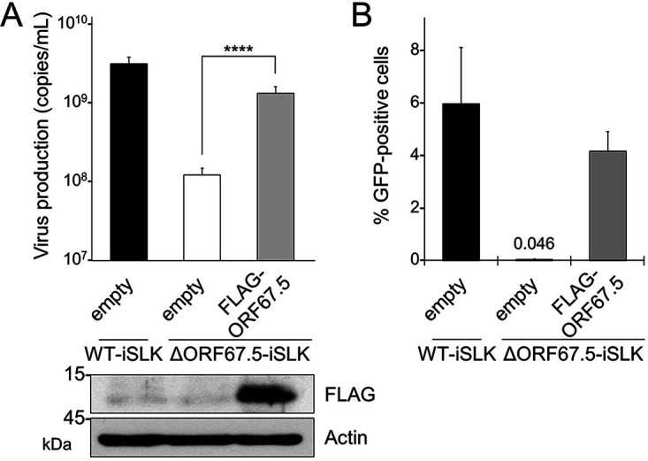
FIG 3 Complementation of ORF67.5-deficient KSHV with exogenous ORF67.5 expression. (A) Exogenous expression of ORF67.5 rescued the reduction in extracellular KSHV production observed in ΔORF67.5-iSLK. ΔORF67.5-iSLK was transiently transfected with empty plasmid (empty) or FLAG-ORF67.5 plasmid and was simultaneously cultured with medium containing Dox and SB for 72 h to induce the lytic phase. Encapsidated KSHV genomes in the culture supernatant were quantified by qPCR. Exogenous ORF67.5 expression was confirmed by Western blotting using an anti-FLAG (FLAG) antibody. An antibody against β-actin (actin) was used as a loading control; ****, P < 0.001. (B) Exogenous expression of ORF67.5 rescued the reduction in infectious virus production observed in ΔORF67.5-iSLK. ΔORF67.5-iSLK was transiently transfected with empty plasmid (empty) or FLAG-ORF67.5 expression plasmid and simultaneously cultured with medium containing Dox and SB for 72 h to induce the lytic phase. Harvested culture supernatants were then used for infection of fresh HEK293T cells. At 24 h postinfection, GFP-positive cells were counted by flow cytometry.
FIG 3 Complementation of ORF67.5-deficient KSHV with exogenous ORF67.5 expression. (A) Exogenous expression of ORF67.5 rescued the reduction in extracellular KSHV production observed in ΔORF67.5-iSLK. ΔORF67.5-iSLK was transiently transfected with empty plasmid (empty) or FLAG-ORF67.5 plasmid and was simultaneously cultured with medium containing Dox and SB for 72 h to induce the lytic phase. Encapsidated KSHV genomes in the culture supernatant were quantified by qPCR. Exogenous ORF67.5 expression was confirmed by Western blotting using an anti-FLAG (FLAG) antibody. An antibody against β-actin (actin) was used as a loading control; ****, P < 0.001. (B) Exogenous expression of ORF67.5 rescued the reduction in infectious virus production observed in ΔORF67.5-iSLK. ΔORF67.5-iSLK was transiently transfected with empty plasmid (empty) or FLAG-ORF67.5 expression plasmid and simultaneously cultured with medium containing Dox and SB for 72 h to induce the lytic phase. Harvested culture supernatants were then used for infection of fresh HEK293T cells. At 24 h postinfection, GFP-positive cells were counted by flow cytometry.CC BY · PubMed Central
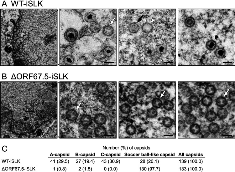
FIG 4 TEM images showing capsids formed in WT-iSLK and ΔORF67.5-iSLK. (A and B) Cells were lytically induced for 48 h, and nuclear viral capsids in WT-iSLK (A) and ΔORF67.5-iSLK (B) were observed by TEM. The black arrowheads, white arrowheads, black arrows, and white arrows indicate C-capsids, A-capsids, B-capsids, and soccer ball-like capsids, respectively. Scale bars, 100 nm. (C) Quantification of each capsid type observed by TEM.
FIG 4 TEM images showing capsids formed in WT-iSLK and ΔORF67.5-iSLK. (A and B) Cells were lytically induced for 48 h, and nuclear viral capsids in WT-iSLK (A) and ΔORF67.5-iSLK (B) were observed by TEM. The black arrowheads, white arrowheads, black arrows, and white arrows indicate C-capsids, A-capsids, B-capsids, and soccer ball-like capsids, respectively. Scale bars, 100 nm. (C) Quantification of each capsid type observed by TEM.CC BY · PubMed Central
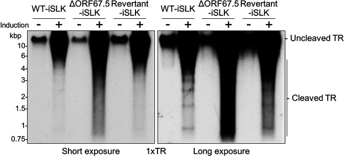
FIG 5 ΔORF67.5-KSHV failed to cleave the TR sites in the viral genome. WT-iSLK, ΔORF67.5-iSLK, and Revertant-iSLK were treated with (+) or without (−) Dox and SB for 72 h to induce the lytic phase. Purified total genomic DNA was digested with EcoRI and SalI and subjected to Southern blotting. Uncleaved and cleaved TRs were detected using a DIG-labeled 1×TR probe, and their migration patterns are denoted to the right of the images; left, short exposure; right, long exposure.
FIG 5 ΔORF67.5-KSHV failed to cleave the TR sites in the viral genome. WT-iSLK, ΔORF67.5-iSLK, and Revertant-iSLK were treated with (+) or without (−) Dox and SB for 72 h to induce the lytic phase. Purified total genomic DNA was digested with EcoRI and SalI and subjected to Southern blotting. Uncleaved and cleaved TRs were detected using a DIG-labeled 1×TR probe, and their migration patterns are denoted to the right of the images; left, short exposure; right, long exposure.CC BY · PubMed Central
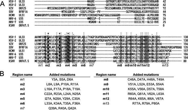
FIG 6 Amino acid sequence alignment between KSHV ORF67.5 and its human herpesviral homologs as well as construction of the ORF67.5 alanine mutants. (A) Amino acid sequences were obtained from the NCBI database (HSV-1 UL33 [GenBank: QAU10292], HSV-2 UL33 [NCBI Reference Sequence: YP_009137185], VZV ORF25 [GenBank: QXM18354], EBV BFRF1A [GenBank: AAA45868], HCMV UL51 [GenBank: QHB20503], HHV-6 U35 [NCBI Reference Sequence: NP_042928], HHV-7 U35 [GenBank: AAC40749], and KSHV ORF67.5 [GenBank: QFU18781]). The raw data used for the alignment were obtained with the Clustal Omega program (EMBL-EBI; https://www.ebi.ac.uk/Tools/msa/clustalo/). The background indicates the degree of homology. The completely matched amino acid residues are shown in white letters with a black background, and similar amino acid residues are shown in black letters with a gray background. The conserved regions (m2 to 6 and m8 to 12) and the nonconserved regions (m1, m7, and m13) among the human herpesviral ORF67.5 homologs are indicated at the bottom of the sequence alignments, and the conserved regions are indicated in bold. (B) The amino acid sequences of the conserved and nonconserved regions of the human herpesviral ORF67.5 homologs that were substituted with alanine in each ORF67.5 mutant. The names of the conserved regions are indicated in bold.
FIG 6 Amino acid sequence alignment between KSHV ORF67.5 and its human herpesviral homologs as well as construction of the ORF67.5 alanine mutants. (A) Amino acid sequences were obtained from the NCBI database (HSV-1 UL33 [GenBank: QAU10292], HSV-2 UL33 [NCBI Reference Sequence: YP_009137185], VZV ORF25 [GenBank: QXM18354], EBV BFRF1A [GenBank: AAA45868], HCMV UL51 [GenBank: QHB20503], HHV-6 U35 [NCBI Reference Sequence: NP_042928], HHV-7 U35 [GenBank: AAC40749], and KSHV ORF67.5 [GenBank: QFU18781]). The raw data used for the alignment were obtained with the Clustal Omega program (EMBL-EBI; https://www.ebi.ac.uk/Tools/msa/clustalo/). The background indicates the degree of homology. The completely matched amino acid residues are shown in white letters with a black background, and similar amino acid residues are shown in black letters with a gray background. The conserved regions (m2 to 6 and m8 to 12) and the nonconserved regions (m1, m7, and m13) among the human herpesviral ORF67.5 homologs are indicated at the bottom of the sequence alignments, and the conserved regions are indicated in bold. (B) The amino acid sequences of the conserved and nonconserved regions of the human herpesviral ORF67.5 homologs that were substituted with alanine in each ORF67.5 mutant. The names of the conserved regions are indicated in bold.CC BY · PubMed Central
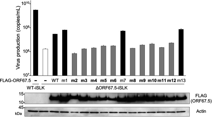
FIG 7 The ORF67.5 conserved region alanine mutants failed to rescue the reduction in virus production from ΔORF67.5-iSLK. To evaluate the contribution of the conserved and nonconserved regions of ORF67.5 to virus production, complementation assays were performed by exogenous ORF67.5 alanine mutant expression in ΔORF67.5-iSLK. ΔORF67.5-iSLK was transiently transfected with FLAG-ORF67.5 WT or FLAG-ORF67.5 alanine mutant expression plasmids (FLAG-ORF67.5 m1 to m13). During transfection, the cells were simultaneously cultured in medium containing Dox and SB for 72 h to induce the lytic phase. Encapsidated KSHV genomes in the culture supernatants were quantified by qPCR. Expression of exogenous FLAG-tagged ORF67.5 WT and mutants was confirmed by Western blotting using an anti-FLAG (FLAG) antibody.
FIG 7 The ORF67.5 conserved region alanine mutants failed to rescue the reduction in virus production from ΔORF67.5-iSLK. To evaluate the contribution of the conserved and nonconserved regions of ORF67.5 to virus production, complementation assays were performed by exogenous ORF67.5 alanine mutant expression in ΔORF67.5-iSLK. ΔORF67.5-iSLK was transiently transfected with FLAG-ORF67.5 WT or FLAG-ORF67.5 alanine mutant expression plasmids (FLAG-ORF67.5 m1 to m13). During transfection, the cells were simultaneously cultured in medium containing Dox and SB for 72 h to induce the lytic phase. Encapsidated KSHV genomes in the culture supernatants were quantified by qPCR. Expression of exogenous FLAG-tagged ORF67.5 WT and mutants was confirmed by Western blotting using an anti-FLAG (FLAG) antibody.CC BY · PubMed Central
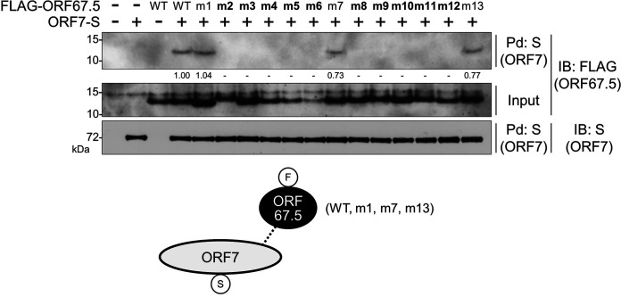
FIG 8 The conserved regions of ORF67.5 were necessary for the interaction of ORF67.5 with ORF7. An expression plasmid encoding FLAG-tagged wild-type ORF67.5 (FLAG-ORF67.5 WT) or FLAG-tagged ORF67.5 mutant (FLAG-ORF67.5 m1 to m13) was cotransfected with an S-tagged ORF7 expression plasmid (ORF7-S) into HEK293T cells. Next, the cell lysates were subjected to pulldown assays (Pd) using S-protein immobilized beads (S) for the precipitation of ORF7-S protein. Any FLAG-ORF67.5 protein that interacted with ORF7 was detected by Western blotting using an anti-FLAG antibody (FLAG). The proteins in parentheses indicate which protein was recognized by the assay. The band intensities of the FLAG-ORF67.5 are shown at the bottom of the image. The intensity value of FLAG-ORF67.5 in the cell transfected with FLAG-ORF67.5 WT and ORF7-S was defined as 1.0. When the intensity value was the less than 0.01, “−” was indicated. The conserved region mutants of ORF67.5 are indicated in bold. Bottom, schematic representation of the results with the protein tags indicated in the circles; IB, immunoblot.
FIG 8 The conserved regions of ORF67.5 were necessary for the interaction of ORF67.5 with ORF7. An expression plasmid encoding FLAG-tagged wild-type ORF67.5 (FLAG-ORF67.5 WT) or FLAG-tagged ORF67.5 mutant (FLAG-ORF67.5 m1 to m13) was cotransfected with an S-tagged ORF7 expression plasmid (ORF7-S) into HEK293T cells. Next, the cell lysates were subjected to pulldown assays (Pd) using S-protein immobilized beads (S) for the precipitation of ORF7-S protein. Any FLAG-ORF67.5 protein that interacted with ORF7 was detected by Western blotting using an anti-FLAG antibody (FLAG). The proteins in parentheses indicate which protein was recognized by the assay. The band intensities of the FLAG-ORF67.5 are shown at the bottom of the image. The intensity value of FLAG-ORF67.5 in the cell transfected with FLAG-ORF67.5 WT and ORF7-S was defined as 1.0. When the intensity value was the less than 0.01, “−” was indicated. The conserved region mutants of ORF67.5 are indicated in bold. Bottom, schematic representation of the results with the protein tags indicated in the circles; IB, immunoblot.CC BY · PubMed Central
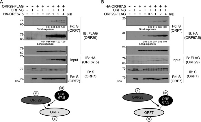
FIG 9 ORF67.5 promoted the interaction between ORF7 and ORF29, and ORF29 increased the interaction between ORF67.5 and ORF7. (A) Expression plasmids encoding ORF29-FLAG and ORF7-S were cotransfected with various amounts of HA-ORF67.5 expression plasmid into HEK293T cells. Next, the cell lysates were subjected to pulldown (Pd) assays using S-protein immobilized beads (S) for the precipitation of ORF7-S protein. Any ORF29-FLAG protein that interacted with ORF7 was detected by Western blotting with an anti-FLAG antibody (FLAG). The proteins in parentheses indicate which protein was recognized by the assay. The band intensities of ORF29-FLAG are shown at the bottom of the image, and the value of ORF29-FLAG in the cell transfected with ORF29-FLAG, ORF7-S, and HA-ORF67.5 (4 μg) is defined as 1.0. When the intensity value was the less than 0.01, “−” was indicated. Bottom, schematic representation of the results with the protein tags indicated in the circles. (B) Expression plasmids encoding HA-ORF67.5 and ORF7-S were cotransfected with various amounts of ORF29-FLAG expression plasmid into HEK293T cells. Next, the cell lysates were subjected to pulldown (Pd) assays using S-protein immobilized beads (S), followed by Western blotting with anti-HA antibody (HA) to detect the ORF7-binding HA-ORF67.5 protein. The band intensities of HA-ORF67.5 are shown at the bottom of the image. The value of HA-ORF67.5 in the cell transfected with HA-ORF67.5, ORF7-S, and ORF29-FLAG (4 μg) was defined as 1.0. When the intensity value was the less than 0.01, “−” was indicated.
FIG 9 ORF67.5 promoted the interaction between ORF7 and ORF29, and ORF29 increased the interaction between ORF67.5 and ORF7. (A) Expression plasmids encoding ORF29-FLAG and ORF7-S were cotransfected with various amounts of HA-ORF67.5 expression plasmid into HEK293T cells. Next, the cell lysates were subjected to pulldown (Pd) assays using S-protein immobilized beads (S) for the precipitation of ORF7-S protein. Any ORF29-FLAG protein that interacted with ORF7 was detected by Western blotting with an anti-FLAG antibody (FLAG). The proteins in parentheses indicate which protein was recognized by the assay. The band intensities of ORF29-FLAG are shown at the bottom of the image, and the value of ORF29-FLAG in the cell transfected with ORF29-FLAG, ORF7-S, and HA-ORF67.5 (4 μg) is defined as 1.0. When the intensity value was the less than 0.01, “−” was indicated. Bottom, schematic representation of the results with the protein tags indicated in the circles. (B) Expression plasmids encoding HA-ORF67.5 and ORF7-S were cotransfected with various amounts of ORF29-FLAG expression plasmid into HEK293T cells. Next, the cell lysates were subjected to pulldown (Pd) assays using S-protein immobilized beads (S), followed by Western blotting with anti-HA antibody (HA) to detect the ORF7-binding HA-ORF67.5 protein. The band intensities of HA-ORF67.5 are shown at the bottom of the image. The value of HA-ORF67.5 in the cell transfected with HA-ORF67.5, ORF7-S, and ORF29-FLAG (4 μg) was defined as 1.0. When the intensity value was the less than 0.01, “−” was indicated.CC BY · PubMed Central
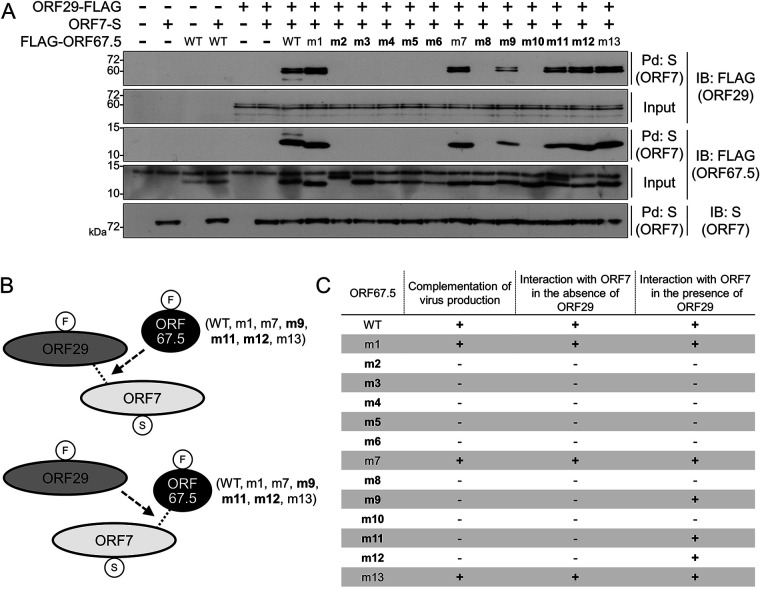
FIG 10 The effects of the conserved and nonconserved region mutants of ORF67.5 on the interaction between ORF7 and ORF29 as well as the effect of ORF29 on the interaction between each ORF67.5 mutant and ORF7. (A) Expression plasmids encoding ORF29-FLAG and ORF7-S were cotransfected with the FLAG-ORF67.5 mutant expression plasmid into HEK293T cells. Next, the cell lysates were subjected to pulldown (Pd) assays using S-protein immobilized beads (S) followed by Western blotting using anti-FLAG or anti-S antibodies (FLAG and S, respectively). Any ORF29-FLAG protein that interacted with ORF7-S in the presence of wild-type (WT) or alanine mutant ORF67.5 (m1 to m13) was detected by Western blotting with anti-FLAG antibody. The proteins in parentheses indicate which protein was recognized by the assay. (B) Schematic representation of the interaction relationship among ORF7, ORF29, and ORF67.5 in this experiment. The protein tags are indicated in the circles, and the conserved region mutants of ORF67.5 are shown in bold. (C) A table summarizing the characteristics of the wild-type (WT) and alanine mutants of ORF67.5 (m1 to m13) revealed by our experiments. The table summarizes the results obtained in Fig. 7 (complementation of virus production), Fig. 8 (the interaction with ORF7 in the absence of ORF29), and Fig. 10A (the interaction with ORF7 in the presence of ORF29). The conserved region mutants in ORF67.5 are indicated in bold.
FIG 10 The effects of the conserved and nonconserved region mutants of ORF67.5 on the interaction between ORF7 and ORF29 as well as the effect of ORF29 on the interaction between each ORF67.5 mutant and ORF7. (A) Expression plasmids encoding ORF29-FLAG and ORF7-S were cotransfected with the FLAG-ORF67.5 mutant expression plasmid into HEK293T cells. Next, the cell lysates were subjected to pulldown (Pd) assays using S-protein immobilized beads (S) followed by Western blotting using anti-FLAG or anti-S antibodies (FLAG and S, respectively). Any ORF29-FLAG protein that interacted with ORF7-S in the presence of wild-type (WT) or alanine mutant ORF67.5 (m1 to m13) was detected by Western blotting with anti-FLAG antibody. The proteins in parentheses indicate which protein was recognized by the assay. (B) Schematic representation of the interaction relationship among ORF7, ORF29, and ORF67.5 in this experiment. The protein tags are indicated in the circles, and the conserved region mutants of ORF67.5 are shown in bold. (C) A table summarizing the characteristics of the wild-type (WT) and alanine mutants of ORF67.5 (m1 to m13) revealed by our experiments. The table summarizes the results obtained in Fig. 7 (complementation of virus production), Fig. 8 (the interaction with ORF7 in the absence of ORF29), and Fig. 10A (the interaction with ORF7 in the presence of ORF29). The conserved region mutants in ORF67.5 are indicated in bold.CC BY · PubMed Central
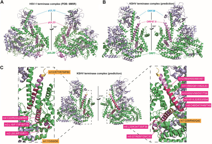
FIG 11 The structure of the HSV-1 terminase complex and the predicted structure model of the KSHV terminase complex. (A) The HSV-1 monomeric terminase complex structure (PDB: 6M5R) (21). Each HSV-1 protein cartoon was colored as follows: HSV-1 pUL28 (green), pUL15 (light blue), and pUL33 (light purple). Unmodelled amino acid residues, expected to comprise the unfolded region of each protein, were listed as follows: HSV-1 pUL28 (aa 268 to 305, 433 to 477, and 652 to 653), pUL15 (aa 178 to 194, 603 to 613, and 686 to 704), and pUL33 (aa 83 to 95). (B) The KSHV monomeric terminase complex was predicted by AlphaFold2. Each KSHV protein cartoon was colored as follows: KSHV ORF7 (green), ORF29 (light blue), and ORF67.5 (light purple). The KSHV terminase complex proteins were colored according to their HSV-1 homologs. (C) Based on the predicted KSHV terminase complex model, the main chain cartoon of the KSHV ORF67.5 regions substituted with alanine in the present study was visualized with rotation. The mutated regions (m1, m7, and m13), which are dispensable for the function of ORF67.5 in virus production, are highlighted in yellow. The mutated regions (m2 to 6 and m8 to 12) that are essential for the function of ORF67.5 in virus production are highlighted in magenta.
FIG 11 The structure of the HSV-1 terminase complex and the predicted structure model of the KSHV terminase complex. (A) The HSV-1 monomeric terminase complex structure (PDB: 6M5R) (21). Each HSV-1 protein cartoon was colored as follows: HSV-1 pUL28 (green), pUL15 (light blue), and pUL33 (light purple). Unmodelled amino acid residues, expected to comprise the unfolded region of each protein, were listed as follows: HSV-1 pUL28 (aa 268 to 305, 433 to 477, and 652 to 653), pUL15 (aa 178 to 194, 603 to 613, and 686 to 704), and pUL33 (aa 83 to 95). (B) The KSHV monomeric terminase complex was predicted by AlphaFold2. Each KSHV protein cartoon was colored as follows: KSHV ORF7 (green), ORF29 (light blue), and ORF67.5 (light purple). The KSHV terminase complex proteins were colored according to their HSV-1 homologs. (C) Based on the predicted KSHV terminase complex model, the main chain cartoon of the KSHV ORF67.5 regions substituted with alanine in the present study was visualized with rotation. The mutated regions (m1, m7, and m13), which are dispensable for the function of ORF67.5 in virus production, are highlighted in yellow. The mutated regions (m2 to 6 and m8 to 12) that are essential for the function of ORF67.5 in virus production are highlighted in magenta.CC BY · PubMed Central

Verification

Claims the paper makes

The claims and statistics of this paper haven’t been checked yet.

Run the check — free, no account needed. It reads the open-access text and checks the sources it cites.

A few free checks a day · papers already verified open instantly · results are public

Self-check

Percentages, sums and reported differences re-computed from the paper’s own numbers.

The self-check needs the paper’s full text.

Citations

Cites

Show all 1 references

Research works printed but not linked: 52

  1. 10.1126/science.7997879 · DOI
  2. Damania B, Cesarman E. 2022. Kaposi’s sarcoma herpesvirus, p 513–572. In Howley PM, Knipe DM (ed), Fields virology, 7th ed. Wolters Kluwer, Philadelphia, PA.
  3. 10.1073/pnas.93.25.14862 · DOI
  4. 10.1182/blood.V88.2.645.bloodjournal882645 · DOI
  5. 10.1182/blood.V86.4.1276.bloodjournal8641276 · DOI
  6. 10.1086/654798 · DOI
  7. 10.1073/pnas.95.18.10866 · DOI
  8. 10.1016/j.coviro.2011.06.003 · DOI
  9. 10.1007/978-3-319-53168-7_6 · DOI
  10. 10.1006/jmbi.1996.0587 · DOI
  11. 10.1038/nsb922 · DOI
  12. 10.1128/mBio.01525-15 · DOI
  13. 10.1128/JVI.10.5.1044-1052.1972 · DOI
  14. 10.1128/JVI.68.6.3702-3712.1994 · DOI
  15. 10.1128/jvi.74.4.1663-1673.2000 · DOI
  16. 10.1016/0092-8674(89)90587-4 · DOI
  17. 10.1016/0092-8674(91)90324-r · DOI
  18. Krug LT, Pellett PE. 2022. The family Herpesviridae: a brief introduction, p 212–234. In Howley PM, Knipe DM (ed), Fields virology, 7th ed. Wolters Kluwer, Philadelphia, PA.
  19. 10.1128/jvi.76.10.4785-4791.2002 · DOI
  20. 10.1128/JVI.02632-13 · DOI
  21. 10.1007/s13238-020-00710-0 · DOI
  22. 10.1128/JVI.00125-06 · DOI
  23. 10.1128/JVI.67.8.4497-4503.1993 · DOI
  24. 10.1128/JVI.68.12.8118-8124.1994 · DOI
  25. 10.1099/0022-1317-71-10-2377 · DOI
  26. 10.1128/JVI.67.6.3470-3480.1993 · DOI
  27. 10.1016/0042-6822(91)90043-b · DOI
  28. 10.1006/viro.1996.0098 · DOI
  29. 10.1128/JVI.05265-11 · DOI
  30. 10.1056/NEJMoa1706640 · DOI
  31. 10.3390/microorganisms9061169 · DOI
  32. 10.1128/jvi.00684-22 · DOI
  33. 10.1021/pr200628s · DOI
  34. 10.1128/JVI.01955-12 · DOI
  35. 10.1128/JVI.02533-12 · DOI
  36. 10.1371/journal.ppat.1003847 · DOI
  37. 10.1128/JVI.00840-18 · DOI
  38. 10.1016/j.jviromet.2011.03.012 · DOI
  39. 10.1128/JVI.01019-12 · DOI
  40. 10.1016/j.jsb.2007.10.016 · DOI
  41. Arvin AM, Abendroth A. 2022. Varicella-zoster virus, p 445–488. In Howley PM, Knipe DM (ed), Fields virology, 7th ed. Wolters Kluwer, Philadelphia, PA.
  42. 10.1038/nature25438 · DOI
  43. 10.1128/JVI.75.6.2866-2878.2001 · DOI
  44. 10.1128/JVI.01313-15 · DOI
  45. 10.1016/j.virol.2021.02.009 · DOI
  46. 10.1128/mBio.01104-14 · DOI
  47. 10.1128/JVI.01048-09 · DOI
  48. 10.1128/AAC.00233-18 · DOI
  49. 10.1007/978-1-60761-652-8_30 · DOI
  50. 10.1128/JVI.01300-19 · DOI
  51. 10.1038/nm829 · DOI
  52. 10.3390/ijms24021238 · DOI

Metadata

Authors
Iwaisako, Y., Watanabe, T., Suzuki, Y., Nakano, T., & Fujimuro, M.
Affiliations
  • Kyoto Pharmaceutical University (JP)
  • University of the Ryukyus (JP)
  • Osaka Medical and Pharmaceutical University (JP)
Corresponding author
Fujimuro, Masahiro
Venue
Journal of Virology
Published
2023-06-29
Volume
97
Issue
6
Pages
e0047523-e0047523
ISSN
  • 0022-538X (ISSN-L)
  • 1098-5514
OpenAlex
W4379375781
Cited by
8
References
1
Citations by year
2026: 4 · 2025: 3 · 2024: 1
FWCI
0.97
Type
journal article
Language
en
Open access
Locations
  • Journal of Virology (publishedVersion, cc-by)
  • bioRxiv (Cold Spring Harbor Laboratory) (acceptedVersion)
  • PubMed Central (publishedVersion, cc-by)
  • PubMed (publishedVersion)
Funders
MEXT | Japan Society for the Promotion of Science (JP22J23607, 18K06642)
Topics
  • Health Sciences › Medicine › Oncology › Viral-associated cancers and disorders
  • Health Sciences › Medicine › Infectious Diseases › Herpesvirus Infections and Treatments
  • Health Sciences › Medicine › Epidemiology › Cytomegalovirus and herpesvirus research
Keywords
Kaposi's sarcoma-associated herpesvirus, ORF7, viral capsid assembly, protein-protein interaction, herpesvirus gene function
MeSH terms
Endodeoxyribonucleases, Humans, Viral Proteins, Virus Replication, Gene Expression Regulation, Viral, Herpesvirus 8, Human

About these checks

Registry, retraction, Cites and statistics results are computed, not judged, with one exception: a printed reference that no index lists is read once by a model before it counts as a citation error. Claims are extracted by a model and kept only when their quote appears verbatim in the text; each cited source is then checked, with a model’s help, for whether it says what the claim relies on. Those verdicts can be wrong — read the claim’s quote and the cited source before relying on one.

Statistics and arithmetic are recomputed, not judged.

Confidence bands are shown only for fields calibrated on our annotated test set. None is calibrated yet, so no band is shown.

What counts as a claim ›How we measure confidence ›How we check this ›

Are you an author, or do you see a problem with this page? Request a review

How to cite

Cite CoChat for the checks on this page, and the paper for its own findings.

This page
CoChat. (2026). Citation check: Kaposi’s Sarcoma-Associated Herpesvirus ORF67.5 Functions as a Component of the Terminase Complex. https://app.cochat.ai/papers/10.1128/jvi.00475-23
The paper
Iwaisako, Y., Watanabe, T., Suzuki, Y., Nakano, T., & Fujimuro, M. (2023). Kaposi’s Sarcoma-Associated Herpesvirus ORF67.5 Functions as a Component of the Terminase Complex. Journal of Virology. https://doi.org/10.1128/jvi.00475-23