

The DOI matches its Crossref record; 55 of the 55 references it cites were found in a registry or an index.
Evaluating the Breadth of Neutralizing Antibody Responses Elicited by Infectious Bursal Disease Virus Genogroup A1 Strains Using a Novel Chicken B-Cell Rescue System and Neutralization Assay
Open accessPublished version of a preprint
Abstract
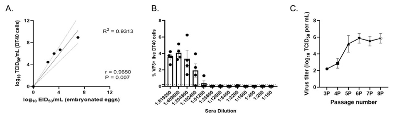
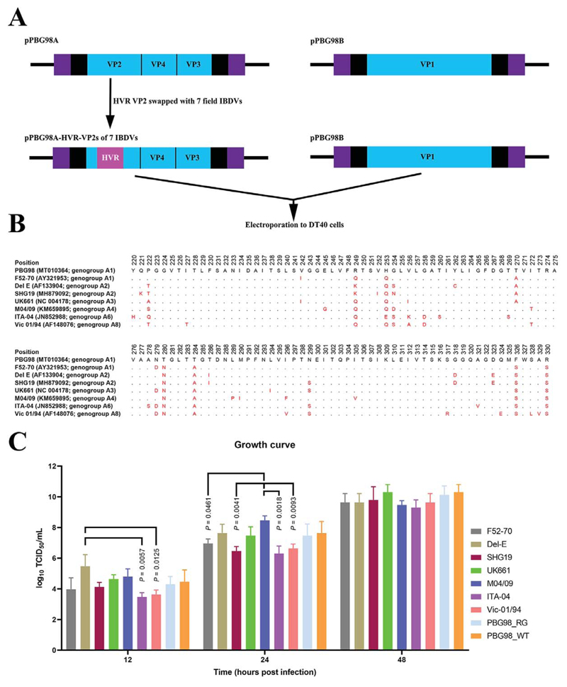
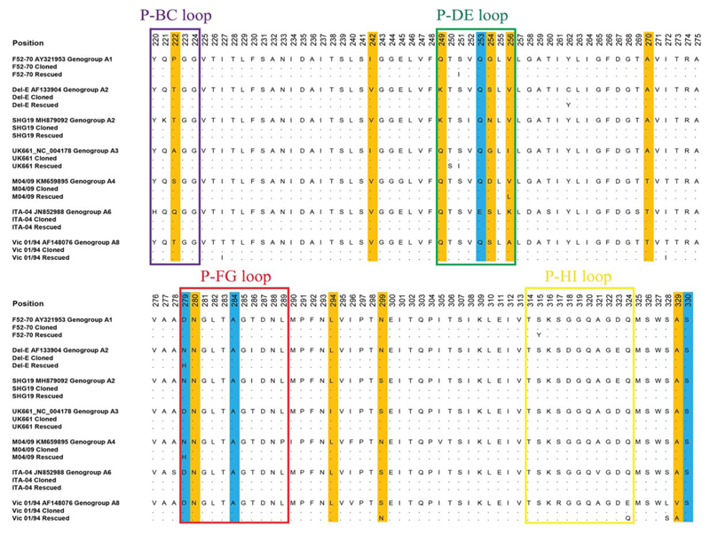
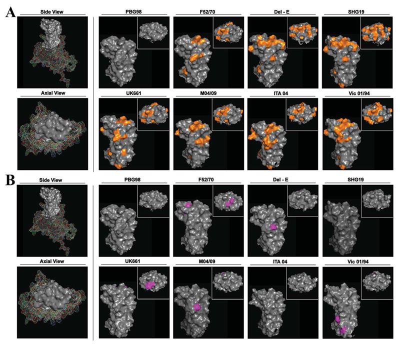
There is a need to evaluate the ability of vaccines to neutralize diverse IBDV genogroups and to better understand the relationship between HVR sequence, structure, and antigenicity. Here, we used a chicken B-cell line to rescue a panel of chimeric IBDVs with the HVR from seven diverse IBDV field strains and to conduct neutralization assays and protein modeling.
Overview
The claims and statistics of this paper haven’t been checked yet.
Run the check — free, no account needed. It reads the open-access text and checks the sources it cites.
Integrity signals
Registry
Cites
Statistics
Figures
Verification
Claims the paper makes
The claims and statistics of this paper haven’t been checked yet.
Run the check — free, no account needed. It reads the open-access text and checks the sources it cites.
Self-check
Percentages, sums and reported differences re-computed from the paper’s own numbers.
The self-check needs the paper’s full text.
Citations
Cites
MEGA7: Molecular Evolutionary Genetics Analysis Version 7.0 for Bigger Datasets
Kumar, S., Stecher, G., & Tamura, K. (2016) · Molecular Biology and Evolution · doi:10.1093/molbev/msw054 · Cited by 44,059
Highly accurate protein structure prediction with AlphaFold
Jumper, J., Evans, R., Pritzel, A., Green, T., Figurnov, M., Ronneberger, O., Tunyasuvunakool, K., Bates, R., Žídek, A., Potapenko, A., et al. (2021) · Nature · doi:10.1038/s41586-021-03819-2 · Cited by 43,764
A SIMPLE METHOD OF ESTIMATING FIFTY PER CENT ENDPOINTS12
REED, L. J., & MUENCH, H. (1938) · American Journal of Epidemiology · doi:10.1093/oxfordjournals.aje.a118408 · Cited by 19,926
Bayliss, C. D., Spies, U., Shaw, K., Peters, R. W., Papageorgiou, A., Muller, H., & Boursnell, M. E. G. (1990) · Journal of General Virology · doi:10.1099/0022-1317-71-6-1303 · Cited by 315
Current status of vaccines against infectious bursal disease
Müller, H., Mundt, E., Eterradossi, N., & Islam, M. R. (2012) · Avian Pathology · doi:10.1080/03079457.2012.661403 · Cited by 248
Fahey, K. J., Erny-Albrecht, K. M., & Crooks, J. (1989) · Journal of General Virology · doi:10.1099/0022-1317-70-6-1473 · Cited by 228
Cell lines derived from avian lymphomas exhibit two distinct phenotypes
Baba, T. W., Giroir, B. P., & Humphries, E. H. (1985) · Virology · doi:10.1016/0042-6822(85)90312-5 · Cited by 209
Clinical Aspects of Immunosuppression in Poultry
Hoerr, F. J. (2010) · Avian Diseases · doi:10.1637/8909-043009-review.1 · Cited by 198
Molecular and Structural Bases for the Antigenicity of VP2 of Infectious Bursal Disease Virus
Letzel, T., Coulibaly, F., Rey, F. A., Delmas, B., Jagt, E., van Loon, A. A. W. M., & Mundt, E. (2007) · Journal of Virology · doi:10.1128/jvi.01501-07 · Cited by 191
Classification of infectious bursal disease virus into genogroups
Michel, L. O., & Jackwood, D. J. (2017) · Archives of Virology · doi:10.1007/s00705-017-3500-4 · Cited by 184
Lejal, N., da Costa, B. R., Delmas, B., & Huet, J. (2000) · Journal of General Virology · doi:10.1099/0022-1317-81-4-983 · Cited by 165
van Loon, A. A. W. M., de Haas, N., Zeyda, I., & Mundt, E. (2002) · Journal of General Virology · doi:10.1099/0022-1317-83-1-121 · Cited by 154
Mundt, E. (1999) · Journal of General Virology · doi:10.1099/0022-1317-80-8-2067 · Cited by 149
Brown, M. D., & Skinner, M. A. (1996) · Virus Research · doi:10.1016/0168-1702(95)01253-2 · Cited by 149
Yamaguchi, T., Ogawa, M., Inoshima, Y., Miyoshi, M., Fukushi, H., & Hirai, K. (1996) · Virology · doi:10.1006/viro.1996.0470 · Cited by 142
Lim, B. L., Cao, Y., Yu, T., & Mo, C. (1999) · Journal of Virology · doi:10.1128/jvi.73.4.2854-2862.1999 · Cited by 140
Snyder, D. B., Lana, D. P., Savage, P. K., Yancey, F. S., Mengel-Whereat, S. A., & Marquardt, W. W. (1988) · Avian Diseases · doi:10.2307/1590924 · Cited by 139
Novel variant strains of infectious bursal disease virus isolated in China
Fan, L., Wu, T., Hussain, A., Gao, Y., Zeng, X., Wang, Y., Gao, L., Li, K., Wang, Y., Liu, C., et al. (2019) · Veterinary Microbiology · doi:10.1016/j.vetmic.2019.01.023 · Cited by 135
Escaffre, O., Nouën, C. L., Amelot, M., Ambroggio, X. I., Ogden, K. M., Guionie, O., Toquin, D., Müller, H., Islam, M. R., & Eterradossi, N. (2012) · Journal of Virology · doi:10.1128/jvi.02360-12 · Cited by 121
A unified genotypic classification of infectious bursal disease virus based on both genome segments
Islam, M. R., Nooruzzaman, M., Rahman, T., Mumu, T. T., Rahman, M. M., Chowdhury, E. H., Eterradossi, N., & Müller, H. (2021) · Avian Pathology · doi:10.1080/03079457.2021.1873245 · Cited by 104
Infectious Bursal Disease Virus: Ribonucleoprotein Complexes of a Double-Stranded RNA Virus
Luque, D., Saugar, I., Rejas, M. T., Carrascosa, J. L., Rodrígúez, J. F., & Castón, J. R. (2008) · Journal of Molecular Biology · doi:10.1016/j.jmb.2008.11.029 · Cited by 97
Activation mechanism of a noncanonical RNA-dependent RNA polymerase
Garriga, D., Navarro, A., Querol‐Audí, J., Abaitua, F., Rodrígúez, J. F., & Verdaguer, N. (2007) · Proceedings of the National Academy of Sciences · doi:10.1073/pnas.0704447104 · Cited by 93
Fan, L., Wu, T., Wang, Y., Hussain, A., Jiang, N., Gao, L., Li, K., Gao, Y., Liu, C., Cui, H., et al. (2019) · Veterinary Microbiology · doi:10.1016/j.vetmic.2019.108507 · Cited by 93
Eterradossi, N., & Saif, Y. Μ. (2013) · doi:10.1002/9781119421481.ch7 · Cited by 84
Vakharia, V. N., Snyder, D. B., He, J., Edwards, G. H., Savage, P. K., & Mengel-Whereat, S. A. (1993) · Journal of General Virology · doi:10.1099/0022-1317-74-6-1201 · Cited by 77
Sapats, S. I., & Ignjatovic, J. (2000) · Archives of Virology · doi:10.1007/s007050050670 · Cited by 70
Infectious Bursal Disease Virus Variant from Commercial Leghorn Pullets
Ismail, N. M., Saif, Y. Μ., Wigle, W. L., HAVENSTEIN, G. B., & Jackson, C. (1990) · Avian Diseases · doi:10.2307/1591345 · Cited by 66
Delgui, L. R., Oña, A., Gutiérrez, S., Luque, D., Navarro, A., Castón, J. R., & Rodrígúez, J. F. (2009) · Virology · doi:10.1016/j.virol.2008.12.036 · Cited by 58
Aliyu, H. B., Bejo, M. H., Omar, A. R., & Ideris, A. (2021) · Frontiers in Veterinary Science · doi:10.3389/fvets.2021.643976 · Cited by 58
Domańska-Blicharz, K., Mató, T., Rivallan, G., Śmietanka, K., Minta, Z., de Boisséson, C., Toquin, D., Lomniczi, B., Palya, V., & Eterradossi, N. (2004) · Archives of Virology · doi:10.1007/s00705-003-0230-6 · Cited by 57
Perozo, F., Villegas, P., Fernandez, R., de la Cruz, J. C. V., & Pritchard, N. (2009) · Avian Diseases · doi:10.1637/8687-31009resnote.1 · Cited by 54
Hitchner, S. B. (1971) · Avian Diseases · doi:10.2307/1588880 · Cited by 54
Lupini, C., Giovanardi, D., Pesente, P., Bonci, M., Felice, V., Rossi, G., Morandini, E., Cecchinato, M., & Catelli, E. (2016) · Avian Pathology · doi:10.1080/03079457.2016.1165792 · Cited by 47
Non-Lytic Egression of Infectious Bursal Disease Virus (IBDV) Particles from Infected Cells
Méndez, F., Romero, N., Cubas-Gaona, L. L., Delgui, L. R., Rodrı́guez, D., & Rodrígúez, J. F. (2017) · PLoS ONE · doi:10.1371/journal.pone.0170080 · Cited by 43
Ye, C., Wang, Y., Zhang, E., Han, X., Yu, Z., & Liu, H. (2017) · Journal of Virology · doi:10.1128/jvi.01345-17 · Cited by 39
Soubiès, S., Courtillon, C., Abed-Zahar, M., Amelot, M., Keïta, A., Broadbent, A., Härtle, S., Kaspers, B., & Eterradossi, N. (2017) · Avian Pathology · doi:10.1080/03079457.2017.1393044 · Cited by 37
Self-citationQuan, R., Zhu, S., Wei, L., Wang, J., Xu, Y., Li, Z., & Liu, J. (2017) · Virology Journal · doi:10.1186/s12985-016-0668-2 · Cited by 35
Lazarus, D. D., Pasmanik‐Chor, M., Gutter, B., Gallili, G. E., Barbakov, M., Krispel, S., & Pitcovski, J. (2008) · Avian Pathology · doi:10.1080/03079450801910206 · Cited by 34
van Hulten, M. C. W., Cruz-Coy, J., Gergen, L., Pouwels, H., ten Dam, G. B., Verstegen, I., de Groof, A., Morsey, M., & Tarpey, I. (2020) · Avian Pathology · doi:10.1080/03079457.2020.1828567 · Cited by 34
Dulwich, K. L., Giotis, E. S., Gray, A. G., Nair, V., Skinner, M. A., & Broadbent, A. (2017) · Journal of General Virology · doi:10.1099/jgv.0.000979 · Cited by 26
Self-citationIgnjatovic, J., & Sapats, S. I. (2002) · Australian Veterinary Journal · doi:10.1111/j.1751-0813.2002.tb11299.x · Cited by 25
Dulwich, K. L., Asfor, A. S., Gray, A. G., Giotis, E. S., Skinner, M. A., & Broadbent, A. (2020) · Frontiers in Cellular and Infection Microbiology · doi:10.3389/fcimb.2020.00315 · Cited by 25
Self-citationRamírez, G., Shivaprasad, H. L., Kim, C.-H., Lillehoj, H. S., Song, H., Osorio, I. G., & Pérez, D. R. (2010) · Avian Diseases · doi:10.1637/8902-042809-reg.1 · Cited by 24
Campbell, E. A., Reddy, V. R. A. P., Gray, A. G., Wells, J., Simpson, J. L., Skinner, M. A., Hawes, P. C., & Broadbent, A. (2020) · Journal of Virology · doi:10.1128/jvi.02107-19 · Cited by 24
Self-citationTerasaki, K., Hirayama, H., Kasanga, C. J., Maw, M. T., Ohya, K., Yamaguchi, T., & Fukushi, H. (2008) · Journal of Veterinary Medical Science · doi:10.1292/jvms.70.407 · Cited by 22
Morla, S., Deka, P., & Kumar, S. (2016) · Microbial Pathogenesis · doi:10.1016/j.micpath.2016.02.004 · Cited by 22
Infectious bursal disease virus persistently infects bursal B-lymphoid DT40 cells
Delgui, L. R., de Llano, D. G., & Rodrígúez, J. F. (2009) · Journal of General Virology · doi:10.1099/vir.0.008870-0 · Cited by 18
Cui, X., Nagesha, H. S., & Holmes, I. H. (2003) · Journal of Virological Methods · doi:10.1016/s0166-0934(03)00049-1 · Cited by 17
Liu, A., Li, H., Qi, X., Wang, Q., Yang, B., Wu, T., Yan, N., Li, Y., Pan, Q., Gao, Y., et al. (2019) · Frontiers in Microbiology · doi:10.3389/fmicb.2019.02225 · Cited by 17
Noor, M., Mahmud, M. S., Ghose, P. R., Roy, U., Nooruzzaman, M., Chowdhury, E. H., Das, P. M., Islam, M. R., & Müller, H. (2013) · Archives of Virology · doi:10.1007/s00705-013-1885-2 · Cited by 16
Olesen, L., Dijkman, R., Koopman, R., van Leeuwen, R., Gardin, Y., Dwars, R. M., de Bruijn, N. D., Boelm, G. J., El-Attrache, J., & de Wit, S. J. (2018) · Avian Pathology · doi:10.1080/03079457.2018.1520388 · Cited by 13
Dulwich, K. L., Asfor, A. S., Gray, A. G., Nair, V., & Broadbent, A. (2018) · Journal of Visualized Experiments · doi:10.3791/58489 · Cited by 12
Self-citationGenome Sequence of a Distinct Infectious Bursal Disease Virus
Tomás, G., Hernández, M. A., Marandino, A. E., Hernández, D., Techera, C., Grecco, S., Panzera, Y., & Pérez, R. (2015) · Genome Announcements · doi:10.1128/genomea.01061-15 · Cited by 7
Cubas-Gaona, L. L., Trombetta, R., Courtillon, C., Li, K., Qi, X., Wang, X., Lotmani, S., Keïta, A., Amelot, M., Eterradossi, N., et al. (2020) · Scientific Reports · doi:10.1038/s41598-020-70095-x · Cited by 6
Durairaj, V., Linnemann, E. G., Icard, A. H., Williams, S. M., Sellers, H. S., & Mundt, E. (2013) · Avian Pathology · doi:10.1080/03079457.2013.793783 · Cited by 4
Metadata
- Authors
- Reddy, V. R. A. P., Nazki, S., Brodrick, A. J., Asfor, A., Urbaniec, J., Morris, Y., & Broadbent, A. J.
- Affiliations
- The Pirbright Institute (GB)
- University of Maryland, College Park (US)
- Corresponding author
- Reddy, Vishwanatha R. A. P.
- Broadbent, Andrew J.
- ORCID
- Venue
- Journal of Virology
- Publisher
- American Society for Microbiology
- Published
- 2022-09-28
- Volume
- 96
- Issue
- 18
- Pages
- e0125522-e0125522
- ISSN
- 0022-538X (ISSN-L)
- 1098-5514
- PMC
- PMC9517715
- OpenAlex
- W4294921245
- Cited by
- 12
- References
- 55
- Citations by year
- 2026: 3 · 2025: 1 · 2024: 2 · 2023: 3 · 2022: 3
- FWCI
- 1.40
- Type
- journal article
- Language
- en
- Open access
- hybrid
- doi.org
- Licence
- cc-by
- doi.org/10.1128/ASMCopyrightv2 (vor)
- journals.asm.org/non-commercial-tdm-license (tdm)
- Locations
- Journal of Virology (publishedVersion, cc-by)
- bioRxiv (Cold Spring Harbor Laboratory) (acceptedVersion, cc-by)
- PubMed Central (publishedVersion)
- PubMed (publishedVersion)
- Funders
- UKRI | Biotechnology and Biological Sciences Research Council (BB/S014594/1, BBS/E/I/00001845, BBS/E/I/00007034, BBS/E/I/00007039)
- Topics
- Life Sciences › Agricultural and Biological Sciences › Animal Science and Zoology › Animal Virus Infections Studies
- Health Sciences › Medicine › Epidemiology › Virology and Viral Diseases
- Life Sciences › Biochemistry, Genetics and Molecular Biology › Molecular Biology › vaccines and immunoinformatics approaches
- Keywords
- infectious bursal disease virus, neutralizing antibodies, antigenic relatedness, vaccine design, immunodominant epitopes, conserved epitopes, protein structure modeling, vaccine screening
- MeSH terms
- Animals, Epitopes, Australia, Chickens, Genotype, Infectious bursal disease virus, Poultry Diseases, Birnaviridae Infections, Antibodies, Neutralizing
About these checks
Registry, retraction, Cites and statistics results are computed, not judged, with one exception: a printed reference that no index lists is read once by a model before it counts as a citation error. Claims are extracted by a model and kept only when their quote appears verbatim in the text; each cited source is then checked, with a model’s help, for whether it says what the claim relies on. Those verdicts can be wrong — read the claim’s quote and the cited source before relying on one.
Statistics and arithmetic are recomputed, not judged.
Confidence bands are shown only for fields calibrated on our annotated test set. None is calibrated yet, so no band is shown.
What counts as a claim ›How we measure confidence ›How we check this ›
Are you an author, or do you see a problem with this page? Request a review
Data sources
Bibliographic data: Semantic Scholar, OpenAlex, Crossref.
Contains information from the Semantic Scholar Academic Graph Datasets, made available under the ODC Attribution License (ODC-BY 1.0).
Semantic Scholar’s data is described in: Kinney, R., et al. (2023). The Semantic Scholar Open Data Platform. arXiv:2301.10140.
