Student or educator? Sign up with your school email and get 6 months of Pro free.
Cited by 1Plant Virus Research StudiesBacteriophages and microbial interactionsPlant and Fungal Interactions Research

The DOI matches its Crossref record; 79 of the 79 references it cites were found in a registry or an index.

A SUMO interacting motif in the replication initiator protein of tomato yellow leaf curl virus is required for viral replication

Open access

Published version of a preprint

Abstract

Circular Rep-encoding single-stranded (CRESS)-DNA viruses comprise a diverse group of viruses that use rolling-circle replication to copy their genomes. They infect organisms in almost all branches of the eukaryotic tree of life. A hallmark of all CRESS-DNA viruses is the presence of a single conserved protein, the replication initiator protein (Rep), that orchestrates viral replication by exploiting the host DNA replication machinery. In the case of the plant-infecting Geminiviridae , this multifunctional protein both recruits the host DNA replication machinery and manipulates post-translational modification including small ubiquitin-like modifier (SUMO) conjugation. In fact, Rep from two different geminiviruses, tomato yellow leaf curl virus (TYLCV; Begomovirus coheni ) and tomato golden mosaic virus (TGMV; B. solanum aureimusivi ), was earlier shown to interact with the SUMO conjugating enzyme SCE1. Here, we demonstrate that TYLCV C1/Rep protein also interacts with Arabidopsis SUMO1 and identify a SUMO interacting motif (SIM) located in the C-terminal SF3 helicase/ATPase domain. Remarkably, a functional SIM proved to be important for the interaction of Rep with both SUMO1 and SCE1. The same motif in the Rep ORF was essential for TYLCV viral replication, disease symptom formation, and systemic movement from an infectious clone, and Rep ATPase activity. Together, our findings thus connect the interaction between Rep and the SUMO machinery with TYLCV viral replication. IMPORTANCE: The identification of a non-canonical SUMO-interacting motif (SIM) within the Rep protein of tomato yellow leaf curl virus (TYLCV) reveals a connection between viral replication and a protein modification, i.e., SUMOylation. Importantly, the motif is conserved between Rep proteins from different geminiviruses. Functionally, the motif was critical for Rep’s interaction with components of the SUMO machinery, for viral DNA replication, and for its ATPase activity. In particular, the third position of the motif was important for each of these activities. We thus uncover a hitherto undescribed mechanism on how geminiviruses recruit the SUMO machinery.

Overview

The claims and statistics of this paper haven’t been checked yet.

Run the check — free, no account needed. It reads the open-access text and checks the sources it cites.

A few free checks a day · papers already verified open instantly · results are public

Integrity signals

Registry

Crossref · 10.1128/jvi.01286-25

Cites

79 of 79 references verified0 citation errors · 0 not checkable

Statistics

Figures

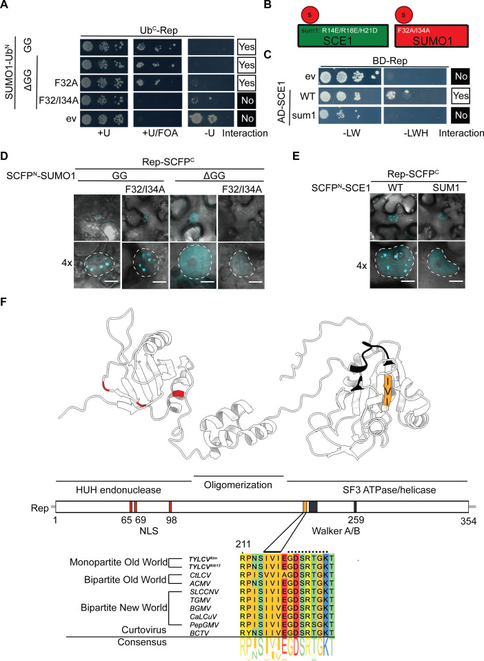
Fig 1 Rep interacts with SUMO1 and SCE1 via a predicted putative SUMO-interacting motif (SIM). (A) Split-ubiquitin Y2H assay between Rep (fused to C-terminal half of ubiquitin, UbC) and different SUMO1 variants (UbN). Positive interactions result in yeast growth on minimal medium (MM) supplemented with Uracil and FOA (+U/FOA) and no growth on MM lacking Uracil (−U). +U, positive control for normal growth. (B) Diagram depicting the SUMO1 and SCE1 variants used in the Y2H and BiFC assays in panels (A) and (D) and (C) and (E), respectively. (C) Gal4 Y2H interaction between RepAlb13 and two SCE1 variants, i.e., WT protein and a variant mutated in the second SUMO binding site (sum1). −LWH, growth when bait and prey interact; −LW, positive control for normal growth. (D, E) Images depict a typical N. benthamiana epidermal cell (top), with a 4× zoom showing only the nucleus (bottom). Dotted lines delineate the nuclei; scale bar, 5 µm. (F) 3D model of Rep generated with AlphaFold2 showing the different domains and motifs. From left to right: HUH endonuclease, central oligomerization domain, and superfamily 3 ATPase/helicase domain. Colored residues depict the different Rep motifs, including the nuclear localization signal (NLS; red), SIM (orange), and Walker A and B motifs (black). Protein sequence alignment of the Rep SIM is shown from different geminiviruses. The shown sequence comprises the residues 211–226. Residues 215–217 of RepAlm and 217–219 of RepAlb13 represent the SIM (continuous line), and residues 219–225 depict the Walker A motif (dashed line).
Fig 1 Rep interacts with SUMO1 and SCE1 via a predicted putative SUMO-interacting motif (SIM). (A) Split-ubiquitin Y2H assay between Rep (fused to C-terminal half of ubiquitin, UbC) and different SUMO1 variants (UbN). Positive interactions result in yeast growth on minimal medium (MM) supplemented with Uracil and FOA (+U/FOA) and no growth on MM lacking Uracil (−U). +U, positive control for normal growth. (B) Diagram depicting the SUMO1 and SCE1 variants used in the Y2H and BiFC assays in panels (A) and (D) and (C) and (E), respectively. (C) Gal4 Y2H interaction between RepAlb13 and two SCE1 variants, i.e., WT protein and a variant mutated in the second SUMO binding site (sum1). −LWH, growth when bait and prey interact; −LW, positive control for normal growth. (D, E) Images depict a typical N. benthamiana epidermal cell (top), with a 4× zoom showing only the nucleus (bottom). Dotted lines delineate the nuclei; scale bar, 5 µm. (F) 3D model of Rep generated with AlphaFold2 showing the different domains and motifs. From left to right: HUH endonuclease, central oligomerization domain, and superfamily 3 ATPase/helicase domain. Colored residues depict the different Rep motifs, including the nuclear localization signal (NLS; red), SIM (orange), and Walker A and B motifs (black). Protein sequence alignment of the Rep SIM is shown from different geminiviruses. The shown sequence comprises the residues 211–226. Residues 215–217 of RepAlm and 217–219 of RepAlb13 represent the SIM (continuous line), and residues 219–225 depict the Walker A motif (dashed line).CC BY · PubMed Central
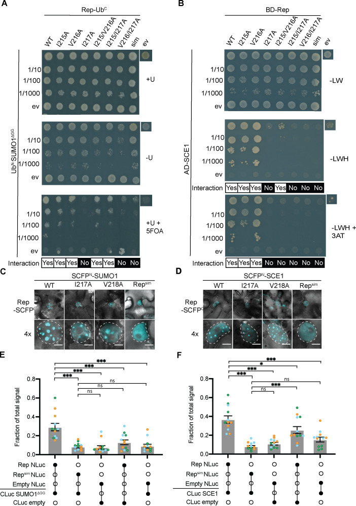
Fig 2 SIM is needed for Rep to interact with both SUMO1 and SCE1. (A) Split-ubiquitin yeast two-hybrid (Y2H) assay between Rep SIM mutants fused to C-terminal half of ubiquitin (UbC) and SUMO1DGG fused to UbN. ev, empty vector control. Top to bottom: tenfold dilution series of the yeast cultures. Similar to the legend of Fig. 1A. (B) Gal4 Y2H assay between Rep SIM variants (GAL4 BD) and SCE1 (GAL4 AD). ev, empty vector control. Top to bottom: tenfold dilution series of the yeast cultures. Similar to the legend of Fig. 1C. (C, D) Bimolecular fluorescence complementation assay showing SUMO1 (C) and SCE1 (D) localization pattern in complex with RepAlb13 and the RepAlb13 SIM variants. Nuclei are outlined by a dashed white line; scale bars represent 5 µm. nls, nuclear localization-deficient variant (43). (E) Split-luciferase complementation assay between different Rep variants and SUMO1DGG. Each data point represents a technical replicate, while colors represent independent biological repeats. X-axis: filled circles represent co-expression of the indicated Rep/SCE1 constructs fused to either the N-terminus (NLuc) or C-terminus (CLuc) of the luciferase. ANOVA followed by a Dunnett multiple comparisons test was performed; ns, non-significant; *P < 0.05; ***P < 0.001; Error bar represents standard error of the mean (SEM) (n = 12). (F) Split-luciferase complementation assay between different Rep variants and SCE1. ANOVA followed by a Dunnett multiple comparisons test was performed. Other details as panel E.
Fig 2 SIM is needed for Rep to interact with both SUMO1 and SCE1. (A) Split-ubiquitin yeast two-hybrid (Y2H) assay between Rep SIM mutants fused to C-terminal half of ubiquitin (UbC) and SUMO1DGG fused to UbN. ev, empty vector control. Top to bottom: tenfold dilution series of the yeast cultures. Similar to the legend of Fig. 1A. (B) Gal4 Y2H assay between Rep SIM variants (GAL4 BD) and SCE1 (GAL4 AD). ev, empty vector control. Top to bottom: tenfold dilution series of the yeast cultures. Similar to the legend of Fig. 1C. (C, D) Bimolecular fluorescence complementation assay showing SUMO1 (C) and SCE1 (D) localization pattern in complex with RepAlb13 and the RepAlb13 SIM variants. Nuclei are outlined by a dashed white line; scale bars represent 5 µm. nls, nuclear localization-deficient variant (43). (E) Split-luciferase complementation assay between different Rep variants and SUMO1DGG. Each data point represents a technical replicate, while colors represent independent biological repeats. X-axis: filled circles represent co-expression of the indicated Rep/SCE1 constructs fused to either the N-terminus (NLuc) or C-terminus (CLuc) of the luciferase. ANOVA followed by a Dunnett multiple comparisons test was performed; ns, non-significant; *P < 0.05; ***P < 0.001; Error bar represents standard error of the mean (SEM) (n = 12). (F) Split-luciferase complementation assay between different Rep variants and SCE1. ANOVA followed by a Dunnett multiple comparisons test was performed. Other details as panel E.CC BY · PubMed Central
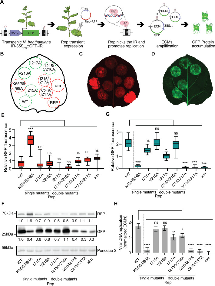
Fig 3 SIM is important for Rep-mediated viral DNA replication in a virus-free system (A) A reporter system to mimic and visualize geminiviral DNA replication in N. benthamiana plants in the absence of the intact virus. Transgenic N. benthamiana expressing IR-35SPro::GFP-IR reporter is agroinfiltrated with a vector to deliver the viral Rep-RFP fusion protein. Upon transient expression, Rep-RFP nicks the DNA nicking at the IR motif and promotes RCR of the nicked DNA, resulting in the formation of circular ECMs. Each ECM contains a single 35Spro::GFP cassette resulting in massive GFP accumulation. (B) Schematic depicting the agroinfiltrated Rep constructs. Clockwise from the bottom left: WT; K65/69/98A (nls), replication-deficient negative control impaired in nuclear localization (43); different SIM mutants, single (I215A, ...), double (I215/V216A, ...), triple residue (sim) mutants, and RFP as negative control. Green and red circles depict replication-competent and replication-deficient Rep variants, respectively. (C, D) Fluorescence images showing accumulation of Rep-RFP and GFP as a proxy for viral replication. (B) The layout of the infiltrated constructs. (E) RFP quantification of fluorescence signal in infiltrated leaf areas. Total fluorescence was normalized per leaf. Kruskal-Wallis test with Dunn’s correction was performed on the pooled data from each replicate. ns, non-significant; *P < 0.05; **P < 0.01; ***P < 0.001. Error bar represents SEM (n = 14). (F) Immunoblot showing both Rep-RFP and GFP levels. Ratios are compared to signal intensity of WT Rep. To demonstrate equal protein loading and normalize the blot signal values, membranes were stained with Ponceau S. (G) Similar to panel E, except that GFP was quantified. Other details are the same as in panel E. (H) Quantification of the ECMs using real-time PCR. ANOVA followed by Dunnett’s multiple comparison test was performed; *P < 0.05; **P < 0.01; ***P < 0.001; ****P < 0.0001. Error bar represents SEM (n = 3).
Fig 3 SIM is important for Rep-mediated viral DNA replication in a virus-free system (A) A reporter system to mimic and visualize geminiviral DNA replication in N. benthamiana plants in the absence of the intact virus. Transgenic N. benthamiana expressing IR-35SPro::GFP-IR reporter is agroinfiltrated with a vector to deliver the viral Rep-RFP fusion protein. Upon transient expression, Rep-RFP nicks the DNA nicking at the IR motif and promotes RCR of the nicked DNA, resulting in the formation of circular ECMs. Each ECM contains a single 35Spro::GFP cassette resulting in massive GFP accumulation. (B) Schematic depicting the agroinfiltrated Rep constructs. Clockwise from the bottom left: WT; K65/69/98A (nls), replication-deficient negative control impaired in nuclear localization (43); different SIM mutants, single (I215A, ...), double (I215/V216A, ...), triple residue (sim) mutants, and RFP as negative control. Green and red circles depict replication-competent and replication-deficient Rep variants, respectively. (C, D) Fluorescence images showing accumulation of Rep-RFP and GFP as a proxy for viral replication. (B) The layout of the infiltrated constructs. (E) RFP quantification of fluorescence signal in infiltrated leaf areas. Total fluorescence was normalized per leaf. Kruskal-Wallis test with Dunn’s correction was performed on the pooled data from each replicate. ns, non-significant; *P < 0.05; **P < 0.01; ***P < 0.001. Error bar represents SEM (n = 14). (F) Immunoblot showing both Rep-RFP and GFP levels. Ratios are compared to signal intensity of WT Rep. To demonstrate equal protein loading and normalize the blot signal values, membranes were stained with Ponceau S. (G) Similar to panel E, except that GFP was quantified. Other details are the same as in panel E. (H) Quantification of the ECMs using real-time PCR. ANOVA followed by Dunnett’s multiple comparison test was performed; *P < 0.05; **P < 0.01; ***P < 0.001; ****P < 0.0001. Error bar represents SEM (n = 3).CC BY · PubMed Central
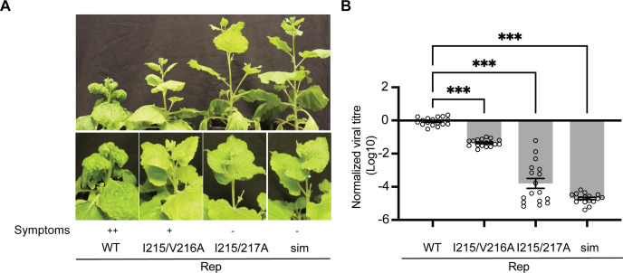
Fig 4 The Rep SIM is crucial for TYLCV to induce disease symptoms and enable viral genome replication in N. benthamiana. (A) 2IR-GFP N. benthamiana plants infected with different TYLCV infectious clones encoding either WT Rep or Rep SIM variants (I215/V126A, I215/I127A, I215/V126A/I127A = sim). TYLCV disease symptoms are indicated: ++, severe symptoms; +, mild symptoms; −, no symptoms. Photos were taken three weeks post-inoculation. (B) Quantification of viral titers using real-time PCR on the extracted DNA from the apical leaves. Levels were normalized to the viral titers by the WT TYLCV (Log10 = 0). ANOVA followed by Dunnett’s multiple comparison test was performed; ***P < 0.0001. Error bar represents SEM (n = 17).
Fig 4 The Rep SIM is crucial for TYLCV to induce disease symptoms and enable viral genome replication in N. benthamiana. (A) 2IR-GFP N. benthamiana plants infected with different TYLCV infectious clones encoding either WT Rep or Rep SIM variants (I215/V126A, I215/I127A, I215/V126A/I127A = sim). TYLCV disease symptoms are indicated: ++, severe symptoms; +, mild symptoms; −, no symptoms. Photos were taken three weeks post-inoculation. (B) Quantification of viral titers using real-time PCR on the extracted DNA from the apical leaves. Levels were normalized to the viral titers by the WT TYLCV (Log10 = 0). ANOVA followed by Dunnett’s multiple comparison test was performed; ***P < 0.0001. Error bar represents SEM (n = 17).CC BY · PubMed Central
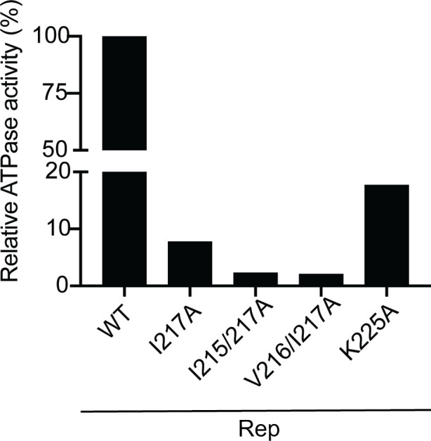
Fig 5 An intact SIM is needed for full Rep ATPase activity. Relative ATPase activity in purified protein fractions corresponding to WT Rep (normalized to 100% activity) versus the indicated Rep SIM mutants on the x-axis. All recombinant proteins were expressed in E. coli. K225A variant: known Rep mutant with lowered ATPase activity (negative control, see reference 20).
Fig 5 An intact SIM is needed for full Rep ATPase activity. Relative ATPase activity in purified protein fractions corresponding to WT Rep (normalized to 100% activity) versus the indicated Rep SIM mutants on the x-axis. All recombinant proteins were expressed in E. coli. K225A variant: known Rep mutant with lowered ATPase activity (negative control, see reference 20).CC BY · PubMed Central

Verification

Claims the paper makes

The claims and statistics of this paper haven’t been checked yet.

Run the check — free, no account needed. It reads the open-access text and checks the sources it cites.

A few free checks a day · papers already verified open instantly · results are public

Self-check

Percentages, sums and reported differences re-computed from the paper’s own numbers.

The self-check needs the paper’s full text.

Citations

Cites

Show all 79 references

Research works printed but not linked: 1

  1. 10.1038/s41592-022-01488-1 · doi:10.1038/s41592-022-01488-1 · Paper page

Metadata

Authors
Gaertner, N. F., Maio, F., Arroyo-Mateos, M., Luna, A. P., Sabarit, B., Kwaaitaal, M., Eltschkner, S., Prins, M., Bejarano, E. R., & van den Burg, H. A.
Affiliations
  • University of Amsterdam (NL)
  • Molecular Plant Pathology, Swammerdam Institute for Life Sciences, research theme Green Life Sciences, Faculty of Science, University of Amsterdam
  • Consejo Superior de Investigaciones Científicas (ES)
  • KeyGene (Netherlands) (NL)
  • Rijk Zwaan (Netherlands) (NL)
Corresponding author
van den Burg, Harrold A.
Venue
Journal of Virology
Published
2025-12-23
Volume
99
Issue
12
Pages
e0128625-e0128625
ISSN
  • 0022-538X (ISSN-L)
  • 1098-5514
OpenAlex
W4416088854
Cited by
1
References
79
Citations by year
2026: 1
FWCI
0.54
Type
journal article
Language
en
Open access
Licence
Locations
  • Journal of Virology (publishedVersion, cc-by)
  • bioRxiv (Cold Spring Harbor Laboratory) (acceptedVersion, cc-by-nc-nd)
  • PubMed (publishedVersion)
  • UvA-DARE (University of Amsterdam) (publishedVersion, cc-by)
  • PubMed Central (publishedVersion, cc-by)
  • DIGITAL.CSIC (Spanish National Research Council (CSIC)) (acceptedVersion, other-oa)
  • DOAJ (DOAJ: Directory of Open Access Journals) (submittedVersion, cc-by-sa)
Funders
  • Top Sector Agri and Food (1409-036, 19157)
  • Ministerio de Cienca y Tecnologia (PID2022-1393760B31)
Topics
  • Life Sciences › Agricultural and Biological Sciences › Plant Science › Plant Virus Research Studies
  • Life Sciences › Immunology and Microbiology › Virology › Bacteriophages and microbial interactions
  • Life Sciences › Immunology and Microbiology › Virology › Plant and Fungal Interactions Research
Keywords
tomato yellow leaf curl virus, geminivirus, replication initiator protein, SUMO-interacting motif, SUMOylation, SUMO1, viral DNA replication, begomovirus, plant-virus interaction
MeSH terms
DNA Replication, DNA Helicases, Plant Diseases, Protein Binding, Nicotiana, Viral Proteins, Virus Replication, Trans-Activators, Arabidopsis, Solanum lycopersicum, Amino Acid Motifs, SUMO-1 Protein, Arabidopsis Proteins, Begomovirus

About these checks

Registry, retraction, Cites and statistics results are computed, not judged, with one exception: a printed reference that no index lists is read once by a model before it counts as a citation error. Claims are extracted by a model and kept only when their quote appears verbatim in the text; each cited source is then checked, with a model’s help, for whether it says what the claim relies on. Those verdicts can be wrong — read the claim’s quote and the cited source before relying on one.

Statistics and arithmetic are recomputed, not judged.

Confidence bands are shown only for fields calibrated on our annotated test set. None is calibrated yet, so no band is shown.

What counts as a claim ›How we measure confidence ›How we check this ›

Are you an author, or do you see a problem with this page? Request a review

How to cite

Cite CoChat for the checks on this page, and the paper for its own findings.

This page
CoChat. (2026). Citation check: A SUMO interacting motif in the replication initiator protein of tomato yellow leaf curl virus is required for viral replication. https://app.cochat.ai/papers/10.1128/jvi.01286-25
The paper
Gaertner, N. F., Maio, F., Arroyo-Mateos, M., Luna, A. P., Sabarit, B., Kwaaitaal, M., Eltschkner, S., Prins, M., Bejarano, E. R., & van den Burg, H. A. (2025). A SUMO interacting motif in the replication initiator protein of tomato yellow leaf curl virus is required for viral replication. Journal of Virology. https://doi.org/10.1128/jvi.01286-25

Data sources

Bibliographic data: Semantic Scholar, OpenAlex, Crossref.

Contains information from the Semantic Scholar Academic Graph Datasets, made available under the ODC Attribution License (ODC-BY 1.0).

Semantic Scholar’s data is described in: Kinney, R., et al. (2023). The Semantic Scholar Open Data Platform. arXiv:2301.10140.