

The DOI matches its Crossref record; 46 of the 46 references it cites were found in a registry or an index.
Characterizing the role of RSD-6 in the biogenesis of virus-derived small interfering RNAs and the modulation of viral pathogenesis
Open accessPublished version of a preprint
Abstract
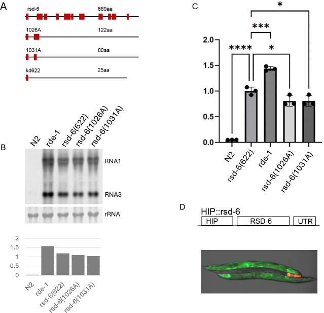
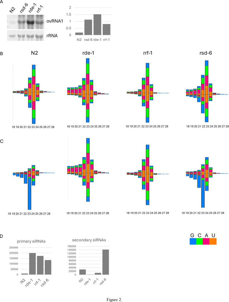
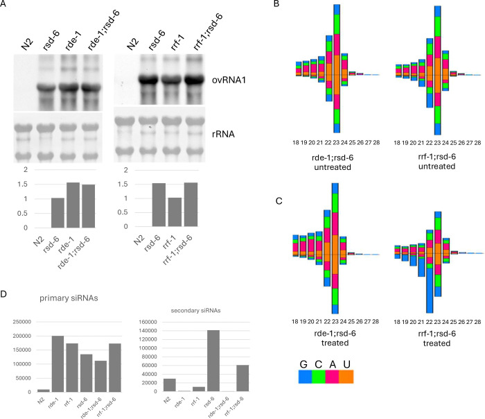
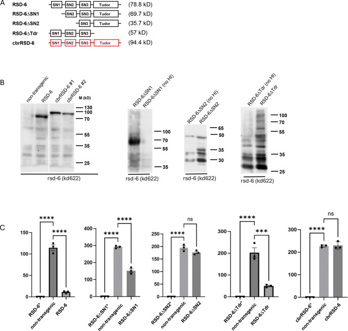
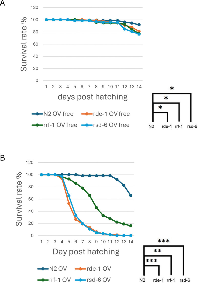
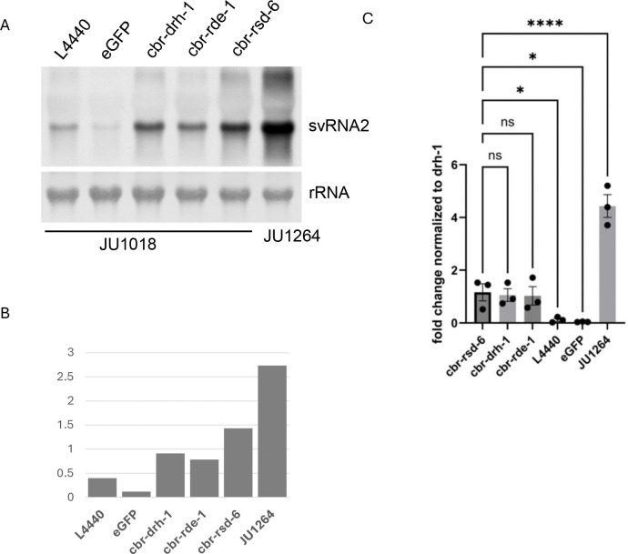
Small interfering RNAs (siRNAs) produced through the processing of viral double-stranded RNAs mediate potent antiviral RNA interference (RNAi) in eukaryotes. In Caenorhabditis elegans , such an antiviral defense is further amplified through the production of secondary siRNAs, yet the mechanisms by which secondary virus-derived siRNAs (vsiRNAs) confer protection remain poorly understood. Here, we characterize the role of rsd-6 , which encodes a Tudor domain protein and plays an important role in antiviral RNAi, in vsiRNA biogenesis and the modulation of viral pathogenesis. Using CRISPR Cas9-generated rsd-6 null mutants, we show that both primary and secondary vsiRNAs accumulate normally in the absence of RSD-6, indicating that it functions downstream of secondary vsiRNA biogenesis. We further showed that secondary vsiRNAs generated in an rrf-1 -independent manner remained detected in the absence of RSD-6 and that viral replication is further enhanced in rrf-1;rsd-6 double mutants compared to rrf-1 single mutants, suggesting a role of rsd-6 in mediating antiviral guided by all secondary vsiRNAs. Consistently, rsd-6 mutants exhibited more severe pathogenesis upon Orsay virus infection compared to rrf-1 mutants, underscoring its role as a major determinant of viral disease outcome. Domain characterization established that the N-terminal tandem domains of RSD-6 are required for antiviral activity, while the C-terminal Tudor domains are dispensable. Functional conservation was confirmed in C. briggsae , where silencing of the rsd-6 homolog enhanced viral replication. Together, our findings identify RSD-6 as a key effector acting downstream of secondary vsiRNA production and highlight its conserved role in modulating viral replication and pathogenesis across Caenorhabditis species. IMPORTANCE: In C. elegans , the RNAi-mediated antiviral defense relies on the production of secondary virus-derived siRNAs (vsiRNAs) to achieve an amplified antiviral effect. However, the mechanism by which these secondary vsiRNAs confer protection remains poorly understood. This is primarily due to the limited number of identified key effector genes. To address this knowledge gap, we profiled vsiRNA biogenesis in loss-of-function mutants and discovered that rsd-6 is dispensable for the production of secondary vsiRNAs, suggesting a role of rsd-6 in mediating antiviral defense downstream of secondary vsiRNA biogenesis. Worm survival assay further confirmed that rsd-6 is a critical modulator of viral pathogenesis and its antiviral function is conserved across Caenorhabditis species. The RSD-6 protein features three N-terminal tandem domains of unknown function and two tandem Tudor domains at its C-terminus. Our domain analyses demonstrated that the N-terminal tandem domains, but not the C-terminal Tudor domains, are essential for antiviral function. The identification of rsd-6 as a key effector gene acting downstream of vsiRNA biogenesis provides a solid foundation for elucidating the mechanism of antiviral RNAi amplification.
Overview
The claims and statistics of this paper haven’t been checked yet.
Run the check — free, no account needed. It reads the open-access text and checks the sources it cites.
Integrity signals
Registry
Cites
Statistics
Figures
Verification
Claims the paper makes
The claims and statistics of this paper haven’t been checked yet.
Run the check — free, no account needed. It reads the open-access text and checks the sources it cites.
Self-check
Percentages, sums and reported differences re-computed from the paper’s own numbers.
The self-check needs the paper’s full text.
Citations
Cites
UCSF Chimera—A visualization system for exploratory research and analysis
Pettersen, E. F., Goddard, T. D., Huang, C. C., Couch, G. S., Greenblatt, D. M., Meng, E. C., & Ferrin, T. E. (2004) · Journal of Computational Chemistry · doi:10.1002/jcc.20084 · Cited by 45,828
Highly accurate protein structure prediction with AlphaFold
Jumper, J., Evans, R., Pritzel, A., Green, T., Figurnov, M., Ronneberger, O., Tunyasuvunakool, K., Bates, R., Žídek, A., Potapenko, A., et al. (2021) · Nature · doi:10.1038/s41586-021-03819-2 · Cited by 43,764
Hierarchical Action and Inhibition of Plant Dicer-Like Proteins in Antiviral Defense
Deleris, A., Gallego‐Bartolomé, J., Bao, J., Kasschau, K. D., Carrington, J. C., & Voinnet, O. (2006) · Science · doi:10.1126/science.1128214 · Cited by 961
Ding, S. (2010) · Nature reviews. Immunology · doi:10.1038/nri2824 · Cited by 884
RNAi-mediated viral immunity requires amplification of virus-derived siRNAs in Arabidopsis thaliana
Wang, X., Wu, Q., Ito, T., Cillo, F., Li, W.-X., Chen, X., Yu, J., & Ding, S. (2009) · Proceedings of the National Academy of Sciences · doi:10.1073/pnas.0904086107 · Cited by 433
Félix, M., Ashe, A., Piffaretti, J., Wu, G. H., Nuez, I., Bélicard, T., Jiang, Y., Zhao, G., Franz, C., Goldstein, L. D., et al. (2011) · PLoS Biology · doi:10.1371/journal.pbio.1000586 · Cited by 417
Díaz‐Pendón, J. A., Li, F., Li, W.-X., & Ding, S. (2007) · The Plant Cell · doi:10.1105/tpc.106.047449 · Cited by 411
Dokshin, G. A., Ghanta, K. S., Piscopo, K. M., & Mello, C. C. (2018) · Genetics · doi:10.1534/genetics.118.301532 · Cited by 403
Wang, X., Jovel, J., Petchthai, U., Wang, Y., Wu, Q., Li, W., Gasciolli, V., Vaucheret, H., & Ding, S. (2011) · The Plant Cell · doi:10.1105/tpc.110.082305 · Cited by 371
Caenorhabditis elegans SID-2 is required for environmental RNA interference
Winston, W. M., Sutherlin, M. E., Wright, A. J., Feinberg, E. H., & Hunter, C. P. (2007) · Proceedings of the National Academy of Sciences · doi:10.1073/pnas.0611282104 · Cited by 329
Ubiquitin-Mediated Response to Microsporidia and Virus Infection in C. elegans
Bakowski, M. A., Desjardins, C. A., Smelkinson, M. G., Dunbar, T. L., López-Moyado, I. F., Rifkin, S. A., Cuomo, C. A., & Troemel, E. R. (2014) · PLoS Pathogens · doi:10.1371/journal.ppat.1004200 · Cited by 232
Ashe, A., Bélicard, T., Pen, J. L., Sarkies, P., Frézal, L., Lehrbach, N. J., Félix, M., & Miska, E. A. (2013) · eLife · doi:10.7554/elife.00994 · Cited by 215
Vasale, J. J., Gu, W., Thivierge, C., Batista, P. J., Claycomb, J. M., Youngman, E. M., Duchaîne, T. F., Mello, C. C., & Conte, D. (2010) · Proceedings of the National Academy of Sciences · doi:10.1073/pnas.0911908107 · Cited by 198
RNA interference-mediated antiviral defense in insects
Gammon, D. B., & Mello, C. C. (2015) · Current Opinion in Insect Science · doi:10.1016/j.cois.2015.01.006 · Cited by 196
Genes Required for Systemic RNA Interference in Caenorhabditis elegans
Tijsterman, M., May, R. C., Simmer, F., Okihara, K. L., & Plasterk, R. H. A. (2004) · Current Biology · doi:10.1016/j.cub.2003.12.029 · Cited by 185
Viral Suppressors of RNA-Based Viral Immunity: Host Targets
Wu, Q., Wang, X., & Ding, S. (2010) · Cell Host & Microbe · doi:10.1016/j.chom.2010.06.009 · Cited by 166
Tudor domain proteins in development
Pek, J. W., Anand, A., & Kai, T. (2012) · Development · doi:10.1242/dev.073304 · Cited by 151
poly(UG)-tailed RNAs in genome protection and epigenetic inheritance
Shukla, A., Yan, J. J., Pagano, D. J., Dodson, A. E., Fei, Y., Gorham, J., Seidman, J. G., Wickens, M., & Kennedy, S. G. (2020) · Nature · doi:10.1038/s41586-020-2323-8 · Cited by 136
A Ribonuclease Coordinates siRNA Amplification and mRNA Cleavage during RNAi
Tsai, H., Chen, C.-C. G., Conte, D., Moresco, J. J., Chaves, D. A., Mitani, S., Yates, J. R., Tsai, M., & Mello, C. C. (2015) · Cell · doi:10.1016/j.cell.2015.01.010 · Cited by 103
Guo, X., Zhang, R., Wang, J., Ding, S., & Lu, R. (2013) · Proceedings of the National Academy of Sciences · doi:10.1073/pnas.1307453110 · Cited by 99
Self-citationVirus-derived siRNAs and piRNAs in immunity and pathogenesis
Ding, S., & Lu, R. (2011) · Current Opinion in Virology · doi:10.1016/j.coviro.2011.10.028 · Cited by 94
Self-citationOrsay, Santeuil and Le Blanc viruses primarily infect intestinal cells in Caenorhabditis nematodes
Franz, C., Renshaw, H., Frézal, L., Jiang, Y., Félix, M., & Wang, D. (2013) · Virology · doi:10.1016/j.virol.2013.09.024 · Cited by 91
Antiviral RNA interference in mammals
Ding, S., Han, Q., Wang, J., & Li, W. (2018) · Current Opinion in Immunology · doi:10.1016/j.coi.2018.06.010 · Cited by 88
The Role of Viruses in Identifying and Analyzing RNA Silencing
Baulcombe, D. C. (2022) · Annual Review of Virology · doi:10.1146/annurev-virology-091919-064218 · Cited by 86
An evolutionarily conserved transcriptional response to viral infection in Caenorhabditis nematodes
Chen, K., Franz, C., Jiang, H., Jiang, Y., & Wang, D. (2017) · BMC Genomics · doi:10.1186/s12864-017-3689-3 · Cited by 78
Zhang, C., Montgomery, T. A., Fischer, S. E. J., et al. (2012) · Current Biology · doi:10.1016/j.cub.2012.04.011 · Cited by 77
Evolution of Susceptibility to Ingested Double-Stranded RNAs in Caenorhabditis Nematodes
Nuez, I., & Félix, M. (2012) · PLoS ONE · doi:10.1371/journal.pone.0029811 · Cited by 76
Tudor domain ERI-5 tethers an RNA-dependent RNA polymerase to DCR-1 to potentiate endo-RNAi
Thivierge, C., Makil, N., Flamand, M. N., Vasale, J. J., Mello, C. C., Wohlschlegel, J. A., Conte, D., & Duchaîne, T. F. (2011) · Nature Structural & Molecular Biology · doi:10.1038/nsmb.2186 · Cited by 72
Packaging of Genomic RNA in Positive-Sense Single-Stranded RNA Viruses: A Complex Story
Comas‐García, M. (2019) · Viruses · doi:10.3390/v11030253 · Cited by 65
Coffman, S. R., Lü, J., Guo, X., Zhong, J., Jiang, H., Broitman-Maduro, G., Li, W.-X., Lu, R., Maduro, M., & Ding, S. (2017) · mBio · doi:10.1128/mbio.00264-17 · Cited by 57
Self-citationJiang, H., Franz, C., Wu, G. H., et al. (2014) · Virology · doi:10.1016/j.virol.2013.12.016 · Cited by 55
Positive-strand RNA virus genome replication organelles: structure, assembly, control
den Boon, J. A., Nishikiori, M., Zhan, H., & Ahlquist, P. (2024) · Trends in Genetics · doi:10.1016/j.tig.2024.04.003 · Cited by 52
Natural Viruses of Caenorhabditis Nematodes
Félix, M., & Wang, D. (2019) · Annual Review of Genetics · doi:10.1146/annurev-genet-112618-043756 · Cited by 51
Sakaguchi, A., Sarkies, P., Simon, M., Doebley, A.-L., Goldstein, L. D., Hedges, A., Ikegami, K., Alvares, S. M., Yang, L., LaRocque, J. R., et al. (2014) · Proceedings of the National Academy of Sciences · doi:10.1073/pnas.1406131111 · Cited by 48
The DEAD Box Helicase RDE-12 Promotes Amplification of RNAi in Cytoplasmic Foci in C. elegans
Yang, H., Vallandingham, J., Shiu, P. K., Li, H., Hunter, C. P., & Mak, H. Y. (2014) · Current Biology · doi:10.1016/j.cub.2014.01.008 · Cited by 41
Characterization of Virus-Encoded RNA Interference Suppressors in Caenorhabditis elegans
Guo, X., & Lu, R. (2013) · Journal of Virology · doi:10.1128/jvi.00148-13 · Cited by 37
Self-citationWang, H., Sundaram, P., Kenjale, H., Grantham, J., & Timmons, L. (2008) · Genetics · doi:10.1534/genetics.107.085472 · Cited by 36
Engineering Recombinant Orsay Virus Directly in the Metazoan Host Caenorhabditis elegans
Jiang, H., Franz, C., & Wang, D. (2014) · Journal of Virology · doi:10.1128/jvi.01630-14 · Cited by 32
Silencing of Host Genes Directed by Virus-Derived Short Interfering RNAs in Caenorhabditis elegans
Guo, X., Li, W.-X., & Lu, R. (2012) · Journal of Virology · doi:10.1128/jvi.01501-12 · Cited by 30
Self-citationGuo, X., Zhang, R., Wang, J., & Lu, R. (2013) · Journal of Virology · doi:10.1128/jvi.01305-13 · Cited by 24
Self-citationProtocol for survival assay of Caenorhabditis elegans to Pseudomonas aeruginosa PA14 infection
Liu, J., Lei, M., Wang, M., Chen, S., & Tu, H. (2024) · STAR Protocols · doi:10.1016/j.xpro.2024.103070 · Cited by 13
Long, T., Meng, F., & Lu, R. (2018) · Journal of Virology · doi:10.1128/jvi.00416-18 · Cited by 12
Self-citationShared and unique mechanisms of RNAi-mediated antiviral immunity in C. elegans
Yan, T., & Lu, R. (2025) · Virology · doi:10.1016/j.virol.2025.110459 · Cited by 8
Self-citationMeng, F., Yan, T., Dahal, K., & Lu, R. (2022) · Virology · doi:10.1016/j.virol.2022.09.013 · Cited by 4
Self-citationXia, M., Yan, T., & Lu, R. (2025) · Virology · doi:10.1016/j.virol.2025.110597 · Cited by 2
Self-citationViral RNA pUGylation promotes antiviral immunity in C. elegans
Lowe, D. D., Shukla, A., & Kennedy, S. G. (2025) · Journal of Virology · doi:10.1128/jvi.01169-25 · Cited by 2
Metadata
- Authors
- Dahal, K., Xia, M., Lu, J., Yan, T., & Lu, R.
- Affiliations
- Louisiana State University (US)
- Department of Neurology, The Taub Institute for Research on Alzheimer's Disease and the Aging Brain, Gertrude H. Sergievsky Center, College of Physicians and Surgeons, Columbia University, The New York Presbyterian Hospital
- Corresponding author
- Lu, Rui
- ORCID
- Lu, Rui
- Venue
- Journal of Virology
- Publisher
- American Society for Microbiology
- Published
- 2026-01-20
- Volume
- 100
- Issue
- 1
- Pages
- e0151625-e0151625
- ISSN
- 0022-538X (ISSN-L)
- 1098-5514
- PMC
- PMC12424992
- OpenAlex
- W4417304987
- Cited by
- 1
- References
- 46
- Citations by year
- 2026: 1
- FWCI
- 0.37
- Type
- journal article
- Language
- en
- Open access
- diamond
- doi.org
- Licence
- Locations
- Journal of Virology (publishedVersion, cc-by)
- bioRxiv (Cold Spring Harbor Laboratory) (acceptedVersion, cc-by-nc-nd)
- PubMed (publishedVersion)
- PubMed Central (publishedVersion, cc-by-nc-nd)
- PubMed Central (publishedVersion, cc-by)
- DOAJ (DOAJ: Directory of Open Access Journals) (submittedVersion, cc-by-sa)
- Funders
- National Institutes of Health (1R01GM119012-01A1, 1R03AI171860-01A1)
- Topics
- Life Sciences › Immunology and Microbiology › Immunology › interferon and immune responses
- Life Sciences › Biochemistry, Genetics and Molecular Biology › Molecular Biology › RNA Interference and Gene Delivery
- Life Sciences › Agricultural and Biological Sciences › Plant Science › Plant Virus Research Studies
- Keywords
- Tudor domain protein, virus-derived small interfering RNAs, antiviral RNA interference, Caenorhabditis elegans, Caenorhabditis briggsae, Orsay virus, viral pathogenesis, viral replication
About these checks
Registry, retraction, Cites and statistics results are computed, not judged, with one exception: a printed reference that no index lists is read once by a model before it counts as a citation error. Claims are extracted by a model and kept only when their quote appears verbatim in the text; each cited source is then checked, with a model’s help, for whether it says what the claim relies on. Those verdicts can be wrong — read the claim’s quote and the cited source before relying on one.
Statistics and arithmetic are recomputed, not judged.
Confidence bands are shown only for fields calibrated on our annotated test set. None is calibrated yet, so no band is shown.
What counts as a claim ›How we measure confidence ›How we check this ›
Are you an author, or do you see a problem with this page? Request a review
Data sources
Bibliographic data: Semantic Scholar, OpenAlex, Crossref.
Contains information from the Semantic Scholar Academic Graph Datasets, made available under the ODC Attribution License (ODC-BY 1.0).
Semantic Scholar’s data is described in: Kinney, R., et al. (2023). The Semantic Scholar Open Data Platform. arXiv:2301.10140.
