Student or educator? Sign up with your school email and get 6 months of Pro free.
Cited by 5Insect and Pesticide ResearchPlant Virus Research StudiesMosquito-borne diseases and control

The DOI matches its Crossref record; 56 of the 56 references it cites were found in a registry or an index.

DWV 3C Protease Uncovers the Diverse Catalytic Triad in Insect RNA Viruses

Open access
Yuan, Xuye, Kadowaki, Tatsuhiko·2022·Microbiology Spectrum

Published version of a preprint

Abstract

The number of managed honey bee ( Apis mellifera ) colonies has considerably declined in many developed countries in the recent years. Deformed wing virus (DWV) vectored by the mites is the major threat to honey bee colonies and health.

Overview

The claims and statistics of this paper haven’t been checked yet.

Run the check — free, no account needed. It reads the open-access text and checks the sources it cites.

A few free checks a day · papers already verified open instantly · results are public

Integrity signals

Registry

Crossref · 10.1128/spectrum.00068-22

Cites

56 of 56 references verified0 citation errors · 0 not checkable

Statistics

Figures

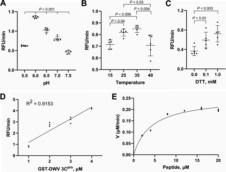
FIG 1 Enzymatic properties of the deformed wing virus (DWV) 3C protease (3Cpro). (A) Optimum pH for protease activity. Multiple comparisons were made by the Tukey-Kramer method and the P-value between pH 6 and other tested pH was < 0.001. (B) Optimum temperature for protease activity. Multiple comparisons were made by the Tukey-Kramer method. Pairs with statistically significant differences are indicated by the P-values. (C) Dithiothreitol (DTT) requirement for protease activity. Multiple comparisons were made by the Dunnett method (one-tailed). Mean values ± standard deviation (SD) (error bars) are shown (n = 5) for A-C. (D) Protease activity with the indicated concentrations of glutathione S-transferase (GST)-DWV 3Cpro protein. (E) Protease activity (V, μM/min) at the indicated peptide substrate concentrations (2-20 μM). Mean values ± SD (error bars) are shown (n = 3) for D and E.
FIG 1 Enzymatic properties of the deformed wing virus (DWV) 3C protease (3Cpro). (A) Optimum pH for protease activity. Multiple comparisons were made by the Tukey-Kramer method and the P-value between pH 6 and other tested pH was < 0.001. (B) Optimum temperature for protease activity. Multiple comparisons were made by the Tukey-Kramer method. Pairs with statistically significant differences are indicated by the P-values. (C) Dithiothreitol (DTT) requirement for protease activity. Multiple comparisons were made by the Dunnett method (one-tailed). Mean values ± standard deviation (SD) (error bars) are shown (n = 5) for A-C. (D) Protease activity with the indicated concentrations of glutathione S-transferase (GST)-DWV 3Cpro protein. (E) Protease activity (V, μM/min) at the indicated peptide substrate concentrations (2-20 μM). Mean values ± SD (error bars) are shown (n = 3) for D and E.CC BY · PubMed Central
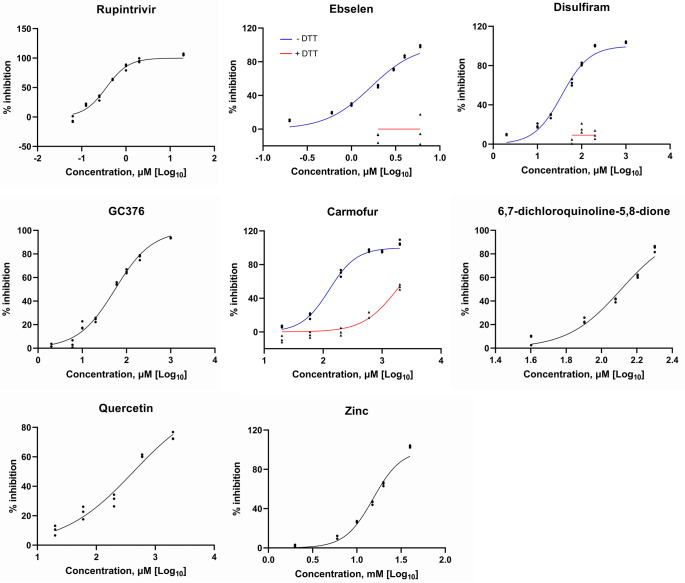
FIG 2 Compounds that inhibit the activity of the deformed wing virus (DWV) 3C protease (3Cpro). Protease activity of DWV 3Cpro in the presence of increasing concentrations of rupintrivir, ebselen, disulfiram, GC376, carmofur, 6,7-dichloroquinoline-5,8-dione, quercetin, and zinc. Ebselen, disulfiram, carmofur were tested in the absence (blue line) or presence (red line) of 2 mM dithiothreitol (DTT). Other compounds were tested in the presence of DTT. All data (n = 3) are shown at the indicated concentrations.
FIG 2 Compounds that inhibit the activity of the deformed wing virus (DWV) 3C protease (3Cpro). Protease activity of DWV 3Cpro in the presence of increasing concentrations of rupintrivir, ebselen, disulfiram, GC376, carmofur, 6,7-dichloroquinoline-5,8-dione, quercetin, and zinc. Ebselen, disulfiram, carmofur were tested in the absence (blue line) or presence (red line) of 2 mM dithiothreitol (DTT). Other compounds were tested in the presence of DTT. All data (n = 3) are shown at the indicated concentrations.CC BY · PubMed Central
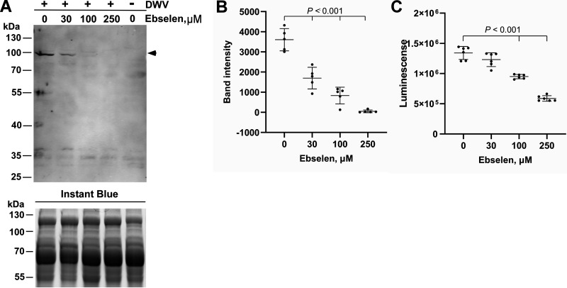
FIG 3 Ebselen suppresses RNA-dependent RNA polymerase (RdRP) synthesis in the deformed wing virus (DWV)-infected pupal head cells. (A) Effect of increasing concentrations of ebselen on RdRP synthesis in DWV-infected (+) pupal head cells. Dimethyl sulfoxide (DMSO) was used as the control. RdRP precursor with 3C protease (97 kDa) is indicated by a black arrowhead. This band was absent in DWV-uninfected (−) cells. The replicate gel was stained with Instant Blue to show that equal amount of protein was applied in each lane. Molecular weight (kDa) of the protein marker is indicated at the left. (B) Band intensity of the RdRP precursor was compared between DMSO and ebselen at the indicated concentrations by the Dunnett method (one-tailed). Mean values ± SD (error bars) are shown (n = 5). (C) Luminescence generated by the luciferase activity dependent on the intracellular ATP level was compared between the pupal head cells treated with DMSO or ebselen at the indicated concentrations by the Dunnett method (one-tailed). There was no significant difference between DMSO and 30 μM ebselen. Mean values ± SD (error bars) are shown (n = 5).
FIG 3 Ebselen suppresses RNA-dependent RNA polymerase (RdRP) synthesis in the deformed wing virus (DWV)-infected pupal head cells. (A) Effect of increasing concentrations of ebselen on RdRP synthesis in DWV-infected (+) pupal head cells. Dimethyl sulfoxide (DMSO) was used as the control. RdRP precursor with 3C protease (97 kDa) is indicated by a black arrowhead. This band was absent in DWV-uninfected (−) cells. The replicate gel was stained with Instant Blue to show that equal amount of protein was applied in each lane. Molecular weight (kDa) of the protein marker is indicated at the left. (B) Band intensity of the RdRP precursor was compared between DMSO and ebselen at the indicated concentrations by the Dunnett method (one-tailed). Mean values ± SD (error bars) are shown (n = 5). (C) Luminescence generated by the luciferase activity dependent on the intracellular ATP level was compared between the pupal head cells treated with DMSO or ebselen at the indicated concentrations by the Dunnett method (one-tailed). There was no significant difference between DMSO and 30 μM ebselen. Mean values ± SD (error bars) are shown (n = 5).CC BY · PubMed Central
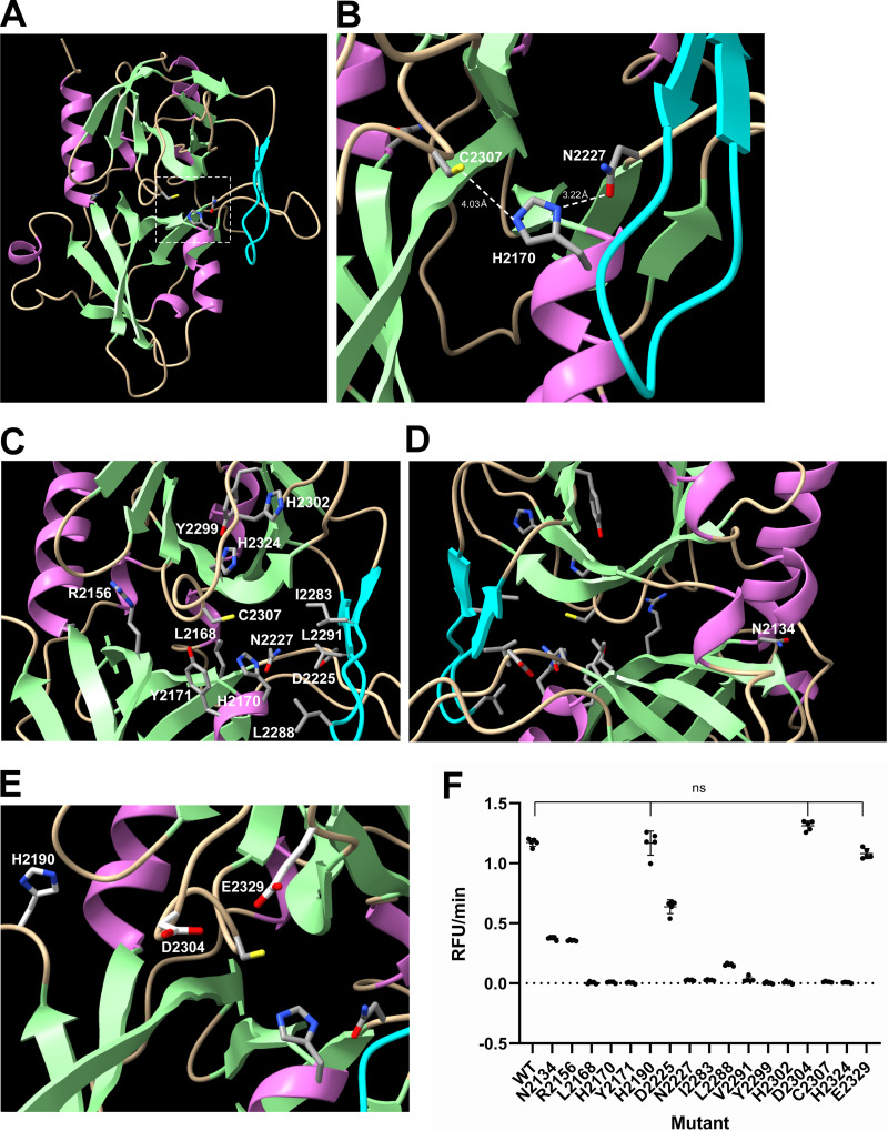
FIG 4 Structure–function analysis of the deformed wing virus (DWV) 3C protease (3Cpro). (A) Structure of DWV 3Cpro predicted by AlphaFold2. The helix, strand, and coil structures are indicated by orchid, pale green, and wheat colors, respectively. β-ribbon is colored by cyan. The catalytic Cys-His-Asn triad is indicated by a square with a white dotted line. (B) Close-up view of the catalytic C2307-H2170-N2227 triad with distance information. (C) Positions of amino acid residues critical for the protease activity. (D) The position of N2134 that was well conserved between 3Cpros of Iflaviruses is shown by rotating the image (C) by 180°. (E) The positions of H2190, D2304, and E2329, which are not essential for the protease activity, are shown together with the catalytic C2307-H2170-N2227 triad. (F) Protease activities of wild type (WT) 3Cpro and alanine substituted mutants. Compared to WT, all mutants showed decreased activities, except H2190A, D2304A, and E2329A (ns), as indicated by the Dunnett test (one-tailed, n = 5).
FIG 4 Structure–function analysis of the deformed wing virus (DWV) 3C protease (3Cpro). (A) Structure of DWV 3Cpro predicted by AlphaFold2. The helix, strand, and coil structures are indicated by orchid, pale green, and wheat colors, respectively. β-ribbon is colored by cyan. The catalytic Cys-His-Asn triad is indicated by a square with a white dotted line. (B) Close-up view of the catalytic C2307-H2170-N2227 triad with distance information. (C) Positions of amino acid residues critical for the protease activity. (D) The position of N2134 that was well conserved between 3Cpros of Iflaviruses is shown by rotating the image (C) by 180°. (E) The positions of H2190, D2304, and E2329, which are not essential for the protease activity, are shown together with the catalytic C2307-H2170-N2227 triad. (F) Protease activities of wild type (WT) 3Cpro and alanine substituted mutants. Compared to WT, all mutants showed decreased activities, except H2190A, D2304A, and E2329A (ns), as indicated by the Dunnett test (one-tailed, n = 5).CC BY · PubMed Central
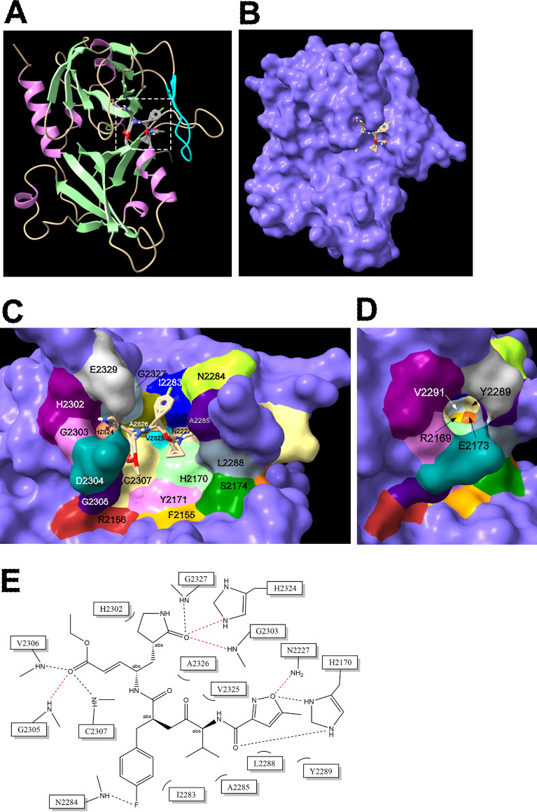
FIG 5 Amino acids interacting with rupintrivir in deformed wing virus (DWV) 3C protease (3Cpro). (A) Overall structure of rupintrivir-bound DWV 3Cpro. Rupintrivir binding site is indicated by a square with white dotted line. (B) Surface view of rupintrivir bound DWV 3Cpro. The binding site of rupintrivir was predicted by the molecular docking tool, AutoDock Vina. (C) Amino acids interacting with rupintrivir as well as the neighboring ones are indicated by different colors. Rupintrivir is also shown. (D) Amino acids in the deep right groove of the substrate-binding domain created by β-ribbon are indicated by rotating the image (C) 45° without rupintrivir. (E) Sixteen amino acids interacting with rupintrivir. Hydrogen bonds are indicated by red dotted lines.
FIG 5 Amino acids interacting with rupintrivir in deformed wing virus (DWV) 3C protease (3Cpro). (A) Overall structure of rupintrivir-bound DWV 3Cpro. Rupintrivir binding site is indicated by a square with white dotted line. (B) Surface view of rupintrivir bound DWV 3Cpro. The binding site of rupintrivir was predicted by the molecular docking tool, AutoDock Vina. (C) Amino acids interacting with rupintrivir as well as the neighboring ones are indicated by different colors. Rupintrivir is also shown. (D) Amino acids in the deep right groove of the substrate-binding domain created by β-ribbon are indicated by rotating the image (C) 45° without rupintrivir. (E) Sixteen amino acids interacting with rupintrivir. Hydrogen bonds are indicated by red dotted lines.CC BY · PubMed Central
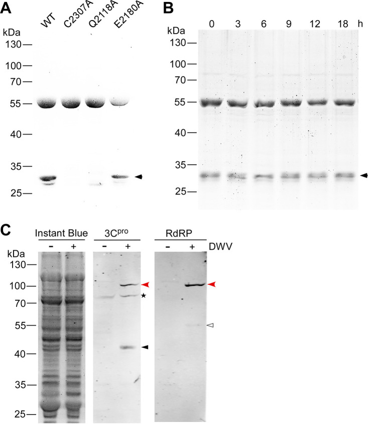
FIG 6 Self-cleavage of the deformed wing virus (DWV) 3C protease (3Cpro) and its synthesis in the virus-infected honey bee cells. (A) Sodium dodecyl sulfate-polyacrylamide gel electrophoresis (SDS-PAGE) of purified wild type (WT) and three mutant (C2307A, Q2118A, and E2180A) glutathione S-transferase (GST)-DWV 3Cpro proteins. The 30 kDa band co-purified with 56 kDa GST-DWV 3Cpro is indicated by a black arrowhead. Molecular weights (kDa) of the protein marker are at the left. (B) SDS-PAGE of purified GST-DWV 3Cpro incubated at 33°C for the indicated time. (C) Western blotting of lysates of the control (−) or DWV-infected (+) honey bee pupal head cells by anti-3Cpro (3Cpro) or anti-RdRP (RdRP) antibody. The replicate gel was stained with Instant Blue to show that equal amount of protein was applied in each lane. Mature 42 kDa 3Cpro and 55 kDa RdRP are indicated by black and white arrowheads, respectively. The 97 kDa precursor of 3Cpro and RdRP (red arrowheads) was detected by both anti-3Cpro and anti-RdRP antibodies. The above-mentioned three bands were specifically present in DWV-infected cells. Asterisk represents the nonspecific band detected by the anti-3Cpro antibody.
FIG 6 Self-cleavage of the deformed wing virus (DWV) 3C protease (3Cpro) and its synthesis in the virus-infected honey bee cells. (A) Sodium dodecyl sulfate-polyacrylamide gel electrophoresis (SDS-PAGE) of purified wild type (WT) and three mutant (C2307A, Q2118A, and E2180A) glutathione S-transferase (GST)-DWV 3Cpro proteins. The 30 kDa band co-purified with 56 kDa GST-DWV 3Cpro is indicated by a black arrowhead. Molecular weights (kDa) of the protein marker are at the left. (B) SDS-PAGE of purified GST-DWV 3Cpro incubated at 33°C for the indicated time. (C) Western blotting of lysates of the control (−) or DWV-infected (+) honey bee pupal head cells by anti-3Cpro (3Cpro) or anti-RdRP (RdRP) antibody. The replicate gel was stained with Instant Blue to show that equal amount of protein was applied in each lane. Mature 42 kDa 3Cpro and 55 kDa RdRP are indicated by black and white arrowheads, respectively. The 97 kDa precursor of 3Cpro and RdRP (red arrowheads) was detected by both anti-3Cpro and anti-RdRP antibodies. The above-mentioned three bands were specifically present in DWV-infected cells. Asterisk represents the nonspecific band detected by the anti-3Cpro antibody.CC BY · PubMed Central

Verification

Claims the paper makes

The claims and statistics of this paper haven’t been checked yet.

Run the check — free, no account needed. It reads the open-access text and checks the sources it cites.

A few free checks a day · papers already verified open instantly · results are public

Self-check

Percentages, sums and reported differences re-computed from the paper’s own numbers.

The self-check needs the paper’s full text.

Citations

Cites

Show all 56 references

Metadata

Authors
Yuan, X., & Kadowaki, T.
Affiliations
Xi’an Jiaotong-Liverpool University (CN)
Corresponding author
Kadowaki, Tatsuhiko
Venue
Microbiology Spectrum
Published
2022-06-29
Volume
10
Issue
3
Pages
e0006822-e0006822
ISSN
2165-0497 (ISSN-L)
OpenAlex
W4280553244
Cited by
5
References
56
Citations by year
2026: 1 · 2024: 1 · 2023: 2 · 2022: 1
FWCI
0.73
Type
journal article
Language
en
Open access
Licence
Locations
  • Microbiology Spectrum (publishedVersion, cc-by)
  • bioRxiv (Cold Spring Harbor Laboratory) (acceptedVersion, cc-by-nc-nd)
  • PubMed Central (publishedVersion, cc-by)
  • PubMed (publishedVersion)
  • DOAJ (DOAJ: Directory of Open Access Journals) (submittedVersion, cc-by-sa)
Funders
  • XJTLU (SURF 2021095)
  • Jinji Lake Double Hundred Talents Programme
Topics
  • Life Sciences › Agricultural and Biological Sciences › Insect Science › Insect and Pesticide Research
  • Life Sciences › Agricultural and Biological Sciences › Plant Science › Plant Virus Research Studies
  • Health Sciences › Medicine › Public Health, Environmental and Occupational Health › Mosquito-borne diseases and control
Keywords
deformed wing virus, catalytic triad, Apis mellifera, colony decline, viral vectors, mites
MeSH terms
3C Viral Proteases, Animals, Bees, Insect Viruses, Insecta, Peptide Hydrolases, RNA, RNA Viruses, Viral Proteins, Polyproteins

About these checks

Registry, retraction, Cites and statistics results are computed, not judged, with one exception: a printed reference that no index lists is read once by a model before it counts as a citation error. Claims are extracted by a model and kept only when their quote appears verbatim in the text; each cited source is then checked, with a model’s help, for whether it says what the claim relies on. Those verdicts can be wrong — read the claim’s quote and the cited source before relying on one.

Statistics and arithmetic are recomputed, not judged.

Confidence bands are shown only for fields calibrated on our annotated test set. None is calibrated yet, so no band is shown.

What counts as a claim ›How we measure confidence ›How we check this ›

Are you an author, or do you see a problem with this page? Request a review

How to cite

Cite CoChat for the checks on this page, and the paper for its own findings.

This page
CoChat. (2026). Citation check: DWV 3C Protease Uncovers the Diverse Catalytic Triad in Insect RNA Viruses. https://app.cochat.ai/papers/10.1128/spectrum.00068-22
The paper
Yuan, X., & Kadowaki, T. (2022). DWV 3C Protease Uncovers the Diverse Catalytic Triad in Insect RNA Viruses. Microbiology Spectrum. https://doi.org/10.1128/spectrum.00068-22

Data sources

Bibliographic data: Semantic Scholar, OpenAlex, Crossref.

Contains information from the Semantic Scholar Academic Graph Datasets, made available under the ODC Attribution License (ODC-BY 1.0).

Semantic Scholar’s data is described in: Kinney, R., et al. (2023). The Semantic Scholar Open Data Platform. arXiv:2301.10140.