

The DOI matches its Crossref record; 49 of the 49 references it cites were found in a registry or an index.
Cysteine-Dependent Conformational Heterogeneity of Shigella flexneri Autotransporter IcsA and Implications of Its Function
Open accessPublished version of a preprint
Abstract
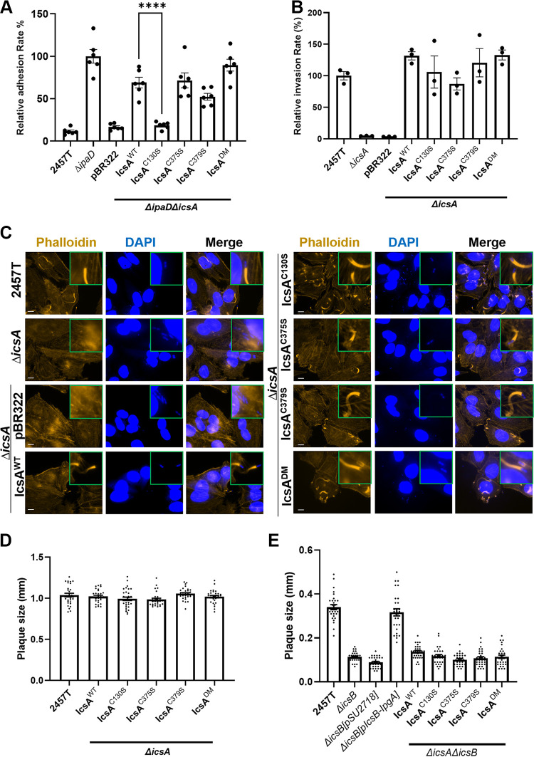
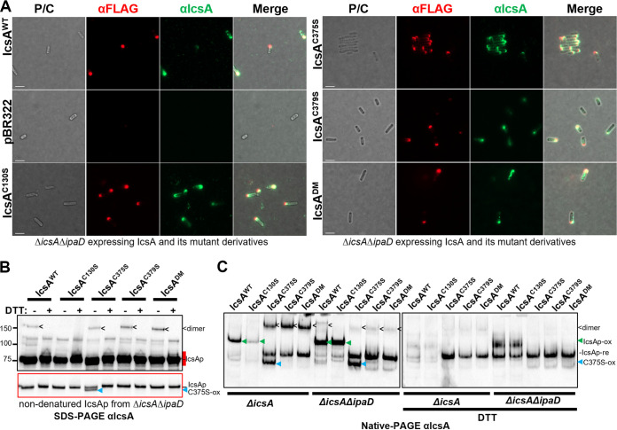
Shigella species are the leading cause of diarrheal-related death globally by causing bacillary dysentery. The surface virulence factor IcsA, which is essential for Shigella pathogenesis, is a unique multifunctional autotransporter that is responsible for cell adhesion, and actin-based motility, yet detailed mechanistic understanding is lacking.
Overview
The claims and statistics of this paper haven’t been checked yet.
Run the check — free, no account needed. It reads the open-access text and checks the sources it cites.
Integrity signals
Registry
Cites
Statistics
Figures
Verification
Claims the paper makes
The claims and statistics of this paper haven’t been checked yet.
Run the check — free, no account needed. It reads the open-access text and checks the sources it cites.
Self-check
Percentages, sums and reported differences re-computed from the paper’s own numbers.
The self-check needs the paper’s full text.
Citations
Cites
Highly accurate protein structure prediction with AlphaFold
Jumper, J., Evans, R., Pritzel, A., Green, T., Figurnov, M., Ronneberger, O., Tunyasuvunakool, K., Bates, R., Žídek, A., Potapenko, A., et al. (2021) · Nature · doi:10.1038/s41586-021-03819-2 · Cited by 43,764
One-step inactivation of chromosomal genes in Escherichia coli K-12 using PCR products
Datsenko, K. A., & Wanner, B. L. (2000) · Proceedings of the National Academy of Sciences · doi:10.1073/pnas.120163297 · Cited by 14,782
UCSF ChimeraX: Structure visualization for researchers, educators, and developers
Pettersen, E. F., Goddard, T. D., Huang, C. C., Meng, E. C., Couch, G. S., Croll, T. I., Morris, J. H., & Ferrin, T. E. (2021) · Protein Science · doi:10.1002/pro.3943 · Cited by 9,514
Transduction of linked genetic characters of the host by bacteriophage P1
Lennox, E. S. (1955) · Virology · doi:10.1016/0042-6822(55)90016-7 · Cited by 2,604
Lugtenberg, B., Meijers, J., Peters, R., van der Hoek, P., & van Alphen, L. (1975) · FEBS Letters · doi:10.1016/0014-5793(75)80272-9 · Cited by 1,416
Type V Protein Secretion Pathway: the Autotransporter Story
Henderson, I. R., Navarro‐García, F., Desvaux, M., Fernandez, R. C., & Ala’Aldeen, D. A. A. (2004) · Microbiology and Molecular Biology Reviews · doi:10.1128/mmbr.68.4.692-744.2004 · Cited by 876
Escape of Intracellular Shigella from Autophagy
Ogawa, M., Yoshimori, T., Suzuki, T., Sagara, H., Mizushima, N., & Sasakawa, C. (2005) · Science · doi:10.1126/science.1106036 · Cited by 828
RF cloning: A restriction-free method for inserting target genes into plasmids
van den Ent, F., & Löwe, J. (2006) · Journal of Biochemical and Biophysical Methods · doi:10.1016/j.jbbm.2005.12.008 · Cited by 640
Kotloff, K. L., Riddle, M. S., Platts-Mills, J. A., Pavlinac, P., & Zaidi, A. K. M. (2018) · The Lancet · doi:10.1016/s0140-6736(17)33296-8 · Cited by 577
Makino, S., Sasakawa, C., Kamata, K., et al. (1986) · Cell · doi:10.1016/0092-8674(86)90880-9 · Cited by 309
Goldberg, M. B., Bârzu, O., Parsot, C., & Sansonetti, P. J. (1993) · Journal of Bacteriology · doi:10.1128/jb.175.8.2189-2196.1993 · Cited by 301
Suzuki, T., Miki, H., Takenawa, T., & Sasakawa, C. (1998) · The EMBO Journal · doi:10.1093/emboj/17.10.2767 · Cited by 228
From self sufficiency to dependence: mechanisms and factors important for autotransporter biogenesis
Leyton, D. L., Rossiter, A. E., & Henderson, I. R. (2012) · Nature Reviews Microbiology · doi:10.1038/nrmicro2733 · Cited by 216
Common structural features of IgA1 protease‐like outer membrane protein autotransporters
Jose, J., Jähnig, F., & Meyer, T. F. (1995) · Molecular Microbiology · doi:10.1111/j.1365-2958.1995.mmi_18020378.x · Cited by 161
Disruption of IcsP, the major Shigella protease that cleaves IcsA, accelerates actin‐based motility
Shere, K. D., Sallustio, S., Manessis, A., D’Aversa, T. G., & Goldberg, M. B. (1997) · Molecular Microbiology · doi:10.1046/j.1365-2958.1997.4681827.x · Cited by 140
Extracellular Transport of VirG Protein in Shigella
Suzuki, T., Lett, M.-C., & Sasakawa, C. (1995) · Journal of Biological Chemistry · doi:10.1074/jbc.270.52.30874 · Cited by 129
Purdy, G. E., Fisher, C. R., & Payne, S. M. (2007) · Journal of Bacteriology · doi:10.1128/jb.00483-07 · Cited by 123
Pope, L. M., Reed, K. E., & Payne, S. M. (1995) · Infection and Immunity · doi:10.1128/iai.63.9.3642-3648.1995 · Cited by 123
Morona, R., Daniels, C. E., & Bosch, L. V. D. (2003) · Microbiology · doi:10.1099/mic.0.26141-0 · Cited by 115
Self-citationLimited tolerance towards folded elements during secretion of the autotransporter Hbp
Jong, W. S. P., ten Hagen‐Jongman, C. M., den Blaauwen, T., Slotboom, D. J., Tame, J. R. H., Wickström, D., Gier, J. D., Otto, B. R., & Luirink, J. (2007) · Molecular Microbiology · doi:10.1111/j.1365-2958.2007.05605.x · Cited by 112
Regulation of O‐antigen chain length is required for Shigella flexneri virulence
Bosch, L. V. D., Manning, P. A., & Morona, R. (1997) · Molecular Microbiology · doi:10.1046/j.1365-2958.1997.2541625.x · Cited by 111
Self-citationJose, J., Krämer, J., Klauser, T., et al. (1996) · Gene · doi:10.1016/0378-1119(96)00343-5 · Cited by 100
Brotcke, A., Goosmann, C., Brinkmann, V., Morona, R., & Zychlinsky, A. (2014) · Cell Host & Microbe · doi:10.1016/j.chom.2014.03.001 · Cited by 94
Self-citationPeriplasmic Transit and Disulfide Bond Formation of the Autotransported Shigella Protein IcsA
Brandon, L. D., & Goldberg, M. B. (2001) · Journal of Bacteriology · doi:10.1128/jb.183.3.951-958.2001 · Cited by 93
Veiga, E., de Lorenzo, V., & Fernández, L. Á. (1999) · Molecular Microbiology · doi:10.1046/j.1365-2958.1999.01571.x · Cited by 90
Shimanovich, A. A., Buskirk, A. D., Heine, S. J., Blackwelder, W. C., Wahid, R., Kotloff, K. L., & Pasetti, M. F. (2016) · Clinical and Vaccine Immunology · doi:10.1128/cvi.00412-16 · Cited by 84
Size and Conformation Limits to Secretion of Disulfide-bonded Loops in Autotransporter Proteins
Leyton, D. L., Sevastsyanovich, Y. R., Browning, D. F., Rossiter, A. E., Wells, T. J., Fitzpatrick, R. E., Overduin, M., Cunningham, A. F., & Henderson, I. R. (2011) · Journal of Biological Chemistry · doi:10.1074/jbc.m111.306118 · Cited by 75
Ogawa, M., Suzuki, T., Tatsuno, I., Abé, H., & Sasakawa, C. (2003) · Molecular Microbiology · doi:10.1046/j.1365-2958.2003.03489.x · Cited by 61
Fu, Z., Thorpe, M., Akula, S., Chahal, G., & Hellman, L. (2018) · Frontiers in Immunology · doi:10.3389/fimmu.2018.02387 · Cited by 59
Miyazaki, H., Yanagida, N., Horinouchi, S., & Beppu, T. (1989) · Journal of Bacteriology · doi:10.1128/jb.171.12.6566-6572.1989 · Cited by 58
Adjacent cysteine residues as a redox switch
Park, C., & Raines, R. T. (2001) · Protein Engineering Design and Selection · doi:10.1093/protein/14.11.939 · Cited by 50
D'Hauteville, H., Dufourcq-Lagelouse, R., Nato, F., & Sansonetti, P. J. (1996) · Infection and Immunity · doi:10.1128/iai.64.2.511-517.1996 · Cited by 48
May, K. L., & Morona, R. (2008) · Journal of Bacteriology · doi:10.1128/jb.00093-08 · Cited by 45
Self-citationSantapaola, D., CHIERICO, F. D., Petrucca, A., Uzzau, S., Casalino, M., Colonna, B., Sessa, R., Berlutti, F., & Nicoletti, M. (2006) · Journal of Bacteriology · doi:10.1128/jb.188.4.1620-1627.2006 · Cited by 44
Ramesh, B., Sendra, V. G., Cirino, P. C., & Varadarajan, N. (2012) · Journal of Biological Chemistry · doi:10.1074/jbc.m112.388199 · Cited by 34
Mauricio, R., Jeffries, C. M., Svergun, D. I., & Deane, J. E. (2016) · Journal of Biological Chemistry · doi:10.1074/jbc.m116.758003 · Cited by 27
Crystal Structure of the Autochaperone Region from the Shigella flexneri Autotransporter IcsA
Kühnel, K., & Diezmann, D. (2011) · Journal of Bacteriology · doi:10.1128/jb.00790-10 · Cited by 24
The virulence domain of Shigella IcsA contains a subregion with specific host cell adhesion function
Qin, J., Doyle, M. T., Tran, E. N. H., & Morona, R. (2020) · PLoS ONE · doi:10.1371/journal.pone.0227425 · Cited by 23
Self-citationIdentification and characterisation of a novel adhesin Ifp in Yersinia pseudotuberculosis
Strong, P. C. R., Hinchliffe, S. J., Patrick, H. L., Atkinson, S., Champion, O. L., & Wren, B. W. (2011) · BMC Microbiology · doi:10.1186/1471-2180-11-85 · Cited by 21
Ndungo, E., Andronescu, L. R., Buchwald, A. G., Lemme‐Dumit, J. M., Mawindo, P., Kapoor, N., Fairman, J., Laufer, M. K., & Pasetti, M. F. (2021) · Frontiers in Immunology · doi:10.3389/fimmu.2021.725129 · Cited by 21
Letley, D. P., Rhead, J. L., Bishop, K., & Atherton, J. C. (2006) · Microbiology · doi:10.1099/mic.0.28548-0 · Cited by 20
Avizonis, D. Z., Farr-Jones, S., Kosen, P. A., & Basus, V. J. (1996) · Journal of the American Chemical Society · doi:10.1021/ja962005v · Cited by 19
Bernard, A., Jessop, T. C., Kumar, P., & Dickenson, N. E. (2017) · Biochemistry · doi:10.1021/acs.biochem.7b00836 · Cited by 19
Leupold, S., Büsing, P., Mas, P. J., Hart, D. J., & Scrima, A. (2017) · Journal of Structural Biology · doi:10.1016/j.jsb.2017.03.003 · Cited by 13
MulticopyicsAis able to suppress the virulence defect caused by thewzzSFmutation inShigella flexneri
Morona, R., & Bosch, L. V. D. (2003) · FEMS Microbiology Letters · doi:10.1016/s0378-1097(03)00217-9 · Cited by 11
Self-citationTeh, M. Y., & Morona, R. (2013) · PLoS ONE · doi:10.1371/journal.pone.0055152 · Cited by 11
Self-citationA small conserved motif supports polarity augmentation of Shigella flexneri IcsA
Doyle, M. T., Morona, R., & Grabowicz, M. (2015) · Microbiology · doi:10.1099/mic.0.000165 · Cited by 10
Self-citationSelf-association of the Shigella flexneri IcsA autotransporter protein
May, K. L., Grabowicz, M., Polyak, S. W., & Morona, R. (2012) · Microbiology · doi:10.1099/mic.0.056465-0 · Cited by 10
Self-citationInvestigation of the IcsA-mediated Shigella flexneri hyper-adherence
Qin, J. (2020) · Adelaide Research & Scholarship (AR&S) (University of Adelaide) · Cited by 1
Self-citation
Metadata
- Authors
- Qin, J., Hong, Y., Morona, R., & Totsika, M.
- Affiliations
- Queensland University of Technology (AU)
- The University of Adelaide (AU)
- Corresponding author
- Qin, Jilong
- Totsika, Makrina
- Venue
- Microbiology Spectrum
- Publisher
- American Society for Microbiology
- Published
- 2022-12-21
- Volume
- 10
- Issue
- 6
- Pages
- e0341022-e0341022
- ISSN
- 2165-0497 (ISSN-L)
- PMC
- PMC9769942
- OpenAlex
- W4308957989
- Cited by
- 6
- References
- 49
- Citations by year
- 2025: 1 · 2024: 2 · 2023: 2 · 2022: 1
- FWCI
- 0.95
- Type
- journal article
- Language
- en
- Open access
- gold
- doi.org
- Licence
- cc-by
- creativecommons.org/licenses/by/4.0/ (vor)
- journals.asm.org/non-commercial-tdm-license (tdm)
- Locations
- Microbiology Spectrum (publishedVersion, cc-by)
- bioRxiv (Cold Spring Harbor Laboratory) (acceptedVersion, cc-by-nc-nd)
- PubMed Central (publishedVersion, cc-by)
- PubMed (publishedVersion)
- ResearchOnline - JCU (James Cook University) (submittedVersion)
- Adelaide Research & Scholarship (AR&S) (University of Adelaide) (submittedVersion, other-oa)
- DOAJ (DOAJ: Directory of Open Access Journals) (submittedVersion, cc-by-sa)
- QUT ePrints (Queensland University of Technology) (submittedVersion)
- Funders
- DHAC | National Health and Medical Research Council (GNT1144046)
- Ramaciotti Foundations (2017HIG0119)
- University of Adelaide
- Topics
- Life Sciences › Immunology and Microbiology › Microbiology › Escherichia coli research studies
- Life Sciences › Biochemistry, Genetics and Molecular Biology › Molecular Biology › Biochemical and Structural Characterization
- Life Sciences › Biochemistry, Genetics and Molecular Biology › Genetics › Bacterial Genetics and Biotechnology
- Keywords
- Shigella flexneri, IcsA, autotransporters, shigellosis, cell adhesion, actin-based motility, protein conformation
- MeSH terms
- Type III Secretion Systems, Type V Secretion Systems, Actins, Bacterial Proteins, Cysteine, DNA-Binding Proteins, Shigella, Shigella flexneri, Transcription Factors, Adhesins, Bacterial, Virulence Factors
About these checks
Registry, retraction, Cites and statistics results are computed, not judged, with one exception: a printed reference that no index lists is read once by a model before it counts as a citation error. Claims are extracted by a model and kept only when their quote appears verbatim in the text; each cited source is then checked, with a model’s help, for whether it says what the claim relies on. Those verdicts can be wrong — read the claim’s quote and the cited source before relying on one.
Statistics and arithmetic are recomputed, not judged.
Confidence bands are shown only for fields calibrated on our annotated test set. None is calibrated yet, so no band is shown.
What counts as a claim ›How we measure confidence ›How we check this ›
Are you an author, or do you see a problem with this page? Request a review
Data sources
Bibliographic data: Semantic Scholar, OpenAlex, Crossref.
Contains information from the Semantic Scholar Academic Graph Datasets, made available under the ODC Attribution License (ODC-BY 1.0).
Semantic Scholar’s data is described in: Kinney, R., et al. (2023). The Semantic Scholar Open Data Platform. arXiv:2301.10140.
