Student or educator? Sign up with your school email and get 6 months of Pro free.
Cited by 15Tuberculosis Research and EpidemiologyHeat shock proteins researchBacterial Genetics and Biotechnology

The DOI matches its Crossref record; 92 of the 92 references it cites were found in a registry or an index.

Interactome Analysis Identifies MSMEI_3879 as a Substrate of Mycolicibacterium smegmatis ClpC1

Open access

Published version of a preprint

Abstract

Drug-resistant tuberculosis infections are a major challenge to global public health. Much effort has been invested in identifying new drug targets in the causative pathogen, Mycobacterium tuberculosis .

Overview

The claims and statistics of this paper haven’t been checked yet.

Run the check — free, no account needed. It reads the open-access text and checks the sources it cites.

A few free checks a day · papers already verified open instantly · results are public

Integrity signals

Registry

Crossref · 10.1128/spectrum.04548-22

Cites

92 of 92 references verified0 citation errors · 0 not checkable

Statistics

Figures

FIG 1 A ClpC1 Walker B double mutant substrate trap was generated in Mycolicibacterium smegmatis (strain ATCC 700084/MC2155). (A) General architecture of the mycobacterial Clp protease. The ATP-dependent unfoldase ClpC1 is responsible for recognizing (via the NTD) and unfolding protein substrates in an ATP-dependent manner (via AAA+ domains D1 and D2), and translocating them into the peptidase barrel composed of catalytic heptamers ClpP1 and ClpP2, wherein degradation occurs. ClpC1EQ associates stably with protein substrates but is unable to unfold or translocate them. (B) Linear sequence models showing the ClpC1 constructs used in this study to identify cellular proteins interacting with specific ClpC1 components such as the NTD and AAA+ core.
FIG 1 A ClpC1 Walker B double mutant substrate trap was generated in Mycolicibacterium smegmatis (strain ATCC 700084/MC2155). (A) General architecture of the mycobacterial Clp protease. The ATP-dependent unfoldase ClpC1 is responsible for recognizing (via the NTD) and unfolding protein substrates in an ATP-dependent manner (via AAA+ domains D1 and D2), and translocating them into the peptidase barrel composed of catalytic heptamers ClpP1 and ClpP2, wherein degradation occurs. ClpC1EQ associates stably with protein substrates but is unable to unfold or translocate them. (B) Linear sequence models showing the ClpC1 constructs used in this study to identify cellular proteins interacting with specific ClpC1 components such as the NTD and AAA+ core.CC BY · PubMed Central
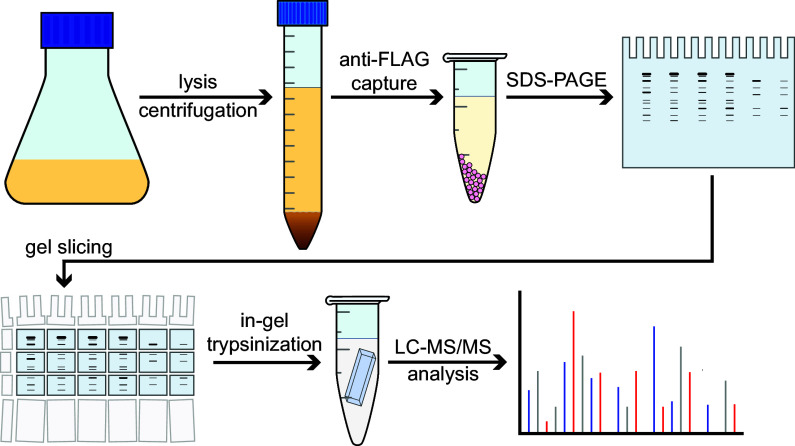
FIG 2 Workflow for identification of ClpC1 interaction partners. Mycolicibacterium smegmatis (ATCC 700084/MC2155) cells were grown up to and A600 of ≈0.6 to 1.0, and ClpC1 expression was induced following addition of 28 mM ε-caprolactam. Identification of the interactome was based on gel electrophoresis, trypsin digestion, and LC-MS/MS run using higher-energy collisional dissociation (HCD) fragmentation. All ClpC1 test construct and control experiments were subjected to the same workflow and were carried out in biological replicates.
FIG 2 Workflow for identification of ClpC1 interaction partners. Mycolicibacterium smegmatis (ATCC 700084/MC2155) cells were grown up to and A600 of ≈0.6 to 1.0, and ClpC1 expression was induced following addition of 28 mM ε-caprolactam. Identification of the interactome was based on gel electrophoresis, trypsin digestion, and LC-MS/MS run using higher-energy collisional dissociation (HCD) fragmentation. All ClpC1 test construct and control experiments were subjected to the same workflow and were carried out in biological replicates.CC BY · PubMed Central
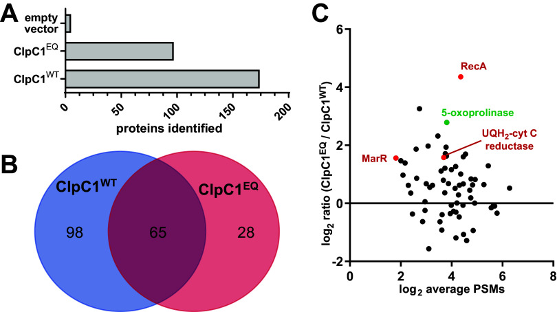
FIG 3 Comparative analysis of the ClpC1EQ and ClpC1WT interactome. (A) Bar graph showing the number of proteins identified in the ClpC1EQ and ClpC1WT data sets; (B) Venn diagram showing the number of Mycolicibacterium smegmatis cellular proteins that interacted with ClpC1EQ and ClpC1WT constructs, respectively. The numbers in panels A and B exclude ClpC1 itself. (C) Volcano plot showing enrichment of proteins captured in the M. smegmatis ClpC1 mutant trap versus wild type by liquid chromatography-tandem mass spectrometry (LC-MS/MS). The y axis shows the log2 ratio of normalized scores in the ClpC1EQ data set to those in the ClpC1WT data set, while the x axis shows log2 average number of PSMs for the proteins. Red circles represent proteins enriched in ClpC1EQ with a P value of <0.05 by Student’s t test.
FIG 3 Comparative analysis of the ClpC1EQ and ClpC1WT interactome. (A) Bar graph showing the number of proteins identified in the ClpC1EQ and ClpC1WT data sets; (B) Venn diagram showing the number of Mycolicibacterium smegmatis cellular proteins that interacted with ClpC1EQ and ClpC1WT constructs, respectively. The numbers in panels A and B exclude ClpC1 itself. (C) Volcano plot showing enrichment of proteins captured in the M. smegmatis ClpC1 mutant trap versus wild type by liquid chromatography-tandem mass spectrometry (LC-MS/MS). The y axis shows the log2 ratio of normalized scores in the ClpC1EQ data set to those in the ClpC1WT data set, while the x axis shows log2 average number of PSMs for the proteins. Red circles represent proteins enriched in ClpC1EQ with a P value of <0.05 by Student’s t test.CC BY · PubMed Central
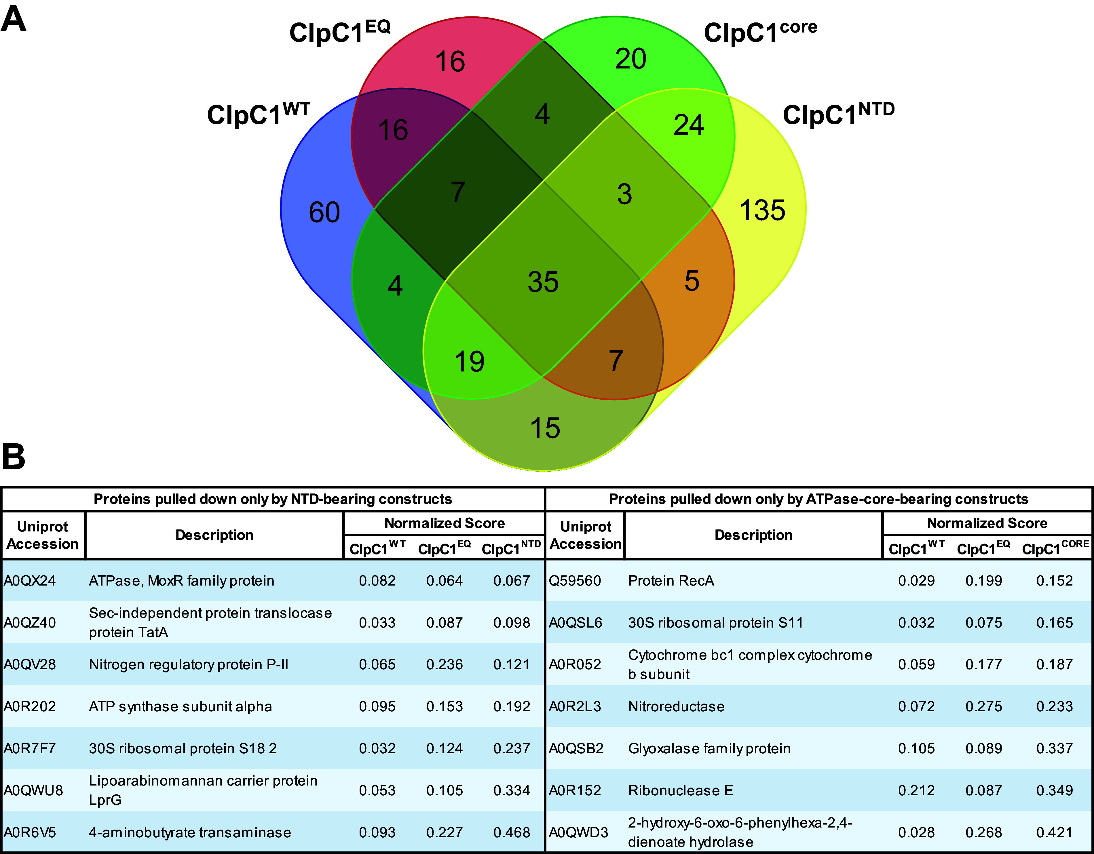
FIG 4 Comparative analysis of the ClpC1 interactome based on the different unfoldase components. (A) A Venn diagram shows protein targets observed to interact with one or more ClpC1 constructs. (B) The tabulated proteins were found to coimmunoprecipitate with all constructs bearing the amino-terminal domain (NTD) or the AAA+ ATPase core. Only 7 proteins were observed to interact with NTD alone. Similarly, as shown on the right only 7 proteins were observed interacting with the core-containing constructs and not in the NTD data sets.
FIG 4 Comparative analysis of the ClpC1 interactome based on the different unfoldase components. (A) A Venn diagram shows protein targets observed to interact with one or more ClpC1 constructs. (B) The tabulated proteins were found to coimmunoprecipitate with all constructs bearing the amino-terminal domain (NTD) or the AAA+ ATPase core. Only 7 proteins were observed to interact with NTD alone. Similarly, as shown on the right only 7 proteins were observed interacting with the core-containing constructs and not in the NTD data sets.CC BY · PubMed Central
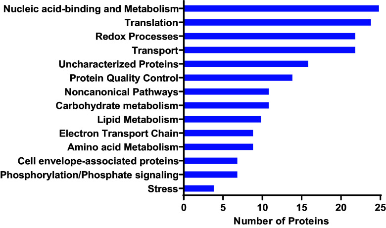
FIG 5 Full-length ClpC1 interacts with diverse groups of proteins in Mycolicibacterium smegmatis. Functional categorization of cellular proteins interacting with ClpC1WT and/or ClpC1EQ is shown. Also shown is the distribution of these proteins by Gene Ontology (GO) annotation based mostly on biological process. Functional classification was performed using Blast2GO and was based on annotation of the closest homologs.
FIG 5 Full-length ClpC1 interacts with diverse groups of proteins in Mycolicibacterium smegmatis. Functional categorization of cellular proteins interacting with ClpC1WT and/or ClpC1EQ is shown. Also shown is the distribution of these proteins by Gene Ontology (GO) annotation based mostly on biological process. Functional classification was performed using Blast2GO and was based on annotation of the closest homologs.CC BY · PubMed Central
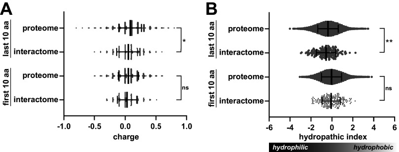
FIG 6 Physicochemical analysis of terminal sequences of full-length ClpC1-interacting proteins. (A) Violin plots illustrate the average charges of the first (N-terminal) and last (C-terminal) amino acids in the ClpC1 interactome and in the entire M. smegmatis proteome. Central lines indicate median values, with error bars indicating 1 SD. (B) Mean hydrophobicity of terminal residues in the ClpC1 interactome and control data sets is shown. For both metrics, the values plotted are the average residue value per terminus. Significance was assessed by two-tailed Welch’s t test. *, P < 0.05; **, P < 0.01; ns, not significant.
FIG 6 Physicochemical analysis of terminal sequences of full-length ClpC1-interacting proteins. (A) Violin plots illustrate the average charges of the first (N-terminal) and last (C-terminal) amino acids in the ClpC1 interactome and in the entire M. smegmatis proteome. Central lines indicate median values, with error bars indicating 1 SD. (B) Mean hydrophobicity of terminal residues in the ClpC1 interactome and control data sets is shown. For both metrics, the values plotted are the average residue value per terminus. Significance was assessed by two-tailed Welch’s t test. *, P < 0.05; **, P < 0.01; ns, not significant.CC BY · PubMed Central
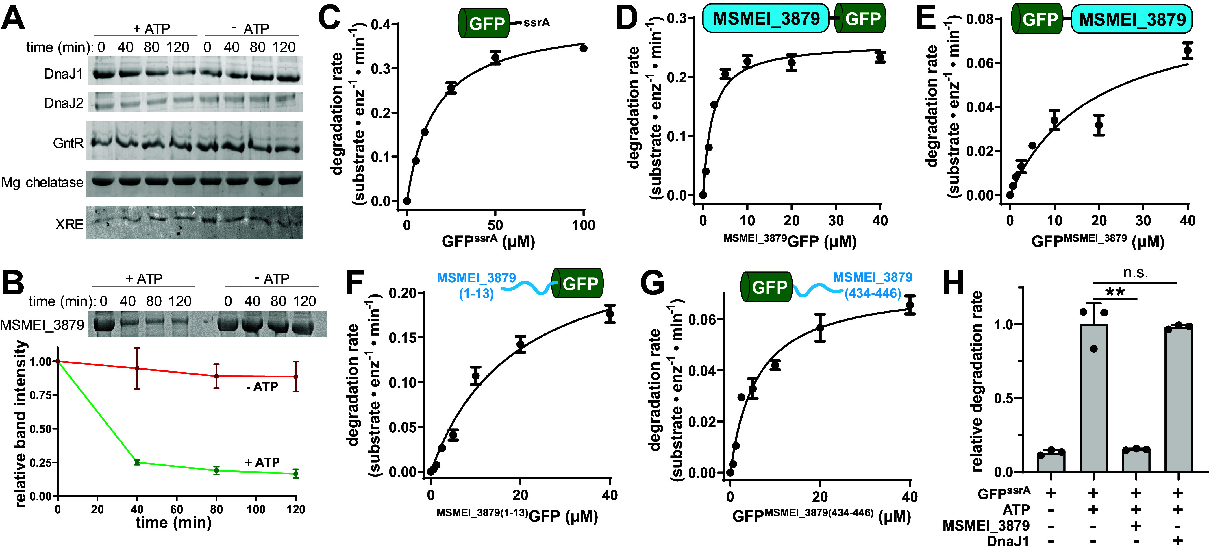
FIG 7 MSMEI_3879 is a substrate of M. smegmatis ClpC1. (A) In vitro degradation assays of DnaJ1, DnaJ2, GntR, magnesium chelatase, and XRE by 1 μM ClpC1 and 1 μM ClpP1P2, monitored by SDS-PAGE. (B) ATP-dependent degradation of 10 μM MSMEI_3879 by ClpC1P1P2 was observed in vitro by SDS-PAGE, with gel densitometry of three replicate assays shown. (C) Michaelis-Menten analysis of GFPssrA proteolysis by 1 μM ClpC1P1P2, as a function of substrate concentration, reveals a kcat of 0.41 ± 0.02 substrate · min−1 · enzyme−1 and a Km of 16.04 ± 1.28 μM. (D) MSMEI_3879GFP was degraded by 1 μM ClpC1P1P2 with a kcat of 0.26 ± 0.01 substrate · min−1 · enzyme−1 and Km of 2.06 ± 0.43 μM. (E) GFPMSMEI_3879 was degraded with a kcat of 0.09 ± 0.02 substrate · min−1 · enzyme−1 and a Km of 18.8 ± 9.6 μM. (F) GFP preceded by the first 13 residues of MSMEI_3879 was degraded with kcat of 0.268 ± 0.033 substrate · min−1 · enzyme−1 and Km of 19.1 ± 4.9 μM. (G) GFP appended with the last 13 residues of MSMEI_3879 was degraded with a kcat of 0.0737 ± 0.0053 substrate · min−1 · enzyme−1 and Km of 5.97 ± 1.3 μM. (H) Degradation of 10 μM GFPssrA by 1 μM ClpC1P1P2 was monitored in the presence and absence of ATP, 10 μM MSMEI_3879, or 10 μM DnaJ1. MSMEI_3879 reduces GFPssrA degradation to the level observed in the absence of ATP. DnaJ1 does not significantly alter the rate of GFPssrA proteolysis. Values are averages of three replicates (n = 3) ± 1 SD. P values were calculated by unpaired two-tailed Student's t test. **, P < 0.01; n.s., not significant.
FIG 7 MSMEI_3879 is a substrate of M. smegmatis ClpC1. (A) In vitro degradation assays of DnaJ1, DnaJ2, GntR, magnesium chelatase, and XRE by 1 μM ClpC1 and 1 μM ClpP1P2, monitored by SDS-PAGE. (B) ATP-dependent degradation of 10 μM MSMEI_3879 by ClpC1P1P2 was observed in vitro by SDS-PAGE, with gel densitometry of three replicate assays shown. (C) Michaelis-Menten analysis of GFPssrA proteolysis by 1 μM ClpC1P1P2, as a function of substrate concentration, reveals a kcat of 0.41 ± 0.02 substrate · min−1 · enzyme−1 and a Km of 16.04 ± 1.28 μM. (D) MSMEI_3879GFP was degraded by 1 μM ClpC1P1P2 with a kcat of 0.26 ± 0.01 substrate · min−1 · enzyme−1 and Km of 2.06 ± 0.43 μM. (E) GFPMSMEI_3879 was degraded with a kcat of 0.09 ± 0.02 substrate · min−1 · enzyme−1 and a Km of 18.8 ± 9.6 μM. (F) GFP preceded by the first 13 residues of MSMEI_3879 was degraded with kcat of 0.268 ± 0.033 substrate · min−1 · enzyme−1 and Km of 19.1 ± 4.9 μM. (G) GFP appended with the last 13 residues of MSMEI_3879 was degraded with a kcat of 0.0737 ± 0.0053 substrate · min−1 · enzyme−1 and Km of 5.97 ± 1.3 μM. (H) Degradation of 10 μM GFPssrA by 1 μM ClpC1P1P2 was monitored in the presence and absence of ATP, 10 μM MSMEI_3879, or 10 μM DnaJ1. MSMEI_3879 reduces GFPssrA degradation to the level observed in the absence of ATP. DnaJ1 does not significantly alter the rate of GFPssrA proteolysis. Values are averages of three replicates (n = 3) ± 1 SD. P values were calculated by unpaired two-tailed Student's t test. **, P < 0.01; n.s., not significant.CC BY · PubMed Central
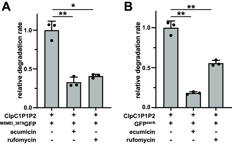
FIG 8 Effect of ClpC1-targeting antibiotics on proteolysis by ClpC1P1P2. Inclusion of 10 μM ECU or RUF inhibits degradation of 10 μM (A) MSMEI_3879GFP and (B) GFPssrA by 1 μM ClpC1P1P2 relative to a nontreatment control. Values are averages from three replicates (n = 3) ± 1 SD. P values were calculated by unpaired two-tailed Student’s t test. * and ** represent P values of <0.05 and 0.01, respectively.
FIG 8 Effect of ClpC1-targeting antibiotics on proteolysis by ClpC1P1P2. Inclusion of 10 μM ECU or RUF inhibits degradation of 10 μM (A) MSMEI_3879GFP and (B) GFPssrA by 1 μM ClpC1P1P2 relative to a nontreatment control. Values are averages from three replicates (n = 3) ± 1 SD. P values were calculated by unpaired two-tailed Student’s t test. * and ** represent P values of <0.05 and 0.01, respectively.CC BY · PubMed Central

Verification

Claims the paper makes

The claims and statistics of this paper haven’t been checked yet.

Run the check — free, no account needed. It reads the open-access text and checks the sources it cites.

A few free checks a day · papers already verified open instantly · results are public

Self-check

Percentages, sums and reported differences re-computed from the paper’s own numbers.

The self-check needs the paper’s full text.

Citations

Cites

Show all 92 references

Research works printed but not linked: 1

  1. WHO. 2021. Global tuberculosis report 2021. World Health Organization, Geneva, Switzerland.

Metadata

Authors
Ogbonna, E. C., Anderson, H. R., Beardslee, P. C., Bheemreddy, P., & Schmitz, K. R.
Affiliations
University of Delaware (US)
Corresponding author
Schmitz, Karl R.
Venue
Microbiology Spectrum
Published
2023-08-17
Volume
11
Issue
4
Pages
e0454822-e0454822
ISSN
2165-0497 (ISSN-L)
OpenAlex
W4381470243
Cited by
15
References
92
Citations by year
2026: 2 · 2025: 3 · 2024: 10
FWCI
3.10
Type
journal article
Language
en
Open access
Licence
Locations
  • Microbiology Spectrum (publishedVersion, cc-by)
  • bioRxiv (Cold Spring Harbor Laboratory) (acceptedVersion, cc-by-nc-nd)
  • PubMed Central (publishedVersion, cc-by)
  • PubMed (publishedVersion)
  • DOAJ (DOAJ: Directory of Open Access Journals) (submittedVersion, cc-by-sa)
Funders
  • National Institute of General Medical Sciences (P20GM104316)
  • HHS | NIH | National Institute of General Medical Sciences (T32GM133395)
Topics
  • Health Sciences › Medicine › Infectious Diseases › Tuberculosis Research and Epidemiology
  • Life Sciences › Biochemistry, Genetics and Molecular Biology › Molecular Biology › Heat shock proteins research
  • Life Sciences › Biochemistry, Genetics and Molecular Biology › Genetics › Bacterial Genetics and Biotechnology
Keywords
Mycolicibacterium smegmatis, protein-protein interaction, protein-protein interaction network, therapeutic target, drug-resistant tuberculosis, Mycobacterium tuberculosis, protein substrates, proteolysis
MeSH terms
Anti-Bacterial Agents, Bacterial Proteins, Heat-Shock Proteins, Humans, Mycobacterium tuberculosis, Tuberculosis, Molecular Chaperones, Mycobacterium smegmatis

About these checks

Registry, retraction, Cites and statistics results are computed, not judged, with one exception: a printed reference that no index lists is read once by a model before it counts as a citation error. Claims are extracted by a model and kept only when their quote appears verbatim in the text; each cited source is then checked, with a model’s help, for whether it says what the claim relies on. Those verdicts can be wrong — read the claim’s quote and the cited source before relying on one.

Statistics and arithmetic are recomputed, not judged.

Confidence bands are shown only for fields calibrated on our annotated test set. None is calibrated yet, so no band is shown.

What counts as a claim ›How we measure confidence ›How we check this ›

Are you an author, or do you see a problem with this page? Request a review

How to cite

Cite CoChat for the checks on this page, and the paper for its own findings.

This page
CoChat. (2026). Citation check: Interactome Analysis Identifies MSMEI_3879 as a Substrate of Mycolicibacterium smegmatis ClpC1. https://app.cochat.ai/papers/10.1128/spectrum.04548-22
The paper
Ogbonna, E. C., Anderson, H. R., Beardslee, P. C., Bheemreddy, P., & Schmitz, K. R. (2023). Interactome Analysis Identifies MSMEI_3879 as a Substrate of Mycolicibacterium smegmatis ClpC1. Microbiology Spectrum. https://doi.org/10.1128/spectrum.04548-22

Data sources

Bibliographic data: Semantic Scholar, OpenAlex, Crossref.

Contains information from the Semantic Scholar Academic Graph Datasets, made available under the ODC Attribution License (ODC-BY 1.0).

Semantic Scholar’s data is described in: Kinney, R., et al. (2023). The Semantic Scholar Open Data Platform. arXiv:2301.10140.