

The DOI matches its Crossref record.
MicroRNA-21-5p Reduces Hypoxia/Reoxygenation-Induced Neuronal Cell Damage through Negative Regulation of CPEB3
Abstract
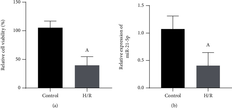
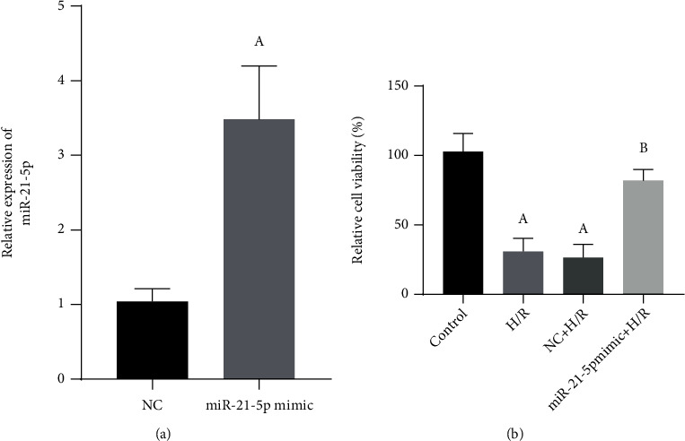
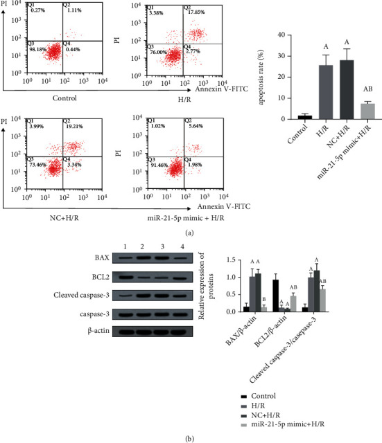
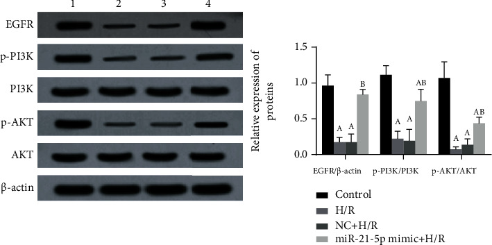
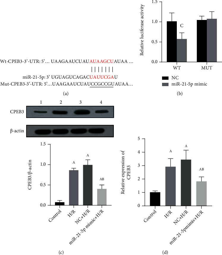
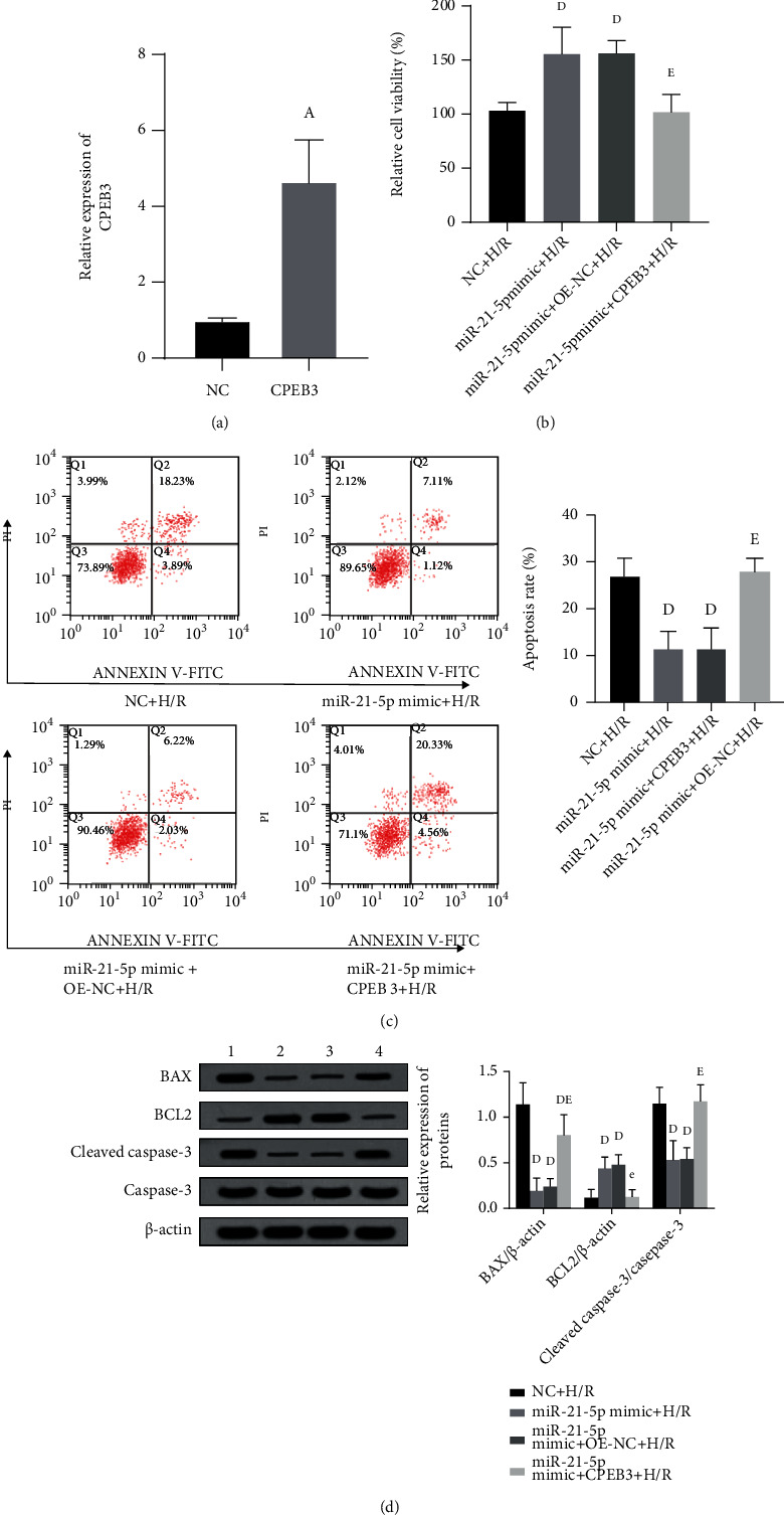
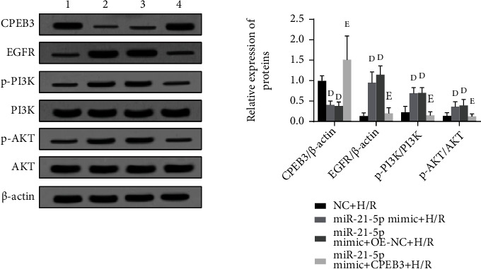
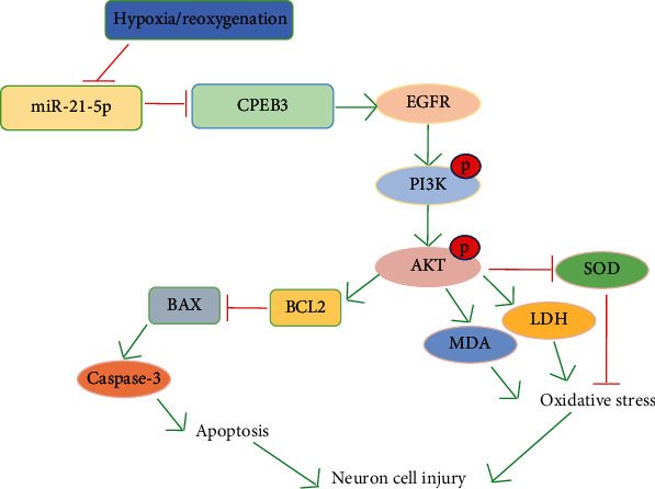
Objectives. To explore the role of microRNA-21-5p (miR-21-5p) in hypoxia/reoxygenation- (H/R-) induced HT22 cell damage. Methods. The hypoxia/reoxygenation (H/R) model was established in mouse neuronal cells HT22. Cell Counting Kit-8 (CCK-8) and qRT-PCR were used to determine the effects of H/R treatment on cell viability and miR-21-5p expression. HT22 cells were transfected with miR-21-5p mimic or negative control (NC) followed by the induction of H/R; cell viability, apoptosis, and SOD, MDA, and LDH activities were detected. Besides, the apoptosis-related proteins including BAX, BCL2, cleaved caspase-3, and caspase-3 as well as proteins of EGFR/PI3K/AKT signaling pathways were measured by Western blot. To verify the target relation between cytoplasmic polyadenylation element binding protein 3 (CPEB3) and miR-21-5p, luciferase reporter gene experiment was performed. After cotransfection with miR-21-5p mimic and CPEB3 plasmids, the reversal effects of CPEB3 on miR-21-5p in H/R damage were studied. Results. H/R treatment could significantly reduce the cell viability ( P < 0.05 ) and miR-21-5p levels ( P < 0.05 ) in HT22 cells. After overexpressing miR-21-5p, cell viability was increased ( P < 0.05 ) under H/R treatment, and the apoptosis rate and the levels of apoptosis-related proteins were suppressed (all P < 0.05 ). Furthermore, SOD activity was increased ( P < 0.05 ), while MDA and LDH activity was decreased (both P < 0.05 ). Besides, miR-21-5p could restore the activation of the EGFR/PI3K/AKT signaling pathway inhibited by H/R treatment (all P < 0.05 ). The luciferase reporter gene experiment verified that CPEB3 is the target of miR-21-5p ( P < 0.05 ). When coexpressing miR-21-5p mimic and CPEB3 in the cells, the protective effects of miR-21-5p under H/R were reversed (all P < 0.05 ), and the activation of the EGFR/PI3K/AKT pathway was also inhibited (all P < 0.05 ). Conclusion. This study showed that miR-21-5p may regulate the EGFR/PI3K/AKT signaling pathway by targeting CPEB3 to reduce H/R-induced cell damage and apoptosis.
Overview
The claims and statistics of this paper haven’t been checked yet.
Run the check — free, no account needed. It reads the open-access text and checks the sources it cites.
Integrity signals
Registry
Cites
Statistics
Figures
Verification
Claims the paper makes
The claims and statistics of this paper haven’t been checked yet.
Run the check — free, no account needed. It reads the open-access text and checks the sources it cites.
Claims other papers make about this one
Each is checked against this paper’s own text: does it say what the citing paper says it does?
The cited source says what the claim attributes to it.
In the citing paper
“Supporting the potential of targeting CPEB3, a decrease in CPEB3 levels has been shown to boost neurotrophic signaling (41), to ameliorate oxidative stress (42), and to increase proteasome activation (43).”
From MicroRNA-92a–CPEB3 axis protects neurons against inflammatory neurodegeneration (2023) · cites this paper as [42]
Self-check
Percentages, sums and reported differences re-computed from the paper’s own numbers.
The self-check needs the paper’s full text.
Citations
Cites
No references could be resolved yet.
Research works printed but not linked: 26
- L. B. Goldstein, 2020, Introduction for focused updates in cerebrovascular disease, Stroke, 51, 708, 10.1161/STROKEAHA.119.024159 · DOI
- 10.1161/STROKEAHA.118.022015 · DOI
- 10.1007/s10571-015-0202-x · DOI
- 10.3390/antiox8100454 · DOI
- 10.1016/j.ejphar.2017.04.002 · DOI
- L. Yang, 2015, MicroRNA-323 regulates ischemia/reperfusion injury-induced neuronal cell death by targeting BRI3, International Journal of Clinical and Experimental Pathology, 8, 10725
- 10.1186/s12974-017-0977-4 · DOI · Paper page
- K. Saliminejad, 2019, An overview of microRNAs: biology, functions, therapeutics, and analysis methods, Journal of cellular physiology, 201, 5451, 10.1002/jcp.27486 · DOI · Paper page
- 10.1177/0300985813502820 · DOI
- 10.1007/978-3-319-22671-2_9 · DOI
- 10.1016/j.yexcr.2017.01.015 · DOI
- 10.5732/cjc.30.0371 · DOI
- 10.1007/s12031-018-1067-5 · DOI
- 10.1007/s00210-014-1006-0 · DOI
- 10.3390/ijms160922368 · DOI
- 10.1016/j.ejphar.2019.01.022 · DOI
- 10.1152/physiolgenomics.00020.2014 · DOI
- 10.1152/physiolgenomics.00130.2010 · DOI
- 10.1007/s13277-016-5035-9 · DOI
- E. Candelario-Jalil, 2009, Injury and repair mechanisms in ischemic stroke: considerations for the development of novel neurotherapeutics, Current Opinion in Investigational Drugs, 10, 644
- 10.2174/1567202612666150311104900 · DOI
- 10.1007/s00232-009-9177-1 · DOI
- 10.1080/02652030500058403 · DOI
- 10.1002/cbf.2977 · DOI
- 10.3892/or.2019.6983 · DOI
- 10.1002/jcb.28528 · DOI
Cited by
MicroRNA-92a–CPEB3 axis protects neurons against inflammatory neurodegeneration
Winkler, I., Engler, J. B., Vieira, V., Bauer, S., Liu, Y.-H., Di Liberto, G., Grochowska, K. M., Wagner, I., Bier, J., Bal, L. C., et al. (2023) · Science Advances · doi:10.1126/sciadv.adi6855 · Cited by 22
Metadata
- Authors
- Wang, B., Yu, P., Lin, W., & Zhai, Z.
- Affiliations
- Department of Neurology, Affiliated Hospital of Weifang Medical College, Weifang, Shandong 261031, China
- Pain Department, Weifang People’s Hospital, Weifang, Shandong 261041, China
- Department of Plastic Surgery, Plastic Surgery Hospital of Weifang Medical University, Weifang, Shandong 261042, China
- Venue
- Analytical Cellular Pathology
- Publisher
- Wiley
- Published
- 2021-12-02
- Volume
- 2021
- Pages
- 1-11
- ISSN
- 2210-7185
- 2210-7177
- PMC
- PMC8660214
- Cited by
- 5
- References
- 0
- Language
- en
- Funders
- Shandong Province Medicine and Health Technology Development Plan (2018WS062)
About these checks
Registry, retraction, Cites and statistics results are computed, not judged, with one exception: a printed reference that no index lists is read once by a model before it counts as a citation error. Claims are extracted by a model and kept only when their quote appears verbatim in the text; each cited source is then checked, with a model’s help, for whether it says what the claim relies on. Those verdicts can be wrong — read the claim’s quote and the cited source before relying on one.
Statistics and arithmetic are recomputed, not judged.
Confidence bands are shown only for fields calibrated on our annotated test set. None is calibrated yet, so no band is shown.
What counts as a claim ›How we measure confidence ›How we check this ›
Are you an author, or do you see a problem with this page? Request a review
Data sources
Bibliographic data: Semantic Scholar, OpenAlex, Crossref.
Contains information from the Semantic Scholar Academic Graph Datasets, made available under the ODC Attribution License (ODC-BY 1.0).
Semantic Scholar’s data is described in: Kinney, R., et al. (2023). The Semantic Scholar Open Data Platform. arXiv:2301.10140.
