

The DOI matches its Crossref record; 4 of the 4 references it cites were found in a registry or an index.
Novel ERBB2 Variant Potentially Associated with Resistance against Anti-HER2 Monoclonal Antibody–Based Therapy in ERBB2 -Amplified Metastatic Colorectal Cancer
Open accessPublished version of a preprint
Abstract
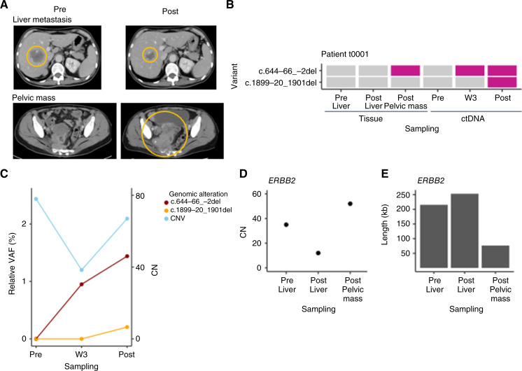
Purpose: HER2-targeted therapies in ERBB2-amplified metastatic colorectal cancer (mCRC) are effective; however, a notable portion of patients do not respond to treatment, and secondary resistance occurs in most patients receiving these treatments. The purpose of this study was to investigate determinants of treatment efficacy and resistance in patients with ERBB2-amplified mCRC who received HER2-targeted therapy by analyzing multiomics data. Experimental Design: We investigated genomic data from a nationwide large cancer genomic screening project, the SCRUM-Japan project. We analyzed paired genome and transcriptome data of tissue and genomic data of ctDNA collected pre- and postprogression in patients enrolled in the related trial, TRIUMPH, in ERBB2-amplified mCRC. Results: In 155 patients with ERBB2-amplified solid tumors who received HER2-targeted therapy based on the SCRUM-Japan project, the objective response rate was 50%, 51%, and 35% in ERBB2 wild-type, variant of unknown significance, and pathogenic variant groups, respectively. In the paired genome and transcriptome data analyses in TRIUMPH, we identified the novel splicing-associated variant c.644-66_-2del in one of the 11 patients with paired whole-exome sequencing and whole-transcriptome sequencing data sets, which lacks the binding domain of pertuzumab, in progressed metastatic tumor as a variant with potential pathogenicity. The time-course ctDNA analysis detected c.644-66_-2del as an acquired variant. Conclusions: This study highlighted the importance of ERBB2 genomic status when evaluating the efficacy of HER2-targeted therapies in ERBB2-amplified mCRC. The identification of a novel splicing-associated variant may provide insights into potential mechanisms of treatment resistance. Furthermore, we demonstrated the utility of ctDNA to follow the acquired genomic status of mCRC tumors.
Overview
The claims and statistics of this paper haven’t been checked yet.
Run the check — free, no account needed. It reads the open-access text and checks the sources it cites.
Integrity signals
Registry
Cites
Statistics
Figures
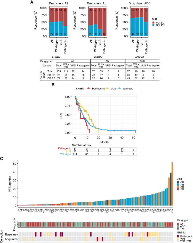
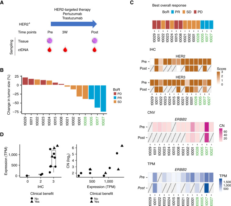
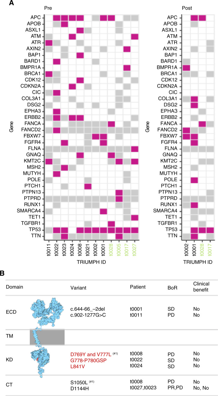
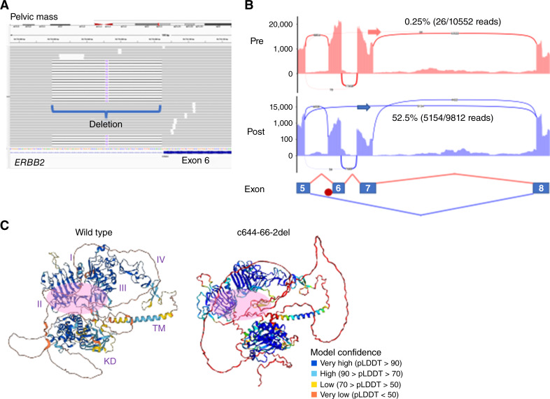
Verification
Claims the paper makes
The claims and statistics of this paper haven’t been checked yet.
Run the check — free, no account needed. It reads the open-access text and checks the sources it cites.
Self-check
Percentages, sums and reported differences re-computed from the paper’s own numbers.
The self-check needs the paper’s full text.
Citations
Cites
Highly accurate protein structure prediction with AlphaFold
Jumper, J., Evans, R., Pritzel, A., Green, T., Figurnov, M., Ronneberger, O., Tunyasuvunakool, K., Bates, R., Žídek, A., Potapenko, A., et al. (2021) · Nature · doi:10.1038/s41586-021-03819-2 · Cited by 43,764
Sartore-Bianchi, A., Trusolino, L., Martino, C., Bencardino, K., Lonardi, S., Bergamo, F., Zagonel, V., Leone, F., Depetris, I., Martinelli, E., et al. (2016) · The Lancet Oncology · doi:10.1016/s1470-2045(16)00150-9 · Cited by 1,016
Adaptive mutability of colorectal cancers in response to targeted therapies
Russo, M., Crisafulli, G., Sogari, A., Reilly, N. M., Arena, S., Lamba, S., Bartolini, A., Amodio, V., Magrì, A., Novara, L., et al. (2019) · Science · doi:10.1126/science.aav4474 · Cited by 433
Strickler, J. H., Cercek, A., Siena, S., André, T., Ng, K., Van Cutsem, E., Wu, C., Paulson, A. S., Hubbard, J. M., Coveler, A. L., et al. (2023) · The Lancet Oncology · doi:10.1016/s1470-2045(23)00150-x · Cited by 259
Research works printed but not linked: 27
- Sawada, 2018, Prognostic and predictive value of HER2 amplification in patients with metastatic colorectal cancer, Clin Colorectal Cancer, 17, 198, 10.1016/j.clcc.2018.05.006 · DOI
- Ross, 2018, Targeting HER2 in colorectal cancer: the landscape of amplification and short variant mutations in ERBB2 and ERBB3, Cancer, 124, 1358, 10.1002/cncr.31125 · DOI
- Strickler, 2022, Diagnosis and treatment of ERBB2-positive metastatic colorectal cancer: a review, JAMA Oncol, 8, 760, 10.1001/jamaoncol.2021.8196 · DOI
- Siena, 2021, Trastuzumab deruxtecan (DS-8201) in patients with HER2-expressing metastatic colorectal cancer (DESTINY-CRC01): a multicentre, open-label, phase 2 trial, Lancet Oncol, 22, 779, 10.1016/S1470-2045(21)00086-3 · DOI
- Meric-Bernstam, 2019, Pertuzumab plus trastuzumab for HER2-amplified metastatic colorectal cancer (MyPathway): an updated report from a multicentre, open-label, phase 2a, multiple basket study, Lancet Oncol, 20, 518, 10.1016/S1470-2045(18)30904-5 · DOI · Paper page
- Nakamura, 2021, Circulating tumor DNA-guided treatment with pertuzumab plus trastuzumab for HER2-amplified metastatic colorectal cancer: a phase 2 trial, Nat Med, 27, 1899, 10.1038/s41591-021-01553-w · DOI · Paper page
- Meric-Bernstam, 2022, Zanidatamab, a novel bispecific antibody, for the treatment of locally advanced or metastatic HER2-expressing or HER2-amplified cancers: a phase 1, dose-escalation and expansion study, Lancet Oncol, 23, 1558, 10.1016/S1470-2045(22)00621-0 · DOI
- Connolly, 2024, Trastuzumab and pertuzumab in patients with non-breast/gastroesophageal HER2-amplified tumors: results from the NCI–MATCH ECOG–ACRIN trial (EAY131) subprotocol J, Clin Cancer Res, 30, 1273, 10.1158/1078-0432.CCR-23-0633 · DOI
- Vaghi, 2023, The predictive role of ERBB2 point mutations in metastatic colorectal cancer: a systematic review, Cancer Treat Rev, 112, 102488, 10.1016/j.ctrv.2022.102488 · DOI
- Yaeger, 2018, Clinical sequencing defines the genomic landscape of metastatic colorectal cancer, Cancer Cell, 33, 125, 10.1016/j.ccell.2017.12.004 · DOI · Paper page
- Cancer Genome Atlas Network, 2012, Comprehensive molecular characterization of human colon and rectal cancer, Nat [Internet], 487, 330, 10.1038/nature11252 · DOI · Paper page
- Nakamura, 2021, SCRUM-Japan GI-SCREEN and MONSTAR-SCREEN: path to the realization of biomarker-guided precision oncology in advanced solid tumors, Cancer Sci, 112, 4425, 10.1111/cas.15132 · DOI
- Fujii, 2020, International harmonization of provisional diagnostic criteria for ERBB2-amplified metastatic colorectal cancer allowing for screening by next-generation sequencing panel, JCO Precis Oncol, 4, 6, 10.1200/PO.19.00154 · DOI
- McKenna, 2010, The genome analysis toolkit: a MapReduce framework for analyzing next-generation DNA sequencing data, Genome Res, 20, 1297, 10.1101/gr.107524.110 · DOI · Paper page
- McLaren, 2016, The ensembl variant effect predictor, Genome Biol, 17, 122, 10.1186/s13059-016-0974-4 · DOI · Paper page
- Talevich, 2016, CNVkit: genome-wide copy number detection and visualization from targeted DNA sequencing, PLoS Comput Biol, 12, e1004873, 10.1371/journal.pcbi.1004873 · DOI
- Tate, 2019, COSMIC: the catalogue of somatic mutations in cancer, Nucleic Acids Res, 47, D941, 10.1093/nar/gky1015 · DOI · Paper page
- Dobin, 2013, STAR: ultrafast universal RNA-seq aligner, Bioinformatics, 29, 15, 10.1093/bioinformatics/bts635 · DOI · Paper page
- Liao, 2014, featureCounts: an efficient general purpose program for assigning sequence reads to genomic features, Bioinformatics, 30, 923, 10.1093/bioinformatics/btt656 · DOI · Paper page
- Garrido-Martín, 2018, ggsashimi: Sashimi plot revised for browser- and annotation-independent splicing visualization, PLoS Comput Biol, 14, e1006360, 10.1371/journal.pcbi.1006360 · DOI
- Shiraishi, 2018, A comprehensive characterization of cis-acting splicing-associated variants in human cancer, Genome Res, 28, 1111, 10.1101/gr.231951.117 · DOI
- Smith, 2020, An ErbB2 splice variant lacking exon 16 drives lung carcinoma, Proc Natl Acad Sci U S A, 117, 20139, 10.1073/pnas.2007474117 · DOI
- Siena, 2021, 386O Exploratory biomarker analysis of DESTINY-CRC01, a phase II, multicenter, open-label study of trastuzumab deruxtecan (T-DXd, DS-8201) in patients (pts) with HER2-expressing metastatic colorectal cancer (mCRC), Ann Oncol, 32, S532, 10.1016/j.annonc.2021.08.908 · DOI
- Siravegna, 2018, Radiologic and genomic evolution of individual metastases during HER2 blockade in colorectal cancer, Cancer Cell, 34, 148, 10.1016/j.ccell.2018.06.004 · DOI
- Hart, 2021, HER2-PI9 and HER2-I12: two novel and functionally active splice variants of the oncogene HER2 in breast cancer, J Cancer Res Clin Oncol, 147, 2893, 10.1007/s00432-021-03689-1 · DOI
- Silipo, 2017, How is Herstatin, a tumor suppressor splice variant of the oncogene HER2, regulated?, RNA Biol, 14, 536, 10.1080/15476286.2016.1267074 · DOI
- Jiao, 2021, HER2 splice site mutation c.1899-1G>A as the potential acquired resistance to trastuzumab in a patient with HER2-positive gastric adenocarcinoma, Oncologist, 26, 717, 10.1002/onco.13799 · DOI
Metadata
- Authors
- Iida, N., Imai, M., Okamoto, W., Kato, T., Esaki, T., Kato, K., Komatsu, Y., Yuki, S., Masuishi, T., Nishina, T., Ebi, H., Taniguchi, H., Nonomura, N., Sunakawa, Y., Shiozawa, M., Yamazaki, K., Boku, S., Bando, H., Shiraishi, Y., Kobayashi, M., Goto, H., Sato, A., Fujii, S., Yoshino, T., & Nakamura, Y.
- Affiliations
- National Cancer Center Hospital East (JP)
- Hiroshima University Hospital (JP)
- Osaka National Hospital (JP)
- National Hospital Organization Kyushu Cancer Center (JP)
- National Cancer Center Hospital (JP)
- Hokkaido University Hospital (JP)
- Aichi Cancer Center (JP)
- Shikoku Cancer Center (JP)
- The University of Osaka (JP)
- St. Marianna University School of Medicine (JP)
- Kanagawa Cancer Center (JP)
- Shizuoka Cancer Center (JP)
- Kansai Medical University Hospital (JP)
- National Cancer Centre Japan (JP)
- National Cancer Center Research Institute (JP)
- Daiichi-Sankyo (Japan) (JP)
- Corresponding author
- Nakamura, Yoshiaki
- ORCID
- Iida, Naoko
- Imai, Mitsuho
- Okamoto, Wataru
- Kato, Takeshi
- Esaki, Taito
- Kato, Ken
- Komatsu, Yoshito
- Yuki, Satoshi
- Masuishi, Toshiki
- Nishina, Tomohiro
- Ebi, Hiromichi
- Taniguchi, Hiroya
- Nonomura, Norio
- Sunakawa, Yu
- Shiozawa, Manabu
- Yamazaki, Kentaro
- Boku, Shogen
- Bando, Hideaki
- Shiraishi, Yuichi
- Kobayashi, Maki
- Goto, Hiroki
- Sato, Akihiro
- Fujii, Satoshi
- Yoshino, Takayuki
- Nakamura, Yoshiaki
- Venue
- Clinical Cancer Research
- Published
- 2024-07-17
- Volume
- 30
- Issue
- 18
- Pages
- 4167-4178
- ISSN
- 1078-0432 (ISSN-L)
- 1557-3265
- PMC
- PMC11393546
- OpenAlex
- W4401715124
- Cited by
- 2
- References
- 4
- Citations by year
- 2026: 1 · 2025: 1
- FWCI
- 0.32
- Type
- journal article
- Language
- en
- Open access
- hybrid
- doi.org
- Licence
- Locations
- Clinical Cancer Research (publishedVersion, cc-by-nc-nd)
- PubMed Central (publishedVersion, cc-by-nc-nd)
- PubMed (publishedVersion)
- Institutional Repositories DataBase (IRDB) (submittedVersion, cc-by-nc-nd)
- Funders
- Japan Agency for Medical Research and Development (16lk0201054h0001, 19ck0106445h0002)
- National Cancer Center Japan (2021-A-06)
- Daiichi Sankyo Company
- Topics
- Health Sciences › Medicine › Oncology › HER2/EGFR in Cancer Research
- Health Sciences › Medicine › Oncology › Colorectal Cancer Treatments and Studies
- Health Sciences › Medicine › Pulmonary and Respiratory Medicine › Lung Cancer Treatments and Mutations
- Keywords
- ERBB2 amplification, metastatic colorectal cancer, HER2-targeted therapy, pertuzumab, circulating tumor DNA, treatment resistance, variants of uncertain significance
- MeSH terms
- Circulating Tumor DNA, Antineoplastic Agents, Immunological, Adult, Aged, Female, Gene Amplification, Humans, Male, Middle Aged, Neoplasm Metastasis, Biomarkers, Tumor, Colorectal Neoplasms, Erb-b2 Receptor Tyrosine Kinases, Drug Resistance, Neoplasm
About these checks
Registry, retraction, Cites and statistics results are computed, not judged, with one exception: a printed reference that no index lists is read once by a model before it counts as a citation error. Claims are extracted by a model and kept only when their quote appears verbatim in the text; each cited source is then checked, with a model’s help, for whether it says what the claim relies on. Those verdicts can be wrong — read the claim’s quote and the cited source before relying on one.
Statistics and arithmetic are recomputed, not judged.
Confidence bands are shown only for fields calibrated on our annotated test set. None is calibrated yet, so no band is shown.
What counts as a claim ›How we measure confidence ›How we check this ›
Are you an author, or do you see a problem with this page? Request a review
