

The DOI matches its Crossref record; 51 of the 51 references it cites were found in a registry or an index.
A Revised Molecular Model of Ovarian Cancer Biomarker CA125 (MUC16) Enabled by Long-read Sequencing
Open accessPublished version of a preprint
Abstract
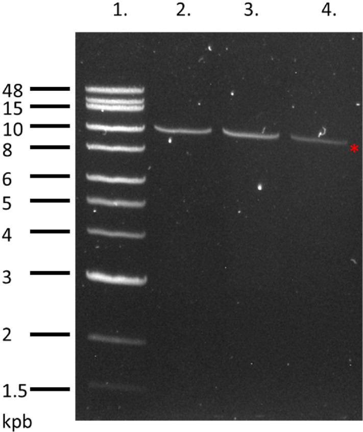
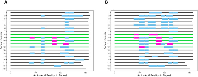
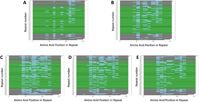
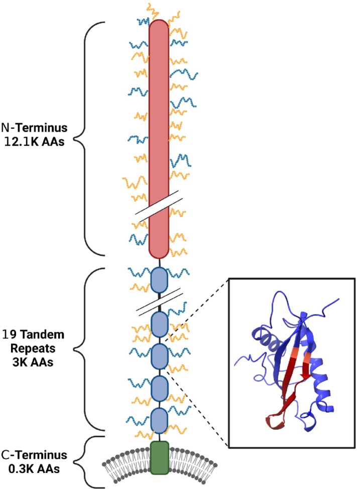
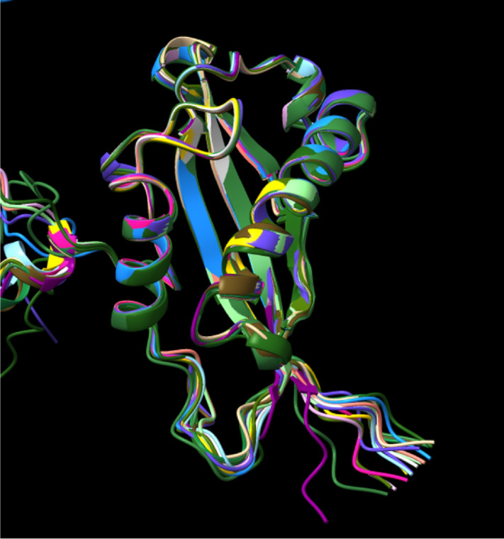
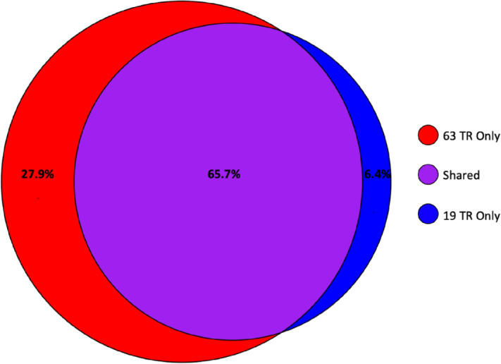
The biomarker CA125, a peptide epitope located in several tandem repeats of the mucin MUC16, is the gold standard for monitoring regression and recurrence of high-grade serous ovarian cancer in response to therapy. However, the CA125 epitope along with several structural features of the MUC16 molecule are ill defined. One central aspect still unresolved is the number of tandem repeats in MUC16 and how many of these repeats contain the CA125 epitope. Studies from the early 2000s assembled short DNA reads to estimate that MUC16 contained 63 repeats. Here, we conduct Nanopore long-read sequencing of MUC16 transcripts from three primary ovarian tumors and established cell lines (OVCAR3, OVCAR5, and Kuramochi) for a more exhaustive and accurate estimation and sequencing of the MUC16 tandem repeats. The consensus sequence derived from these six sources was confirmed by proteomics validation and agrees with recent additions to the NCBI database. We propose a model of MUC16 containing 19—not 63—tandem repeats. In addition, we predict the structure of the tandem repeat domain using the deep learning algorithm, AlphaFold. The predicted structure displays an SEA domain and unstructured linker region rich in proline, serine, and threonine residues in all 19 tandem repeats. These studies now pave the way for a detailed characterization of the CA125 epitope. Sequencing and modeling of the MUC16 tandem repeats along with their glycoproteomic characterization, currently underway in our laboratories, will help identify novel epitopes in the MUC16 molecule that improve on the sensitivity and clinical utility of the current CA125 assay. Significance: Despite its crucial role in clinical management of ovarian cancer, the exact molecular sequence and structure of the biomarker, CA125, are not defined. Here, we combine long-read sequencing, mass spectrometry, and in silico modeling to provide the foundational dataset for a more complete characterization of the CA125 epitope.
Overview
The claims and statistics of this paper haven’t been checked yet.
Run the check — free, no account needed. It reads the open-access text and checks the sources it cites.
Integrity signals
Registry
Cites
Statistics
Figures

Verification
Claims the paper makes
The claims and statistics of this paper haven’t been checked yet.
Run the check — free, no account needed. It reads the open-access text and checks the sources it cites.
Self-check
Percentages, sums and reported differences re-computed from the paper’s own numbers.
The self-check needs the paper’s full text.
Citations
Cites
R: A Language and Environment for Statistical Computing
R Core Team (2025) · doi:10.32614/r.manuals · Cited by 352,181
Highly accurate protein structure prediction with AlphaFold
Jumper, J., Evans, R., Pritzel, A., Green, T., Figurnov, M., Ronneberger, O., Tunyasuvunakool, K., Bates, R., Žídek, A., Potapenko, A., et al. (2021) · Nature · doi:10.1038/s41586-021-03819-2 · Cited by 43,764
ggplot2: Elegant Graphics for Data Analysis
Ginestet, C. E. (2011) · Journal of the Royal Statistical Society Series A (Statistics in Society) · doi:10.1111/j.1467-985x.2010.00676_9.x · Cited by 14,531
UniProt: the Universal Protein Knowledgebase in 2023
The UniProt Consortium, Bateman, A., Martin, M.-J., Orchard, S., Magrane, M., Ahmad, S., Alpi, E., Bowler-Barnett, E. H., Britto, R., Bye-A-Jee, H., et al. (2023) · Nucleic Acids Research · doi:10.1093/nar/gkac1052 · Cited by 7,210
dplyr: A Grammar of Data Manipulation
Wickham, H., François, R., Henry, L., Müller, K., & Vaughan, D. (2014) · doi:10.32614/cran.package.dplyr · Cited by 3,612
The complete sequence of a human genome
Nurk, S., Koren, S., Rhie, A., Rautiainen, M., Bzikadze, A. V., Mikheenko, A., Vollger, M. R., Altemose, N., Uralsky, L., Gershman, A., et al. (2022) · Science · doi:10.1126/science.abj6987 · Cited by 3,136
I-TASSER server: new development for protein structure and function predictions
Yang, J., & Zhang, Y. (2015) · Nucleic Acids Research · doi:10.1093/nar/gkv342 · Cited by 2,552
A Radioimmunoassay Using a Monoclonal Antibody to Monitor the Course of Epithelial Ovarian Cancer
Bast, R. C., Klug, T. L., John, E. S., Jenison, E. L., Niloff, J. M., Lazarus, H., Berkowitz, R. S., Leavitt, T. J., Griffiths, C. T., Parker, L., et al. (1983) · New England Journal of Medicine · doi:10.1056/nejm198310133091503 · Cited by 2,135
Reactivity of a monoclonal antibody with human ovarian carcinoma.
Bast, R. C., Feeney, M., Lazarus, H., Nadler, L. M., Colvin, R. B., & KNAPP, R. C. (1981) · Journal of Clinical Investigation · doi:10.1172/jci110380 · Cited by 1,661
Zhang, J., Xin, L., Shan, B., Chen, W., Xie, M., Yuen, D., Zhang, W., Zhang, Z., Lajoie, G., & Ma, B. (2011) · Molecular & Cellular Proteomics · doi:10.1074/mcp.m111.010587 · Cited by 1,120
Charif, D., & Lobry, J. R. (2007) · Biological and medical physics series · doi:10.1007/978-3-540-35306-5_10 · Cited by 959
CA125 and Ovarian Cancer: A Comprehensive Review
Charkhchi, P., Cybulski, C., Gronwald, J., Wong, F. O., Narod, S. A., & Akbari, M. R. (2020) · Cancers · doi:10.3390/cancers12123730 · Cited by 669
Molecular Cloning of the CA125 Ovarian Cancer Antigen
Yin, B. W. T., & Lloyd, K. O. (2001) · Journal of Biological Chemistry · doi:10.1074/jbc.m103554200 · Cited by 625
Zhang, C., Freddolino, L., & Zhang, Y. (2017) · Nucleic Acids Research · doi:10.1093/nar/gkx366 · Cited by 606
Zheng, W. H., Zhang, C., Li, Y., Pearce, R., Bell, E. W., & Zhang, Y. (2021) · Cell Reports Methods · doi:10.1016/j.crmeth.2021.100014 · Cited by 594
Binding of Ovarian Cancer Antigen CA125/MUC16 to Mesothelin Mediates Cell Adhesion
Rump, A., Morikawa, Y., Tanaka, M., Minami, S., Umesaki, N., Takeuchi, M., & Miyajima, A. (2004) · Journal of Biological Chemistry · doi:10.1074/jbc.m312372200 · Cited by 567
MUC16 (CA125): tumor biomarker to cancer therapy, a work in progress
Felder, M. A. R., Kapur, A., Bosquet, J. G., Horibata, S., Heintz, J. A., Albrecht, R. M., Fass, L., kaur, J., Hu, K. S., Shojaei, H., et al. (2014) · Molecular Cancer · doi:10.1186/1476-4598-13-129 · Cited by 543
Self-citationKarst, S. M., Ziels, R. M., Kirkegaard, R. H., Sørensen, E. A., McDonald, D. T., Zhu, Q., Knight, R. D., & Albertsen, M. (2021) · Nature Methods · doi:10.1038/s41592-020-01041-y · Cited by 433
Gubbels, J. A. A., Belisle, J. A., Onda, M., Rancourt, C., Migneault, M., Ho, M., Bera, T. K., Connor, J. P., Sathyanarayana, B. K., Lee, B., et al. (2006) · Molecular Cancer · doi:10.1186/1476-4598-5-50 · Cited by 423
Self-citationThe CA 125 Gene: An Extracellular Superstructure Dominated by Repeat Sequences
Brien, T. J. O. R., Beard, J. B., Underwood, L. J., Dennis, R. A., Santin, A. D., & York, L. (2001) · Tumor Biology · doi:10.1159/000050638 · Cited by 300
Ovarian cancer antigen CA125 is encoded by the MUC16 mucin gene
Yin, B. W. T., Dnistrian, A. M., & Lloyd, K. O. (2002) · International Journal of Cancer · doi:10.1002/ijc.10250 · Cited by 239
Johnson, S. W., Laub, P. B., Beesley, J. S., Ozols, R. F., & Hamilton, T. C. (1997) · PubMed · Cited by 178
Brien, T. J. O. R., Beard, J. B., Underwood, L. J., & Shigemasa, K. (2002) · Tumor Biology · doi:10.1159/000064032 · Cited by 141
Huang, C., Liu, Z., Li, X., Xia, Y., Huang, J., Luo, H., Zong, Z., & Zhu, Z. (2019) · Frontiers in Oncology · doi:10.3389/fonc.2019.01159 · Cited by 111
Reducing Ovarian Cancer Mortality Through Early Detection: Approaches Using Circulating Biomarkers
Trinidad, C. V., Tetlow, A. L., Bantis, L. E., & Godwin, A. K. (2020) · Cancer Prevention Research · doi:10.1158/1940-6207.capr-19-0184 · Cited by 96
Maeda, T., Inoue, M., Koshiba, S., Yabuki, T., Aoki, M., Nunokawa, E., Seki, E., Matsuda, T., Motoda, Y., Kobayashi, A., et al. (2004) · Journal of Biological Chemistry · doi:10.1074/jbc.m309417200 · Cited by 86
Tripathi, P., Kumar, A., Sachan, M., Gupta, S., & Nara, S. (2020) · Biosensors and Bioelectronics · doi:10.1016/j.bios.2020.112368 · Cited by 86
CA 125 and the detection of recurrent ovarian cancer
Bast, R. C. (2010) · Cancer · doi:10.1002/cncr.25203 · Cited by 73
Huang, C., Jiang, J., Chang, S., Lin, J., & Yang, S.-H. (2016) · Medicine · doi:10.1097/md.0000000000005177 · Cited by 63
Characterization of Binding Epitopes of CA125 Monoclonal Antibodies
Marcos-Silva, L., Narimatsu, Y., Halim, A., Campos, D., Yang, Z., Tarp, M. A., Pereira, P. J. B., Mandel, U., Bennett, E., Vakhrushev, S. Y., et al. (2014) · Journal of Proteome Research · doi:10.1021/pr500215g · Cited by 61
Coelho, R., Marcos-Silva, L., Ricardo, S. A. V., Ponte, F., Costa, A., Lopes, J. M., & David, L. (2017) · Expert Review of Anticancer Therapy · doi:10.1080/14737140.2018.1418326 · Cited by 53
The Potential Role of MUC16 (CA125) Biomarker in Lung Cancer: A Magic Biomarker but with Adversity
Saad, . M., Tourky, G. F., Al-Kuraishy, H. M., Al-Gareeb, A. I., Khattab, A. M., Elmasry, S. A., Alsayegh, A. A., Hakami, Z. H., Alsulimani, A., Sabatier, J., et al. (2022) · Diagnostics · doi:10.3390/diagnostics12122985 · Cited by 52
Scoville, D. J., Uhm, T. K. B., Shallcross, J. A., & Whelan, R. J. (2017) · Journal of Nucleic Acids · doi:10.1155/2017/9879135 · Cited by 40
Self-citationDeciphering the Molecular Nature of Ovarian Cancer Biomarker CA125
Weiland, F., Martin, K., Oehler, M. K., & Hoffmann, P. (2012) · International Journal of Molecular Sciences · doi:10.3390/ijms130810568 · Cited by 36
Methods for Identification of CA125 from Ovarian Cancer Ascites by High Resolution Mass Spectrometry
Weiland, F., Fritz, K., Oehler, M. K., & Hoffmann, P. (2012) · International Journal of Molecular Sciences · doi:10.3390/ijms13089942 · Cited by 34
Blayney, J. K., Davison, T. S., McCabe, N., Walker, S. M., Keating, K., Delaney, T., Greenan, C., Williams, A. R. W., McCluggage, W. G., Capes‐Davis, A., et al. (2016) · Nucleic Acids Research · doi:10.1093/nar/gkw578 · Cited by 33
Bressan, A., Bozzo, F., Maggi, C. A., & Binaschi, M. (2013) · Disease Markers · doi:10.1155/2013/917898 · Cited by 30
Conflicting Views on the Molecular Structure of the Cancer Antigen CA125/MUC16
Bouanène, H., & Miled, A. (2010) · Disease Markers · doi:10.1155/2010/918457 · Cited by 28
White, B., Patterson, M., Karnwal, S., & Brooks, C. L. (2022) · Proteins Structure Function and Bioinformatics · doi:10.1002/prot.26303 · Cited by 28
Aithal, A., Junker, W. M., Kshirsagar, P., Das, S., Kaur, S., Orzechowski, C., Gautam, S. K., Jahan, R., Sheinin, Y. M., Lakshmanan, I., et al. (2018) · PLoS ONE · doi:10.1371/journal.pone.0193907 · Cited by 27
Huang, J., Liu, H., Hu, T., Zhang, Z., He, X., Mo, T., Wen, X., Lan, P., Lian, L., & Wu, X. (2022) · Gastroenterology report · doi:10.1093/gastro/goac020 · Cited by 25
Research progress of CA125 in endometriosis: Teaching an old dog new tricks
Chen, Y., Pan, M., Zuo, Y., Yang, B., & Wang, S. (2022) · Gynecology and Obstetrics Clinical Medicine · doi:10.1016/j.gocm.2022.10.006 · Cited by 22
Schuster-Little, N., Fritz-Klaus, R., Etzel, M. R., Patankar, N., Javeri, S., Patankar, M. S., & Whelan, R. J. (2020) · The Analyst · doi:10.1039/d0an01701a · Cited by 17
Self-citationWarren, D. J., Nustad, K., Beard, J. B., & Brien, T. J. O. R. (2009) · Tumor Biology · doi:10.1159/000209988 · Cited by 14
Weaver, S. D., Schuster-Little, N., & Whelan, R. J. (2022) · Analytical Methods · doi:10.1039/d1ay02145a · Cited by 12
Self-citationYue, E., Yang, G., Yao, Y., Wang, G., Mohanty, A. R., Fan, F., Zhao, L., Zhang, Y., Mirzapoiazova, T., Walser, T. C., et al. (2021) · Cancers · doi:10.3390/cancers13174265 · Cited by 11
Editorial: Long-read sequencing—Pitfalls, benefits and success stories
Wohlers, I., Garg, S., & Hehir‐Kwa, J. Y. (2023) · Frontiers in Genetics · doi:10.3389/fgene.2022.1114542 · Cited by 11
Berman, Z. T., Moore, L. J., Knudson, K. E., & Whelan, R. J. (2010) · Tumor Biology · doi:10.1007/s13277-010-0062-4 · Cited by 9
Self-citationGeneration and characterization of a monoclonal antibody to the cytoplasmic tail of MUC16
Gipson, I. K., Mandel, U., Menon, B. B., Michaud, S., Tisdale, A. S., Campos, D., & Clausen, H. (2017) · Glycobiology · doi:10.1093/glycob/cwx054 · Cited by 7
Individual recombinant repeats of MUC16 display variable binding to CA125 antibodies
Wang, C.-W., Hanson, E. K., Minkoff, L., & Whelan, R. J. (2023) · Cancer Biomarkers · doi:10.3233/cbm-220191 · Cited by 7
Self-citationLin, H., Hu, R., Chen, H.-C., Lin, C., Lee, C., & Chou, C. (2023) · Cancers · doi:10.3390/cancers15030813 · Cited by 6
Metadata
- Authors
- Wang, C.-W., Weaver, S. D., Boonpattrawong, N., Schuster-Little, N., Patankar, M., & Whelan, R. J.
- Affiliations
- University of Kansas (US)
- University of Notre Dame (US)
- University of Wisconsin–Madison (US)
- Corresponding author
- Boonpattrawong, Nicha
- ORCID
- Venue
- Cancer Research Communications
- Published
- 2024-01-31
- Volume
- 4
- Issue
- 1
- Pages
- 253-263
- ISSN
- 2767-9764 (ISSN-L)
- PMC
- PMC10829539
- OpenAlex
- W4390703493
- Cited by
- 12
- References
- 51
- Citations by year
- 2026: 3 · 2025: 6 · 2024: 3
- FWCI
- 4.73
- Type
- journal article
- Language
- en
- Open access
- gold
- doi.org
- Licence
- Locations
- Cancer Research Communications (publishedVersion, cc-by)
- PubMed Central (publishedVersion, cc-by)
- PubMed (publishedVersion)
- Funders
- HHS | NIH | National Cancer Institute (R21CA267532)
- Tell Every Amazing Lady About Ovarian Cancer Louisa M. McGregor Ovarian Cancer Foundation (n/a)
- HHS | NIH | National Institute of General Medical Sciences (T32GM075762)
- Veterans Administration Medical Center (5I01BX005627)
- Diane Lindstrom Funds (n/a)
- Topics
- Health Sciences › Medicine › Oncology › Ovarian cancer diagnosis and treatment
- Life Sciences › Biochemistry, Genetics and Molecular Biology › Molecular Biology › Glycosylation and Glycoproteins Research
- Physical Sciences › Chemistry › Organic Chemistry › Carbohydrate Chemistry and Synthesis
- Keywords
- CA125, ovarian cancer biomarker, tandem repeats, long-read sequencing, nanopore sequencing, AlphaFold structure prediction, SEA domain, mucin structure, high-grade serous ovarian cancer, glycoproteomics, OVCAR3
- MeSH terms
- Epitopes, Female, Humans, Membrane Proteins, Models, Molecular, Ovarian Neoplasms, Biomarkers, Tumor, Apoptosis, CA-125 Antigen, Cell Line, Tumor
About these checks
Registry, retraction, Cites and statistics results are computed, not judged, with one exception: a printed reference that no index lists is read once by a model before it counts as a citation error. Claims are extracted by a model and kept only when their quote appears verbatim in the text; each cited source is then checked, with a model’s help, for whether it says what the claim relies on. Those verdicts can be wrong — read the claim’s quote and the cited source before relying on one.
Statistics and arithmetic are recomputed, not judged.
Confidence bands are shown only for fields calibrated on our annotated test set. None is calibrated yet, so no band is shown.
What counts as a claim ›How we measure confidence ›How we check this ›
Are you an author, or do you see a problem with this page? Request a review
Data sources
Bibliographic data: Semantic Scholar, OpenAlex, Crossref.
Contains information from the Semantic Scholar Academic Graph Datasets, made available under the ODC Attribution License (ODC-BY 1.0).
Semantic Scholar’s data is described in: Kinney, R., et al. (2023). The Semantic Scholar Open Data Platform. arXiv:2301.10140.
