

The DOI matches its Crossref record; 25 of the 25 references it cites were found in a registry or an index.
High efficient isolation and systematic identification of human adipose-derived mesenchymal stem cells
Open accessAbstract
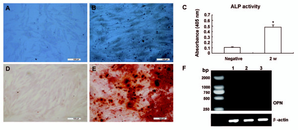
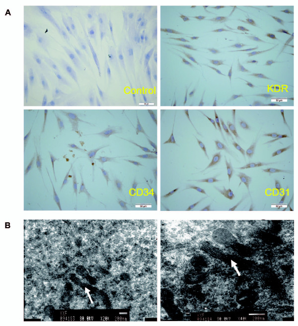
BACKGROUND: Developing efficient methods to isolate and identify human adipose-derived mesenchymal stem cells (hADSCs) remains to be one of the major challenges in tissue engineering. METHODS: We demonstrate here a method by isolating hADSCs from abdominal subcutaneous adipose tissue harvested during caesarian section. The hADSCs were isolated from human adipose tissue by collagenase digestion and adherence to flasks. RESULTS: The yield reached around 1 × 10(6) hADSCs per gram adipose tissue. The following comprehensive identification and characterization illustrated pronounced features of mesenchymal stem cells (MSCs). The fibroblast-like hADSCs exhibited typical ultrastructure details for vigorous cell activities. Karyotype mapping showed normal human chromosome. With unique immunophenotypes they were positive for CD29, CD44, CD73, CD105 and CD166, but negative for CD31, CD34, CD45 and HLA-DR. The growth curve and cell cycle analysis revealed high capability for self-renewal and proliferation. Moreover, these cells could be functionally induced into adipocytes, osteoblasts, and endothelial cells in the presence of appropriate conditioned media. CONCLUSION: The data presented here suggest that we have developed high efficient isolation and cultivation methods with a systematic strategy for identification and characterization of hADSCs. These techniques will be able to provide safe and stable seeding cells for research and clinical application.
Overview
The claims and statistics of this paper haven’t been checked yet.
Run the check — free, no account needed. It reads the open-access text and checks the sources it cites.
Integrity signals
Registry
Cites
Statistics
Figures
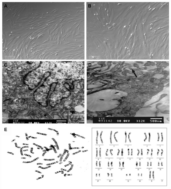
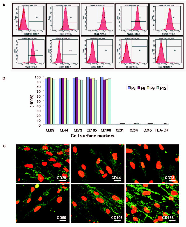
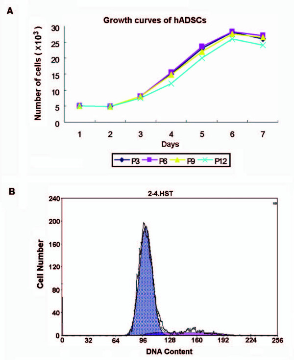
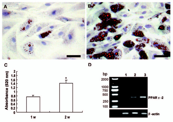
Verification
Claims the paper makes
The claims and statistics of this paper haven’t been checked yet.
Run the check — free, no account needed. It reads the open-access text and checks the sources it cites.
Claims other papers make about this one
Each is checked against this paper’s own text: does it say what the citing paper says it does?
The cited source says what the claim attributes to it.
In the citing paper
“These findings confirmed differentiation capacities and phenotypes of hADSCs, in agreement with the literature [41].”
Self-check
Percentages, sums and reported differences re-computed from the paper’s own numbers.
The self-check needs the paper’s full text.
Citations
Cites
2 retracted
RETRACTED ARTICLE: Pluripotency of mesenchymal stem cells derived from adult marrow
Jiang, Y., Jahagirdar, B., Reinhardt, R. L., Schwartz, R. E., Keene, C. D., Ortiz-Gonzalez, X. R., Reyes, M., Lenvik, T. R., Lund, T. C., Blackstad, M., et al. (2002) · Nature · doi:10.1038/nature00870 · Cited by 5,523
RetractedInduced Endothelial Cells Enhance Osteogenesis and Vascularization of Mesenchymal Stem Cells
Tao, J., Sun, Y., Wang, Q., & Liu, C. (2009) · Cells Tissues Organs · doi:10.1159/000218139 · Cited by 29
RetractedMultilineage potential of adult human mesenchymal stem cells
Pittenger, M. F., Mackay, A. M., Beck, S. C., Jaiswal, R. K., Douglas, R., Mosca, J. D., Moorman, M. A., Simonetti, D. W., Craig, S., & Marshak, D. R. (1999) · Science · doi:10.1126/science.284.5411.143 · Cited by 21,052
Multilineage Cells from Human Adipose Tissue: Implications for Cell-Based Therapies
Zuk, P. A., Zhu, M., Mizuno, H., Huang, J., Futrell, J. W., Katz, A. J., Benhaim, P., Lorenz, H. P., & Hedrick, M. H. (2001) · Tissue Engineering · doi:10.1089/107632701300062859 · Cited by 7,956
Human Adipose Tissue Is a Source of Multipotent Stem Cells
Zuk, P. A., Zhu, M., Ashjian, P. H., Ugarte, D. A. D., Huang, J. I., Mizuno, H., Alfonso, Z. C., Fraser, J. K., Benhaim, P., & Hedrick, M. H. (2002) · Molecular Biology of the Cell · doi:10.1091/mbc.e02-02-0105 · Cited by 6,762
Kern, S., Eichler, H., Stoeve, J., Klüter, H., & Bieback, K. (2006) · STEM CELLS · doi:10.1634/stemcells.2005-0342 · Cited by 3,370
Mesenchymal stem cells: clinical applications and biological characterization
Barry, F., & Murphy, M. J. (2004) · The International Journal of Biochemistry & Cell Biology · doi:10.1016/j.biocel.2003.11.001 · Cited by 1,669
Multipotential differentiation of adipose tissue-derived stem cells
Strem, B. M., Hicok, K. C., Zhu, M., Wulur, I. H., Alfonso, Z., Schreiber, R. E., Fraser, J. K., & Hedrick, M. H. (2005) · The Keio Journal of Medicine · doi:10.2302/kjm.54.132 · Cited by 946
Yoshimura, H., Muneta, T., Nimura, A., Yokoyama, A., Koga, H., & Sekiya, I. (2006) · Cell and Tissue Research · doi:10.1007/s00441-006-0308-z · Cited by 734
Yield of human adipose-derived adult stem cells from liposuction aspirates
Aust, L. E., Devlin, B. H., Foster, S. J., Halvorsen, Y.-D., Hicok, K. C., du Laney, T., Sen, A., Willingmyre, G. D., & Gimble, J. M. (2004) · Cytotherapy · doi:10.1080/14653240310004539 · Cited by 707
Cited by
Sorted by citations
Kang, Y., Xu, C., Meng, L., Dong, X., Qi, M., & Jiang, D. (2022) · Bioactive materials · doi:10.1016/j.bioactmat.2022.02.012 · Cited by 253
Lv, Q., Deng, J., Chen, Y., Wang, Y., Liu, B., & Liu, J. (2020) · Molecular Pharmaceutics · doi:10.1021/acs.molpharmaceut.0c00177 · Cited by 229
Priglinger, E., Steffenhagen, C., Wurzer, C., Gabriel, C., Redl, H. R., & Wolbank, S. (2015) · Cell Regeneration · doi:10.1186/s13619-015-0020-0 · Cited by 200
Bone matrix production in hydroxyapatite-modified hydrogels suitable for bone bioprinting
Wenz, A., Borchers, K., Tovar, G. E. M., & Kluger, P. J. (2017) · Biofabrication · doi:10.1088/1758-5090/aa91ec · Cited by 161
Impact of Age on Human Adipose Stem Cells for Bone Tissue Engineering
Dufrane, D. (2017) · Cell Transplantation · doi:10.1177/0963689717721203 · Cited by 141
Metadata
- Authors
- Yang, X.-F., He, X., He, J., Zhang, L.-H., Su, X.-J., Dong, Z.-Y., Xu, Y.-J., Li, Y., & Li, Y.-L.
- Affiliations
- Jilin University (CN)
- Corresponding author
- He, Xu
- Li, Yan
- Venue
- Journal of Biomedical Science
- Published
- 2011-08-19
- Volume
- 18
- Issue
- 1
- Pages
- 59-59
- ISSN
- 1021-7770 (ISSN-L)
- 1423-0127
- PMC
- PMC3175156
- OpenAlex
- W2087905124
- Cited by
- 112
- References
- 25
- Citations by year
- 2026: 1 · 2025: 6 · 2024: 1 · 2023: 2 · 2022: 7 · 2021: 7 · 2020: 8 · 2019: 6 · 2018: 17 · 2017: 9 · 2016: 9 · 2015: 12 · 2014: 8 · 2013: 15 · 2012: 4
- FWCI
- 4.37
- Type
- journal article
- Language
- en
- Open access
- diamond
- doi.org
- Licence
- cc-by
- Locations
- Journal of Biomedical Science (publishedVersion, cc-by)
- Europe PMC (PubMed Central) (publishedVersion, cc-by)
- PubMed Central (publishedVersion, cc-by)
- PubMed (publishedVersion)
- DOAJ (DOAJ: Directory of Open Access Journals) (submittedVersion)
- Funders
- National Natural Science Foundation of China (863 Program)
- National High-tech Research and Development Program
- Topics
- Life Sciences › Biochemistry, Genetics and Molecular Biology › Cell Biology › Mesenchymal stem cell research
- Life Sciences › Biochemistry, Genetics and Molecular Biology › Molecular Biology › Pluripotent Stem Cells Research
- Health Sciences › Medicine › Physiology (medical) › Adipose Tissue and Metabolism
- Keywords
- human adipose-derived mesenchymal stem cells, adipose tissue harvesting, collagenase digestion, cell isolation, immunophenotyping, cell cycle analysis, self-renewal, multilineage differentiation, adipogenesis, bone formation, endothelial differentiation, karyotype
- MeSH terms
- Adult, Cell Cycle, Cell Differentiation, Cell Separation, Female, Humans, Immunohistochemistry, Karyotyping, Immunophenotyping, Collagenases, Fluorescent Antibody Technique, Indirect, Tissue Engineering, Microscopy, Electron, Transmission, Cell Proliferation, Subcutaneous Fat, Mesenchymal Stem Cells, Real-Time Polymerase Chain Reaction
About these checks
Registry, retraction, Cites and statistics results are computed, not judged, with one exception: a printed reference that no index lists is read once by a model before it counts as a citation error. Claims are extracted by a model and kept only when their quote appears verbatim in the text; each cited source is then checked, with a model’s help, for whether it says what the claim relies on. Those verdicts can be wrong — read the claim’s quote and the cited source before relying on one.
Statistics and arithmetic are recomputed, not judged.
Confidence bands are shown only for fields calibrated on our annotated test set. None is calibrated yet, so no band is shown.
What counts as a claim ›How we measure confidence ›How we check this ›
Are you an author, or do you see a problem with this page? Request a review
