

The DOI matches its Crossref record.
Global regulation of gene expression in response to cysteine availability in Clostridium perfringens
Abstract
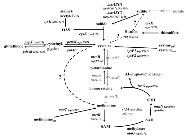
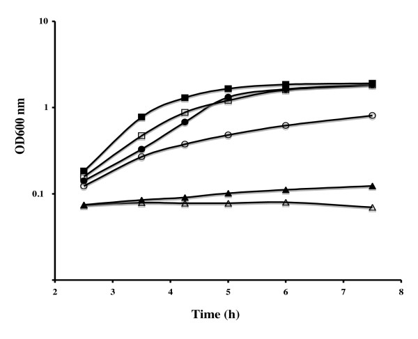
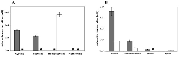
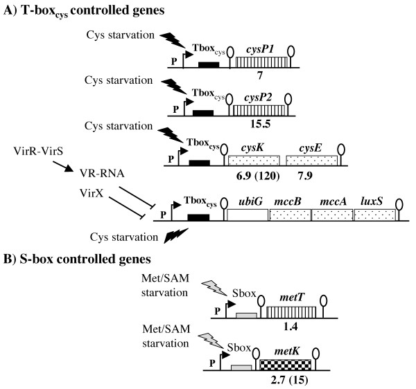
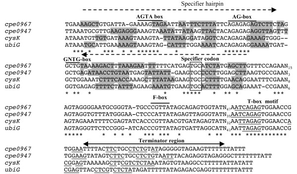
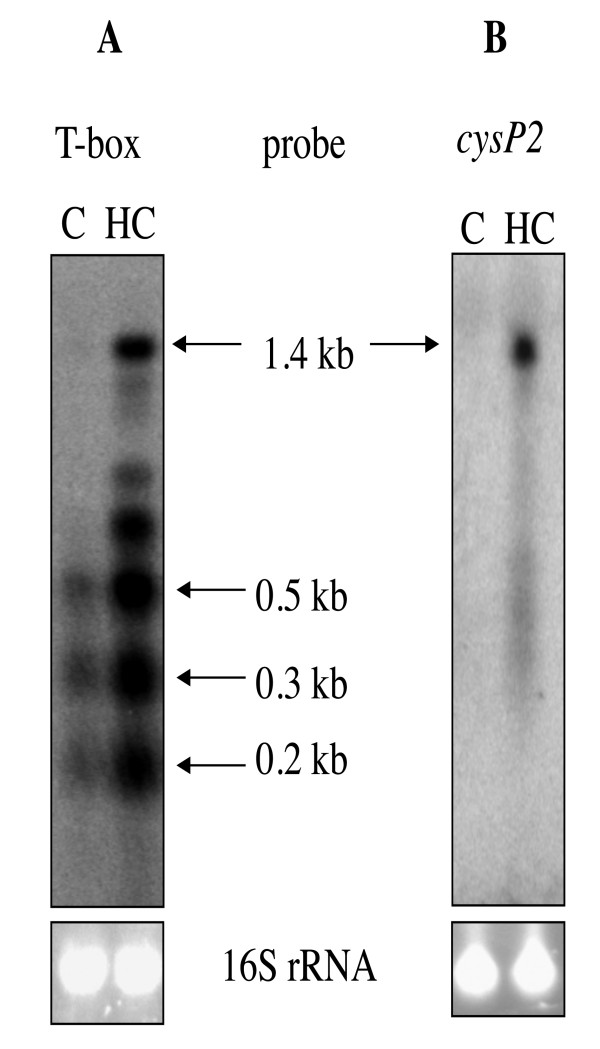
Background: Cysteine has a crucial role in cellular physiology and its synthesis is tightly controlled due to its reactivity. However, little is known about the sulfur metabolism and its regulation in clostridia compared with other firmicutes. In Clostridium perfringens, the two-component system, VirR/VirS, controls the expression of the ubiG operon involved in methionine to cysteine conversion in addition to the expression of several toxin genes. The existence of links between the C. perfringens virulence regulon and sulfur metabolism prompted us to analyze this metabolism in more detail. Results: We first performed a tentative reconstruction of sulfur metabolism in C. perfringens and correlated these data with the growth of strain 13 in the presence of various sulfur sources. Surprisingly, C. perfringens can convert cysteine to methionine by an atypical still uncharacterized pathway. We further compared the expression profiles of strain 13 after growth in the presence of cystine or homocysteine that corresponds to conditions of cysteine depletion. Among the 177 genes differentially expressed, we found genes involved in sulfur metabolism and controlled by premature termination of transcription via a cysteine specific T-box system (cysK-cysE, cysP1 and cysP2) or an S-box riboswitch (metK and metT). We also showed that the ubiG operon was submitted to a triple regulation by cysteine availability via a T-box system, by the VirR/VirS system via the VR-RNA and by the VirX regulatory RNA. In addition, we found that expression of pfoA (theta-toxin), nagL (one of the five genes encoding hyaluronidases) and genes involved in the maintenance of cell redox status was differentially expressed in response to cysteine availability. Finally, we showed that the expression of genes involved in [Fe-S] clusters biogenesis and of the ldh gene encoding the lactate dehydrogenase was induced during cysteine limitation. Conclusion: Several key functions for the cellular physiology of this anaerobic bacterium were controlled in response to cysteine availability. While most of the genes involved in sulfur metabolism are regulated by premature termination of transcription, other still uncharacterized mechanisms of regulation participated in the induction of gene expression during cysteine starvation.
Overview
The claims and statistics of this paper haven’t been checked yet.
Run the check — free, no account needed. It reads the open-access text and checks the sources it cites.
Integrity signals
Registry
Cites
Statistics
Figures
Verification
Claims the paper makes
The claims and statistics of this paper haven’t been checked yet.
Run the check — free, no account needed. It reads the open-access text and checks the sources it cites.
Claims other papers make about this one
Each is checked against this paper’s own text: does it say what the citing paper says it does?
In the citing paper
“In PG cultures, the majority of abundant Firmicutes were related to clostridial species (Figure 6), such as the non-sulfate-reducing bacterium, Clostridium perfringens, which may use thiosulfate and sulfite as electron acceptors (Fuchs and Bonde, 1957; André et al., 2010), or the potential sulfate, thiosulfate- or sulfite-reducer, Brassicibacter thermophilus (Wang et al., 2015).”
From Microbial Diversity in Sulfate-Reducing Marine Sediment Enrichment Cultures Associated with Anaerobic Biotransformation of Coastal Stockpiled Phosphogypsum (Sfax, Tunisia) (2017) · cites this paper as André et al., 2010
Self-check
Percentages, sums and reported differences re-computed from the paper’s own numbers.
The self-check needs the paper’s full text.
Citations
Cites
No references could be resolved yet.
Research works printed but not linked: 63
- Ayala-Castro C, Saini A, Outten FW: Fe-S cluster assembly pathways in bacteria. Microbiol Mol Biol Rev. 2008, 72 (1): 110-125. 10.1128/MMBR.00034-07. · DOI
- Masip L, Veeravalli K, Georgiou G: The many faces of glutathione in bacteria. Antioxid Redox Signal. 2006, 8 (5-6): 753-762. 10.1089/ars.2006.8.753. · DOI
- Newton GL, Rawat M, La Clair JJ, Jothivasan VK, Budiarto T, Hamilton CJ, Claiborne A, Helmann JD, Fahey RC: Bacillithiol is an antioxidant thiol produced in Bacilli. Nat Chem Biol. 2009, 5 (9): 625-627. 10.1038/nchembio.189. · DOI
- Zeller T, Klug G: Thioredoxins in bacteria: functions in oxidative stress response and regulation of thioredoxin genes. Naturwissenschaften. 2006, 93 (6): 259-266. 10.1007/s00114-006-0106-1. · DOI
- Xavier KB, Bassler BL: LuxS quorum sensing: more than just a numbers game. Current Opinion in Microbiology. 2003, 6 (2): 191-197. 10.1016/S1369-5274(03)00028-6. · DOI
- Soutourina O, Martin-Verstraete I: Global regulatory network of sulfur metabolism in Bacillus subtilis. Global regulatory networks in Bacillus subtilis. Edited by: Fujita Y. 2007, Transworld research network, 111-141.
- van der Ploeg JR, Barone M, Leisinger T: Functional analysis of the Bacillus subtilis cysK and cysJI genes. FEMS Microbiol Lett. 2001, 201 (1): 29-35. 10.1016/S0378-1097(01)00225-7. · DOI
- Hullo MF, Auger S, Soutourina O, Barzu O, Yvon M, Danchin A, Martin-Verstraete I: Conversion of methionine to cysteine in Bacillus subtilis and its regulation. J Bacteriol. 2007, 189 (1): 187-197. 10.1128/JB.01273-06. · DOI
- Rodionov DA, Vitreschak AG, Mironov AA, Gelfand MS: Comparative genomics of the methionine metabolism in Gram-positive bacteria: a variety of regulatory systems. Nucleic Acids Res. 2004, 32 (11): 3340-3353. 10.1093/nar/gkh659. · DOI
- Grundy FJ, Henkin TM: The T box and S box transcription termination control systems. Front Biosci. 2003, 8 (1): 20-31. 10.2741/908. · DOI
- Gutierrez-Preciado A, Henkin TM, Grundy FJ, Yanofsky C, Merino E: Biochemical features and functional implications of the RNA-based T-box regulatory mechanism. Microbiol Mol Biol Rev. 2009, 73 (1): 36-61. 10.1128/MMBR.00026-08. · DOI
- Burguière P, Fert J, Guillouard I, Auger S, Danchin A, Martin-Verstraete I: Regulation of the Bacillus subtilis ytmI operon, involved in sulfur metabolism. J Bacteriol. 2005, 187 (17): 6019-6030. 10.1128/JB.187.17.6019-6030.2005. · DOI
- Guillouard I, Auger S, Hullo MF, Chetouani F, Danchin A, Martin-Verstraete I: Identification of Bacillus subtilis CysL, a regulator of the cysJI operon, which encodes sulfite reductase. J Bacteriol. 2002, 184 (17): 4681-4689. 10.1128/JB.184.17.4681-4689.2002. · DOI
- Sperandio B, Gautier C, Pons N, Ehrlich DS, Renault P, Guedon E: Three paralogous LysR-type transcriptional regulators control sulfur amino acid supply in Streptococcus mutans. J Bacteriol. 2010, 192 (13): 3464-3473. 10.1128/JB.00119-10. · DOI
- Sperandio B, Gautier C, McGovern S, Ehrlich DS, Renault P, Martin-Verstraete I, Guedon E: Control of methionine synthesis and uptake by MetR and homocysteine in Streptococcus mutans. J Bacteriol. 2007, 189 (19): 7032-7044. 10.1128/JB.00703-07. · DOI
- Even S, Burguière P, Auger S, Soutourina O, Danchin A, Martin-Verstraete I: Global control of cysteine metabolism by CymR in Bacillus subtilis. J Bacteriol. 2006, 188 (6): 2184-2197. 10.1128/JB.188.6.2184-2197.2006. · DOI
- Soutourina O, Poupel O, Coppée JY, Danchin A, Msadek T, Martin-Verstraete I: CymR, the master regulator of cysteine metabolism in Staphylococcus aureus, controls host sulfur source utilization and plays a role in biofilm formation. Mol Microbiol. 2009, 73 (2): 194-211. 10.1111/j.1365-2958.2009.06760.x. · DOI
- Tanous C, Soutourina O, Raynal B, Hullo MF, Mervelet P, Gilles AM, Noirot P, Danchin A, England P, Martin-Verstraete I: The CymR Regulator in Complex with the Enzyme CysK Controls Cysteine Metabolism in Bacillus subtilis. J Biol Chem. 2008, 283 (51): 35551-35560. 10.1074/jbc.M805951200. · DOI
- Andre G, Even S, Putzer H, Burguiere P, Croux C, Danchin A, Martin-Verstraete I, Soutourina O: S-box and T-box riboswitches and antisense RNA control a sulfur metabolic operon of Clostridium acetobutylicum. Nucleic Acids Res. 2008, 36 (18): 5955-5969. 10.1093/nar/gkn601. · DOI
- Rood JI: Virulence genes of Clostridium perfringens. Annu Rev Microbiol. 1998, 52: 333-360. 10.1146/annurev.micro.52.1.333. · DOI
- Shimizu T, Ohtani K, Hirakawa H, Ohshima K, Yamashita A, Shiba T, Ogasawara N, Hattori M, Kuhara S, Hayashi H: Complete genome sequence of Clostridium perfringens, an anaerobic flesh-eater. Proc Natl Acad Sci USA. 2002, 99 (2): 996-1001. 10.1073/pnas.022493799. · DOI
- BaThein W, Lyristis M, Ohtani K, Nisbet IT, Hayashi H, Rood JI, Shimizu T: The virR/virS locus regulates the transcription of genes encoding extracellular toxin production in Clostridium perfringens. J Bacteriol. 1996, 178 (9): 2514-2520. · DOI
- Shimizu T, Shima K, Yoshino K, Yonezawa K, Hayashi H: Proteome and transcriptome analysis of the virulence genes regulated by the VirR/VirS system in Clostridium perfringens. J Bacteriol. 2002, 184 (10): 2587-2594. 10.1128/JB.184.10.2587-2594.2002. · DOI
- Cheung JK, Rood JI: The VirR response regulator from Clostridium perfringens binds independently to two imperfect direct repeats located upstream of the pfoA promoter. J Bacteriol. 2000, 182 (10): 2992-2992. 10.1128/JB.182.10.2992-2992.2000. · DOI
- Okumura K, Ohtani K, Hayashi H, Shimizu T: Characterization of genes regulated directly by the VirR/VirS system in Clostridium perfringens. J Bacteriol. 2008, 190 (23): 7719-7727. 10.1128/JB.01573-07. · DOI
- Shimizu T, Yaguchi H, Ohtani K, Banu S, Hayashi H: Clostridial VirR/VirS regulon involves a regulatory RNA molecule for expression of toxins. Mol Microbiol. 2002, 43 (1): 257-265. 10.1046/j.1365-2958.2002.02743.x. · DOI
- Ohtani K, Bhowmik SK, Hayashi H, Shimizu T: Identification of a novel locus that regulates expression of toxin genes in Clostridium perfringens. FEMS Microbiol Lett. 2002, 209 (1): 113-118. 10.1111/j.1574-6968.2002.tb11118.x. · DOI
- Banu S, Ohtani K, Yaguchi H, Swe T, Cole ST, Hayashi H, Shimizu T: Identification of novel VirR/VirS-regulated genes in Clostridium perfringens. Mol Microbiol. 2000, 35 (4): 854-864. 10.1046/j.1365-2958.2000.01760.x. · DOI
- Ohtani K, Takamura H, Yaguchi H, Hayashi H, Shimizu T: Genetic analysis of the ycgJmetB-cysK-ygaG operon negatively regulated by the VirR/VirS system in Clostridium perfringens. Microbiol and Immunol. 2000, 44 (6): 525-528. · DOI
- Sebald M, Costilow RN: Minimal growth requirements for Clostridium perfringens and isolation of auxotrophic mutants. Appl Microbiol. 1975, 29 (1): 1-6. · DOI
- Guerlava P, Izac V, Tholozan JL: Comparison of different methods of cell lysis and protein measurements in Clostridium perfringens: application to the cell volume determination. Curr Microbiol. 1998, 36 (3): 131-135. 10.1007/PL00006756. · DOI
- Livak KJ, Schmittgen TD: Analysis of relative gene expression data using real-time quantitative PCR and the 2(-Delta Delta C(T)) Method. Methods. 2001, 25 (4): 402-408. 10.1006/meth.2001.1262. · DOI · Paper page
- Rouillard JM, Zuker M, Gulari E: OligoArray 2.0: design of oligonucleotide probes for DNA microarrays using a thermodynamic approach. Nucleic Acids Res. 2003, 31 (12): 3057-3062. 10.1093/nar/gkg426. · DOI
- Breitling R, Armengaud P, Amtmann A, Herzyk P: Rank products: a simple, yet powerful, new method to detect differentially regulated genes in replicated microarray experiments. FEBS Lett. 2004, 573 (1-3): 83-92. 10.1016/j.febslet.2004.07.055. · DOI
- Smyth GK, Speed T: Normalization of cDNA microarray data. Methods. 2003, 31 (4): 265-273. 10.1016/S1046-2023(03)00155-5. · DOI · Paper page
- Benjamini Y, Hochberg Y: Controlling the false discovery rate: a practical and powerful approach to multiple testing. J Roy Statist Soc Ser. 1995, 289-300. 57 · DOI · Paper page
- Nygard O, Vollset SE, Refsum H, Brattstrom L, Ueland PM: Total homocysteine and cardiovascular disease. J Intern Med. 1999, 246 (5): 425-454. 10.1046/j.1365-2796.1999.00512.x. · DOI
- Kredich NM: Biosynthesis of Cysteine. Escherichia coli and Salmonella, cellular and molecular biology. Edited by: Neidhardt FC. 1996, Washington, D.C.: ASM Press, 514-527. Second
- Kino K, Kuratsu S, Noguchi A, Kokubo M, Nakazawa Y, Arai T, Yagasaki M, Kirimura K: Novel substrate specificity of glutathione synthesis enzymes from Streptococcus agalactiae and Clostridium acetobutylicum. Biochem Biophys Res Commun. 2007, 352 (2): 351-359. 10.1016/j.bbrc.2006.11.016. · DOI
- Baudouin-Cornu P, Surdin-Kerjan Y, Marliere P, Thomas D: Molecular evolution of protein atomic composition. Science. 2001, 293 (5528): 297-300. 10.1126/science.1061052. · DOI
- Mazel D, Marliere P: Adaptive eradication of methionine and cysteine from cyanobacterial light-harvesting proteins. Nature. 1989, 341 (6239): 245-248. 10.1038/341245a0. · DOI
- Vitreschak AG, Mironov AA, Lyubetsky VA, Gelfand MS: Comparative genomic analysis of T-box regulatory systems in bacteria. RNA. 2008, 14 (4): 717-735. 10.1261/rna.819308. · DOI
- Wels M, Kormelink TG, Kleerebezem M, Siezen RJ, Francke C: An in silico analysis of T-box regulated genes and T-box evolution in prokaryotes, with emphasis on prediction of substrate specificity of transporters. BMC Genomics. 2008, 9: 330-10.1186/1471-2164-9-330. · DOI
- Even S, Pellegrini O, Zig L, Labas V, Vinh J, Brechemmier-Baey D, Putzer H: Ribonucleases J1 and J2: two novel endoribonucleases in B. subtilis with functional homology to E. coli RNase E. Nucleic Acids Res. 2005, 33 (7): 2141-2152. 10.1093/nar/gki505. · DOI
- Burguiere P, Auger S, Hullo MF, Danchin A, Martin-Verstraete I: Three different systems participate in L-cystine uptake in Bacillus subtilis. J Bacteriol. 2004, 186 (15): 4875-4884. 10.1128/JB.186.15.4875-4884.2004. · DOI
- Ohtani K, Hayashi H, Shimizu T: The luxS gene is involved in cell-cell signalling for toxin production in Clostridium perfringens. Mol Microbiol. 2002, 44 (1): 171-179. 10.1046/j.1365-2958.2002.02863.x. · DOI
- Mehta PK, Christen P: The molecular evolution of pyridoxal-5'-phosphate-dependent enzymes. Adv Enzymol Relat Areas Mol Biol. 2000, 74: 129-184.
- Ohtani K, Hirakawa H, Tashiro K, Yoshizawa S, Kuhara S, Shimizu T: Identification of a two-component VirR/VirS regulon in Clostridium perfringens. Anaerobe. 2010, 16 (3): 258-264. 10.1016/j.anaerobe.2009.10.003. · DOI
- Harrison G, Curle C, Laishley EJ: Purification and characterization of an inducible dissimilatory type sulfite reductase from Clostridium pasteurianum. Arch Microbiol. 1984, 138 (1): 72-78. 10.1007/BF00425411. · DOI
- Schwartz CJ, Giel JL, Patschkowski T, Luther C, Ruzicka FJ, Beinert H, Kiley PJ: IscR, an Fe-S cluster-containing transcription factor, represses expression of Escherichia coli genes encoding Fe-S cluster assembly proteins. Proc Natl Acad Sci USA. 2001, 98 (26): 14895-14900. 10.1073/pnas.251550898. · DOI
- Vinella D, Brochier-Armanet C, Loiseau L, Talla E, Barras F: Iron-sulfur (Fe/S) protein biogenesis: phylogenomic and genetic studies of A-type carriers. PLoS Genet. 2009, 5 (5): e1000497-10.1371/journal.pgen.1000497. · DOI
- Giel JL, Rodionov D, Liu M, Blattner FR, Kiley PJ: IscR-dependent gene expression links iron-sulphur cluster assembly to the control of O2-regulated genes in Escherichia coli. Mol Microbiol. 2006, 60 (4): 1058-1075. 10.1111/j.1365-2958.2006.05160.x. · DOI
- Meyer J: Clostridial iron-sulphur proteins. J Mol Microbiol Biotechnol. 2000, 2 (1): 9-14.
- Gheshlaghi R, Scharer JM, Moo-Young M, Chou CP: Metabolic pathways of clostridia for producing butanol. Biotechnol Adv. 2009, 27 (6): 764-781. 10.1016/j.biotechadv.2009.06.002. · DOI
- Bahl H, Gottwald M, Kuhn A, Rale V, Andersch W, Gottschalk G: Nutritional Factors Affecting the Ratio of Solvents Produced by Clostridium acetobutylicum. Appl Environ Microbiol. 1986, 52 (1): 169-172. · DOI
- Kawasaki S, Sakai Y, Takahashi T, Suzuki I, Niimura Y: O2 and reactive oxygen species detoxification complex, composed of O2-responsive NADH:rubredoxin oxidoreductase-flavoprotein A2-desulfoferrodoxin operon enzymes, rubperoxin, and rubredoxin, in Clostridium acetobutylicum. Appl Environ Microbiol. 2009, 75 (4): 1021-1029. 10.1128/AEM.01425-08. · DOI
- Riebe O, Fischer RJ, Bahl H: Desulfoferrodoxin of Clostridium acetobutylicum functions as a superoxide reductase. FEBS Lett. 2007, 581 (29): 5605-5610. 10.1016/j.febslet.2007.11.008. · DOI
- Jean D, Briolat V, Reysset G: Oxidative stress response in Clostridium perfringens. Microbiology. 2004, 150 (Pt 6): 1649-1659. 10.1099/mic.0.27017-0. · DOI
- Hillmann F, Riebe O, Fischer RJ, Mot A, Caranto JD, Kurtz DM, Bahl H: Reductive dioxygen scavenging by flavo-diiron proteins of Clostridium acetobutylicum. FEBS Lett. 2009, 583 (1): 241-245. 10.1016/j.febslet.2008.12.004. · DOI
- Riebe O, Fischer RJ, Wampler DA, Kurtz DM, Bahl H: Pathway for H2O2 and O2 detoxification in Clostridium acetobutylicum. Microbiology. 2009, 155 (Pt 1): 16-24. 10.1099/mic.0.022756-0. · DOI
- Newton GL, Arnold K, Price MS, Sherrill C, Delcardayre SB, Aharonowitz Y, Cohen G, Davies J, Fahey RC, Davis C: Distribution of thiols in microorganisms: Mycothiol is a major thiol in most actinomycetes. J Bacteriol. 1996, 178 (7): 1990-1995. · DOI · Paper page
- Toledano MB, Kumar C, Le Moan N, Spector D, Tacnet F: The system biology of thiol redox system in Escherichia coli and yeast: differential functions in oxidative stress, iron metabolism and DNA synthesis. FEBS Lett. 2007, 581 (19): 3598-3607. 10.1016/j.febslet.2007.07.002. · DOI
- Park S, Imlay JA: High levels of intracellular cysteine promote oxidative DNA damage by driving the fenton reaction. J Bacteriol. 2003, 185 (6): 1942-1950. 10.1128/JB.185.6.1942-1950.2003. · DOI
Cited by
Sorted by citations
Saujet, L., Monot, M., Dupuy, B., Soutourina, O., & Martin-Verstraete, I. (2011) · Journal of Bacteriology · doi:10.1128/jb.00272-11 · Cited by 173
Self-citationZouch, H., Karray, F., Armougom, F., Chifflet, S., Hirschler-Réa, A., Kharrat, H., Kamoun, L., Ben Hania, W., Ollivier, B., Sayadi, S., et al. (2017) · Frontiers in Microbiology · doi:10.3389/fmicb.2017.01583 · Cited by 37
Metadata
- Authors
- André, G., Haudecoeur, E., Monot, M., Ohtani, K., Shimizu, T., Dupuy, B., & Martin-Verstraete, I.
- Venue
- BMC Microbiology
- Published
- 2010-09-07
- Volume
- 10
- Issue
- 1
- ISSN
- 1471-2180
- PMC
- PMC2940859
- Cited by
- 24
- References
- 0
- Language
- en
About these checks
Registry, retraction, Cites and statistics results are computed, not judged, with one exception: a printed reference that no index lists is read once by a model before it counts as a citation error. Claims are extracted by a model and kept only when their quote appears verbatim in the text; each cited source is then checked, with a model’s help, for whether it says what the claim relies on. Those verdicts can be wrong — read the claim’s quote and the cited source before relying on one.
Statistics and arithmetic are recomputed, not judged.
Confidence bands are shown only for fields calibrated on our annotated test set. None is calibrated yet, so no band is shown.
What counts as a claim ›How we measure confidence ›How we check this ›
Are you an author, or do you see a problem with this page? Request a review
