

The DOI matches its Crossref record; 189 of the 189 references it cites were found in a registry or an index.
Cell wall structure and function in lactic acid bacteria
Open accessAbstract
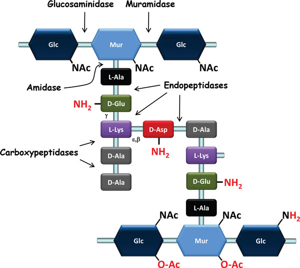
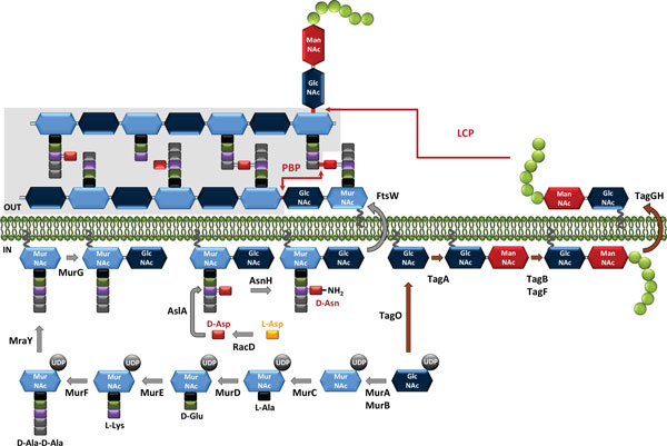
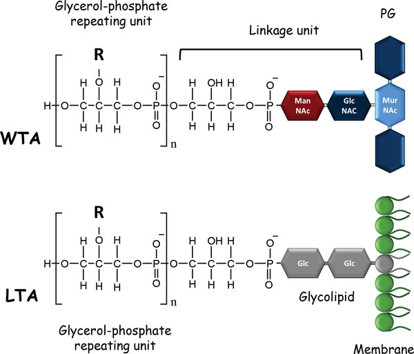
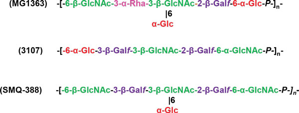
The cell wall of Gram-positive bacteria is a complex assemblage of glycopolymers and proteins. It consists of a thick peptidoglycan sacculus that surrounds the cytoplasmic membrane and that is decorated with teichoic acids, polysaccharides, and proteins. It plays a major role in bacterial physiology since it maintains cell shape and integrity during growth and division; in addition, it acts as the interface between the bacterium and its environment. Lactic acid bacteria (LAB) are traditionally and widely used to ferment food, and they are also the subject of more and more research because of their potential health-related benefits. It is now recognized that understanding the composition, structure, and properties of LAB cell walls is a crucial part of developing technological and health applications using these bacteria. In this review, we examine the different components of the Gram-positive cell wall: peptidoglycan, teichoic acids, polysaccharides, and proteins. We present recent findings regarding the structure and function of these complex compounds, results that have emerged thanks to the tandem development of structural analysis and whole genome sequencing. Although general structures and biosynthesis pathways are conserved among Gram-positive bacteria, studies have revealed that LAB cell walls demonstrate unique properties; these studies have yielded some notable, fundamental, and novel findings. Given the potential of this research to contribute to future applied strategies, in our discussion of the role played by cell wall components in LAB physiology, we pay special attention to the mechanisms controlling bacterial autolysis, bacterial sensitivity to bacteriophages and the mechanisms underlying interactions between probiotic bacteria and their hosts.
Overview
The claims and statistics of this paper haven’t been checked yet.
Run the check — free, no account needed. It reads the open-access text and checks the sources it cites.
Integrity signals
Registry
Cites
Statistics
Figures
Verification
Claims the paper makes
The claims and statistics of this paper haven’t been checked yet.
Run the check — free, no account needed. It reads the open-access text and checks the sources it cites.
Claims other papers make about this one
Each is checked against this paper’s own text: does it say what the citing paper says it does?
The check wasn’t confident either way, so it gives no verdict — never disputed.
In the citing paper
“α-Glucans and β-fructans are then produced through glucansucrases and fructansucrase, respectively.”
From Lactic Acid Bacteria Exopolysaccharides Producers: A Sustainable Tool for Functional Foods (2021) · cites this paper as [14]
Experimental — accuracy not yet measured
Self-check
Percentages, sums and reported differences re-computed from the paper’s own numbers.
The self-check needs the paper’s full text.
Citations
Cites
Peptidoglycan types of bacterial cell walls and their taxonomic implications
SCHLEIFER, K. H., & Kandler, O. (1972) · Bacteriological Reviews · doi:10.1128/br.36.4.407-477.1972 · Cited by 4,203
Comparative genomics of the lactic acid bacteria.
Makarova, K., Slesarev, A., Wolf, Y., Sorokin, A., Mirkin, B., Koonin, E., Pavlov, A., Pavlova, N., Karamychev, V., Polouchine, N., et al. (2006) · Proceedings of the National Academy of Sciences · doi:10.1073/pnas.0607117103 · Cited by 1,391
Navarre, W. W., & Schneewind, O. (1999) · Microbiology and Molecular Biology Reviews · doi:10.1128/mmbr.63.1.174-229.1999 · Cited by 1,391
From the regulation of peptidoglycan synthesis to bacterial growth and morphology
Typas, A., Banzhaf, M., Gross, C. A., & Vollmer, W. (2011) · Nature Reviews Microbiology · doi:10.1038/nrmicro2677 · Cited by 1,338
The penicillin-binding proteins: structure and role in peptidoglycan biosynthesis
Sauvage, E., Kerff, F., Terrak, M., Ayala, J. A., & Charlier, P. (2008) · FEMS Microbiology Reviews · doi:10.1111/j.1574-6976.2008.00105.x · Cited by 1,322
Lactic acid bacteria of foods and their current taxonomy
Stiles, M. E., & Holzapfel, W. H. (1997) · International Journal of Food Microbiology · doi:10.1016/s0168-1605(96)01233-0 · Cited by 1,100
Host interactions of probiotic bacterial surface molecules: comparison with commensals and pathogens
Lebeer, S., Vanderleyden, J., & De Keersmaecker, S. C. J. (2010) · Nature Reviews Microbiology · doi:10.1038/nrmicro2297 · Cited by 1,062
Neuhaus, F. C., & BADDILEY, J. (2003) · Microbiology and Molecular Biology Reviews · doi:10.1128/mmbr.67.4.686-723.2003 · Cited by 1,035
Wall Teichoic Acids of Gram-Positive Bacteria
Brown, S., Santa Maria, J. P., & Walker, S. (2013) · Annual Review of Microbiology · doi:10.1146/annurev-micro-092412-155620 · Cited by 966
Bacterial peptidoglycan (murein) hydrolases
Vollmer, W., Joris, B., Charlier, P., & Foster, S. J. (2008) · FEMS Microbiology Reviews · doi:10.1111/j.1574-6976.2007.00099.x · Cited by 880
Research works printed but not linked: 1
- 10.1016/S0168-6445(98)00042-4 · DOI
Cited by
Sorted by citations
Items 301–400 of 410
Wu, B., Zhao, A., Chen, W., Zhou, Y., Zhang, W., Zhang, Y., Hou, Q., Yao, N., Zhang, S., Duan, J., et al. (2025) · Frontiers in Microbiology · doi:10.3389/fmicb.2025.1689735 · Cited by 4
Sasahara, H., Aso, H., Niitsuma, F., Masaki, A., Arai, T., Yamaguchi, S., Jeong, H., Ikeda, S., Tanimachi, H., Hayashida, H., et al. (2025) · Journal of Functional Foods · doi:10.1016/j.jff.2025.107053 · Cited by 2
Kahraman, H. A., Deveci, M., Gürsel, E. N., Gacar, A., & Öztürk, N. (2026) · European Food Research and Technology · doi:10.1007/s00217-025-04988-4 · Cited by 3
Sharif, S., Azizan, K. A., & Baharum, S. N. (2017) · Annals of Microbiology · doi:10.1007/s13213-017-1268-0 · Cited by 2
Pan, W., Zhao, Z., Wu, J., Fan, Q., Huang, H., He, R., Shen, H., Zhao, Z., Feng, S., Gan, G., et al. (2022) · International Journal of Molecular Sciences · doi:10.3390/ijms231911466 · Cited by 2
Enumeration and characterization of bacteria from civet gastrointestinal tract
Muzaifa, M., Hasni, D., Abubakar, Y., Febriani, F., & Abubakar, A. (2021) · IOP Conference Series Earth and Environmental Science · doi:10.1088/1755-1315/667/1/012002 · Cited by 2
Balaraman, R. P., Mendel, J., Flores, L., & Choudhary, M. (2021) · Nanotechnology in the life sciences · doi:10.1007/978-3-030-65792-5_15 · Cited by 2
Escobedo, S., de Pipaon, M. P., Rendueles, C., Rodrı́guez, A., & Martı́nez, B. (2023) · Frontiers in Microbiology · doi:10.3389/fmicb.2023.1106049 · Cited by 2
Ambrosio, C. M. S. (2020) · Digital Library of Theses and Dissertations (Universidade de São Paulo) · doi:10.11606/t.11.2020.tde-18052020-151325 · Cited by 2
Fellner, M., Rankin, J. A., Hu, J. P., & Hausinger, R. P. (2017) · Encyclopedia of Inorganic and Bioinorganic Chemistry · doi:10.1002/9781119951438.eibc2508 · Cited by 2
Al-Mujahidy, S. M. J., Ikeo, K., Saito, K., Sina, A. A. I., & Uddin, M. E. (2023) · Preprints.org · doi:10.20944/preprints202311.1374.v1 · Cited by 2
Scornec, H., Palud, A., Pédron, T., Wheeler, R., Petitgonnet, C., Boneca, I. G., Cavin, J., Sansonetti, P. J., & Licandro, H. (2020) · Frontiers in Microbiology · doi:10.3389/fmicb.2020.00156 · Cited by 1
Metal oxide–based nanocomposites: greener synthesis routes and their potentiality
Kumari, V., Kumari, K., & Rana, R. K. (2023) · Elsevier eBooks · doi:10.1016/b978-0-323-85241-8.00011-6 · Cited by 1
Nguyen, P.-T., & Thanh, N. H. (2023) · TRA VINH UNIVERSITY JOURNAL OF SCIENCE · doi:10.35382/tvujs.13.1.2023.1685 · Cited by 1
Rahmi, F., Ridhana, F., & Muzaifa, M. (2023) · AGROINTEK · doi:10.21107/agrointek.v17i3.15691 · Cited by 1
Ferreira, T. H., Maximiano, P., & Simões, P. N. (2024) · Food Research International · doi:10.1016/j.foodres.2024.115475 · Cited by 1
Glycophenotyping of mutants of Lacticaseibacillus paracasei by lectin microarray
Suzuki, E., Serata, M., Sako, T., Sato, S., Iino, T., Tateno, H., & Hirabayashi, J. (2025) · Applied and Environmental Microbiology · doi:10.1128/aem.01707-24 · Cited by 1
Zhu, X., Chen, Q., & Liu, D. H. (2025) · ChemistrySelect · doi:10.1002/slct.202502328 · Cited by 1
Cell Surface and Outer Membrane
Barton, L. L. (2025) · Springer eBooks · doi:10.1007/978-3-031-91320-4_4 · Cited by 1
Alvarez-Villagómez, K. G., Balleza, D., Peña-Montes, C., Fonseca-Barrera, I. D. C., & Mendoza-García, P. G. (2025) · Frontiers in Microbiology · doi:10.3389/fmicb.2025.1728482 · Cited by 1
Zhang, K., Lu, M., Fu, S., Jiang, C., Dong, X., Liu, T., Chen, Y., Li, X., Xu, S., Su, H., et al. (2026) · Biofilm · doi:10.1016/j.bioflm.2026.100346 · Cited by 1
Çolak, B., Barut, D., Yaman, B. N., Altıntaş, Ö. E., Çabuk, A., & Çelik, P. A. (2026) · Current Microbiology · doi:10.1007/s00284-026-04763-y · Cited by 1
Fukuchi, M., Arai, N., Honda, S., & Tominaga, Y. (2026) · Cell Biochemistry and Biophysics · doi:10.1007/s12013-026-02057-5 · Cited by 1
Neef, J., van Dijl, J. M., & Buist, G. (2026) · New Biotechnology · doi:10.1016/j.nbt.2026.04.001 · Cited by 1
Grigore‐Gurgu, L., Cotârleț, M., Sergentu, A. C., Oprișanu, M., Măntăilă, S., CANTARAGIU, A.-M., Iosifescu, C., Botezatu, A. V. D., Aprodu, I., & Bahrim, G. E. (2026) · Applied Food Research · doi:10.1016/j.afres.2026.102237 · Cited by 1
Proteomic Analysis of Lactococcus lactis FM03 Under Prolonged Alkaline Stress
Bendig, T. A. L., Abee, T., Boeren, S. A., Smid, E. J., & van Mastrigt, O. (2026) · Environmental Microbiology · doi:10.1111/1462-2920.70381 · Cited by 1
AcmA-mediated non-GMO probiotic engineering for functional jujube juice fermentation
Peng, K., Tian, G., Mao, K., Wang, X., Zhao, F., & Sang, Y. (2026) · Food Research International · doi:10.1016/j.foodres.2026.120580 · Cited by 1
Progress Towards Engineering Microbial Surfaces to Degrade Biomass
Huang, G. L., & Clubb, R. (2017) · InTech eBooks · doi:10.5772/65509 · Cited by 1
Cui, L., Lin, S., Yi, J., Liu, X., Hao, L., Ji, Y., Lu, L., Ji, Z., Kang, Q., & Lu, J. (2020) · Journal of Food Science · doi:10.1111/1750-3841.15148 · Cited by 1
ICT1 deficiency leads to reduced oxygen resistance due to the cell wall damage in S. cerevisiae
Zhu, H., Wang, M., Zhou, H., & Cai, H. J. (2022) · Genes & Genomics · doi:10.1007/s13258-021-01208-7 · Cited by 1
Hasani, A., Dehghani, L., Soltani, E., & Leylabadlo, H. E. (2021) · Pharmaceutical Sciences · doi:10.34172/ps.2021.78 · Cited by 1
Studies on the Mode of Action of Bacteriocin Produced by Lactobacillus fermentum CrT21
(2015) · International Journal of Science and Research (IJSR) · doi:10.21275/v5i5.nov163177 · Cited by 1
ZHI, C. K., Lani, M. N., Hamzah, Y., Ahmad, F. T., & UBAIDILLAH, N. H. N. (2021) · Universiti Malaysia Terengganu Journal of Undergraduate Research · doi:10.46754/umtjur.v3i4.244 · Cited by 1
Biofilm mediated bioremediation and other applications
Sridharan, R., & Krishnaswamy, V. G. (2021) · Elsevier eBooks · doi:10.1016/b978-0-323-85657-7.00018-3 · Cited by 1
Joly, A., & Leulier, F. (2023) · médecine/sciences · doi:10.1051/medsci/2023119 · Cited by 1
Jyakhwo, S., Bocharova, V., Serov, N., Dmitrenko, A., & Vinogradov, V. V. (2024) · Advanced Materials Technologies · doi:10.1002/admt.202400458 · Cited by 1
Verma, M. K., Kesarwani, S., Singh, V., Vamanu, E., & Mishra, V. (2024) · Environmental Research Communications · doi:10.1088/2515-7620/ad85c6 · Cited by 1
Zulu, N. X., Kunadu, A. P., Kayitesi, E., & Dlamini, B. C. (2024) · Journal of Food Processing and Preservation · doi:10.1155/jfpp/7030605 · Cited by 2
Kajitani, K., & Takahashi, S. (2021) · Microbiology Resource Announcements · doi:10.1128/mra.00568-21 · Cited by 1
Structure and Function Relationships of Exopolysaccharides Produced by Lactic Acid Bacteria
Chen, X. (2017) · ERA: Education and Research Archive (University of Alberta) · doi:10.7939/r3445hq7c · Cited by 1
Exploitation of Exopolysaccharides from Lactic Acid Bacteria
TSUDA, H. (2020) · Wiley eBooks · doi:10.1002/9783527818310.ch4 · Cited by 1
Matos, R. C., Nikolopoulos, N., Perrier, Q., Robert, X., Gueguen‐Chaignon, V., Hirayama, H., Leulier, F., Grangeasse, C., Guérardel, Y., & Ravaud, S. (2026) · bioRxiv (Cold Spring Harbor Laboratory) · doi:10.64898/2026.03.19.713020 · Cited by 1
Meng, L. (2016) · UKnowledge (University of Kentucky) · doi:10.13023/etd.2016.001
Kylä-Nikkilä, K. (2016) · Työväentutkimus Vuosikirja
Analysis of Enterococcus faecalis mutants affected in septum cleavage during cell division
Elsarmane, F. (2016) · White Rose eTheses Online (University of Leeds, The University of Sheffield, University of York)
Отримання та характеристика продуктів деструкції пептидоглікану Lactobacillus acidophilus K 3111
Капустян, А. І., & Cherno, N. (2018) · Науковий вісник Львівського національного університету ветеринарної медицини та біотехнологій імені С.З. Ґжицького · doi:10.15421/nvlvet8526
OBTAINING AND CHARACTERISTIC OF MUROPEPTIDES OF PROBIOTIC CULTURES CELL WALLS
Капустян, А. І., & Cherno, N. (2020) · Food Science and Technology · doi:10.15673/fst.v14i1.1661
Jiang, Y. (2020) · Advancements in Case Studies · doi:10.31031/aics.2020.02.000538
Gene expression patterns of the female genital tract and immunomodulation by Lactobacillus species
Abrahams, A. G. (2020) · Open University of Cape Town (University of Cape Town)
The Roles of Lactic Acid Bacteria for Control of Fungal Growth and Mycotoxins
Kim, J., & Lee, H. (2020) · JoLS Journal of Life Sciences · doi:10.5352/jls.2020.30.12.1128
Factors Influencing Aggregation and Adhesion of Oral Lactobacillus gasseri
Plansangket, L., Teanpaisan, R., & Piwat, S. (2021) · Walailak Journal of Science and Technology (WJST) · doi:10.48048/wjst.2021.6950
Biological Decontamination of Aflatoxins
de Oliveira, C. A. F., de França, M. M., Corassin, C. H., & Muaz, K. (2021) · Springer eBooks · doi:10.1007/978-3-030-85762-2_12
Lactic acid bacteria in food quality enrichment
Satpute, S., Nawani, N. N., Sharma, D., & Junnarkar, M. V. (2022) · Elsevier eBooks · doi:10.1016/b978-0-323-89875-1.00014-6
Genetic Tools for the Enhancement of Probiotic Properties
Ortiz-Velez, L., & Britton, R. A. (2018) · ASM Press eBooks · doi:10.1128/9781555819705.ch16
КОМБІНОВАНИЙ МЕТОД ДЕЗІНТЕГРАЦІЇ МІКРОБІАЛЬНОЇ БІОМАСИ
Капустян, А. І., & Cherno, N. (2018) · Scientific Works · doi:10.15673/swonaft.v81i2.897
Biosys-LiDeOGraM: A visual analytics framework for interactive modelling of multiscale biosystemsPreprint
Perrot, N., Layec, S., Tonda, A. P., Boukhelifa, N., Fonseca, F., & Lutton, É. (2023) · bioRxiv (Cold Spring Harbor Laboratory) · doi:10.1101/2023.06.23.546209
Clinical Utility of Probiotics Therapy in Managing Mycotoxin Illness.
LaTouche, L. (2023) · PubMed Central
Díaz, D. P. M., Gutiérrez-Miceli, F. A., Ventura-Canseco, L. M. C., Abud‐Archila, M., Valdez, B. S., Ruiz, M. C. M., & Luján-Hidalgo, M. C. (2023) · BIOtecnia · doi:10.18633/biotecnia.v25i3.1950
Ghoul, M. (2024) · Wiley eBooks · doi:10.1002/9781394312337.refs2
Yang, H.-J., Zhang, Z.-L., Chen, Z.-Q., Cheng, X., Wang, J., Yan, L.-B., Gao, X.-J., Lin, X.-J., & Lian, Y.-Y. (2024) · Journal of Carbohydrate Chemistry · doi:10.1080/07328303.2024.2410794
Study of Bacillus cereus as an Effective Multi-Type A Trichothecene Inactivator
García-García, F. A., Cristiani‐Urbina, E., Morales-Barrera, L., Rodríguez‐Peña, N., Hernández‐Portilla, L. B., Campos, J. E., & Flores-Ortíz, C. M. (2024) · Microorganisms · doi:10.3390/microorganisms12112236
Biodegradation of mycotoxins using postbiotics
Hiththatiyage, R., Wickramasinghe, M. A., Rathnayaka, I., Dias, C., Rambodagedara, S., Jayawardana, B. C., & Liyanage, R. (2025) · Elsevier eBooks · doi:10.1016/b978-0-443-22188-0.00040-1
Biological Approach for Lead (Pb) Removal from Meat and Meat Products in Bangladesh
Maisa, N. S., Hoque, S. B., & Toushik, S. H. (2025) · Processes · doi:10.3390/pr13072018
Reactivity of Carbohydrate Phosphodiesters, Potential Targets of Antibacterial Agents
Sillanpää, E. R. J., Lönnberg, T., & Mikkola, S. (2025) · Chemistry & Biodiversity · doi:10.1002/cbdv.202501852
Shanumuganandam, P., & Thangavelu, S. (2025) · Research Square · doi:10.21203/rs.3.rs-7440837/v1
Chaston, J. M. (2025) · Applied and Environmental Microbiology · doi:10.1128/aem.01491-25
Gram-positive bacterial cell wall components inhibit herpes simplex virus infectionPreprint
Adams, A. N. D., Griffin, L. E., Burnie, J., Powers, J., Causey, A., Glick, V. J., Gavitt, M., Richmond-Buccola, D., Kim, C. H., Ahmad, M., et al. (2025) · bioRxiv (Cold Spring Harbor Laboratory) · doi:10.1101/2025.11.02.686169
Ando, M., Hata, C., Nishizaki, H., Togao, M., & Kawakami, K. (2025) · Microbiology Research · doi:10.3390/microbiolres16110234 · Cited by 1
Alisoltani, A., Manhanzva, M. T., Potgieter, M., Balle, C., Bell, L., Ross, E. J., Iranzadeh, A., du Plessis, M., Radzey, N., McDonald, Z., et al. (2020) · Figshare · doi:10.6084/m9.figshare.13270780
Alisoltani, A., Manhanzva, M. T., Potgieter, M., Balle, C., Bell, L., Ross, E. J., Iranzadeh, A., du Plessis, M., Radzey, N., McDonald, Z., et al. (2020) · Figshare · doi:10.6084/m9.figshare.13270783
Weng, L., Wu, L., Guo, R., Ye, J., Liang, W., Wu, W., Chen, L., & Yang, D. (2022) · Figshare · doi:10.6084/m9.figshare.20423794
Paquete-Ferreira, J., Romão, M. J., & Santos‐Silva, T. (2025) · Royal Society of Chemistry eBooks · doi:10.1039/9781837676101-00154
Moganti, L. K., & Dutta, D. (2026) · Lecture notes in civil engineering · doi:10.1007/978-981-95-3775-4_4
Csarmann, K., Jung, F., Baumann, A., Simbrunner, B., Schweiger, K., Burger, K., Staltner, R., Hellerbrand, C., & Bergheim, I. (2026) · European Journal of Nutrition · doi:10.1007/s00394-026-03931-x
Kim, Y. S., Kim, Y., Kim, J. Y., Lee, S. H., Roh, S. W., Lee, D. Y., Oh, Y. J., & Whon, T. W. (2026) · Food Research International · doi:10.1016/j.foodres.2026.119034
Gomand, F. (2019) · theses.fr (ABES) · doi:10.70675/8ab9d06fz722ez46eezbe0ezb450beb59680 · Cited by 1
Identification des interactions positives entre bactéries lactiques en fermentation de jus de soja
Harlé, O. (2020) · theses.fr (ABES) · doi:10.70675/c2570d6ezd936z44b0za264zf7ca78861867
Coutino, M. D. L. T. (2022) · theses.fr (ABES) · doi:10.70675/70da2432z1664z41eaz9cabz768ea551c125 · Cited by 1
Lactobacillus paragasseri ameliorates age-dependent thermotaxis decline in Caenorhabditis elegans
Tanaka, M., Tsukada, S., Jang, M., Aleogho, B. M., Mori, I., & Noma, K. (2026) · Scientific Reports · doi:10.1038/s41598-026-48002-7
Zhang, C., Xing, S., Hao, Z., He, K., Pan, J., Pan, Y., He, D., Wu, H., Ma, X., & Yuan, L. (2026) · LWT · doi:10.1016/j.lwt.2026.119446
Khajavi, M. Z., Nikiforov, A., Morent, R., Fraeye, I., Meulenaer, B. D., Devlieghere, F., Ragaert, P., & Geyter, N. D. (2026) · Food Research International · doi:10.1016/j.foodres.2026.119404
Jiang, X., Risbo, J., & Ye, Y. (2026) · Food Chemistry · doi:10.1016/j.foodchem.2026.149723
Staphylococcus aureus activates dendrite elongation in dendritic cells
Kobata, K., Furuta, K., Ikeya, Y., Chishaki, Y., Ishikawa, K., & Kaito, C. (2026) · Biochemical and Biophysical Research Communications · doi:10.1016/j.bbrc.2026.154001
Tobías‐Soria, D., Chacón-García, K. F., Pérez-Vega, S. B., Gutiérrez‐Méndez, N., de la Cueva, S. C., & Salmerón, I. (2026) · Processes · doi:10.3390/pr14121880
Li, L., Zheng, Y., Lin, J., Mei, B., Li, Y., Li, H., & Yi, L. (2026) · Postharvest Biology and Technology · doi:10.1016/j.postharvbio.2026.114515
Xiao, H., Kajdi, K., Wimmer, R., & Bang‐Berthelsen, C. H. (2026) · Microorganisms · doi:10.3390/microorganisms14061358
Liu, Y., Zhang, Y., Zheng, J., Zhao, Y., Zhang, J. C., Sun, Z., Zhu, Q., Liu, Z., Liu, H., & Guo, Q. M. (2026) · Journal of Agricultural and Food Chemistry · doi:10.1021/acs.jafc.6c00977
Hristovski, K., Massadeh, R. J. M. A., Süli, B. K., Micevic, S. S., Ziane, S. R., Brezovcsik, G., Kiss, Z., Hitka, G., Tran, A. M., Bujna, E., et al. (2026) · Applied Sciences · doi:10.3390/app16136491
Onwuakor, C. E., & Ngene, A. C. (2026) · Frontiers in Antibiotics · doi:10.3389/frabi.2026.1907088
Jo, S.-H., Hong, N.-Y., Choi, B.-G., Chang, J., Seo, S., 이가영, Ahn, H. Y., Song, J.-H., Kim, H. T., Yang, Y.-H., et al. (2026) · Foods · doi:10.3390/foods15152736
Aladhadh, M. A., Abou-Elazm, F. I., Ahmed, R. M., Elsanhoty, R. M., Al‐Saman, M. A., & Mabrouk, S. S. (2026) · Microorganisms · doi:10.3390/microorganisms14081710
Maher, A., Rył, A., & Nowak, A. (2026) · Probiotics and Antimicrobial Proteins · doi:10.1007/s12602-026-11157-w
Metadata
- Authors
- Chapot-Chartier, M.-P., & Kulakauskas, S.
- Affiliations
- AgroParisTech (FR)
- ORCID
- Kulakauskas, Saulius
- Venue
- Microbial Cell Factories
- Published
- 2014
- Volume
- 13
- Issue
- Suppl 1
- Pages
- S9
- ISSN
- 1475-2859 (ISSN-L)
- PMC
- PMC4155827
- OpenAlex
- W2130696295
- Cited by
- 410
- References
- 189
- Citations by year
- 2026: 42 · 2025: 40 · 2024: 30 · 2023: 44 · 2022: 36 · 2021: 49 · 2020: 61 · 2019: 25 · 2018: 25 · 2017: 25 · 2016: 23 · 2015: 7 · 2014: 3
- Type
- conference abstract
- Language
- en
- Open access
- gold
- doi.org
- Licence
- cc-by
- Locations
- Microbial Cell Factories (publishedVersion, cc-by)
- PubMed Central (publishedVersion, cc-by)
- PubMed (publishedVersion)
- Funders
- Agence Nationale de la Recherche (ANR-11-BSV8-0004, ANR-11-BSV8-004-01)
- Institut National de la Recherche Agronomique
- Topics
- Life Sciences › Agricultural and Biological Sciences › Food Science › Probiotics and Fermented Foods
- Life Sciences › Biochemistry, Genetics and Molecular Biology › Genetics › Bacterial Genetics and Biotechnology
- Life Sciences › Biochemistry, Genetics and Molecular Biology › Molecular Biology › Biochemical and Structural Characterization
- Keywords
- lactic acid bacteria, bacterial cell wall, peptidoglycan, teichoic acid, cell wall polysaccharides, cell wall proteins, cell wall biosynthesis, bacterial autolysis, bacteriophage sensitivity, Gram-positive bacteria, food fermentation
- MeSH terms
- Acetylation, Bacterial Proteins, Cell Wall, Lactobacillaceae, Lipopolysaccharides, Peptidoglycan, Polysaccharides, Bacterial, Teichoic Acids, Host-Pathogen Interactions
About these checks
Registry, retraction, Cites and statistics results are computed, not judged, with one exception: a printed reference that no index lists is read once by a model before it counts as a citation error. Claims are extracted by a model and kept only when their quote appears verbatim in the text; each cited source is then checked, with a model’s help, for whether it says what the claim relies on. Those verdicts can be wrong — read the claim’s quote and the cited source before relying on one.
Statistics and arithmetic are recomputed, not judged.
Confidence bands are shown only for fields calibrated on our annotated test set. None is calibrated yet, so no band is shown.
What counts as a claim ›How we measure confidence ›How we check this ›
Are you an author, or do you see a problem with this page? Request a review
