

The DOI matches its Crossref record; 88 of the 88 references it cites were found in a registry or an index.
Comparative analysis of Klebsiella pneumoniae genomes identifies a phospholipase D family protein as a novel virulence factor
Open accessAbstract
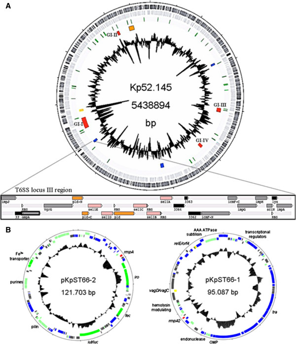
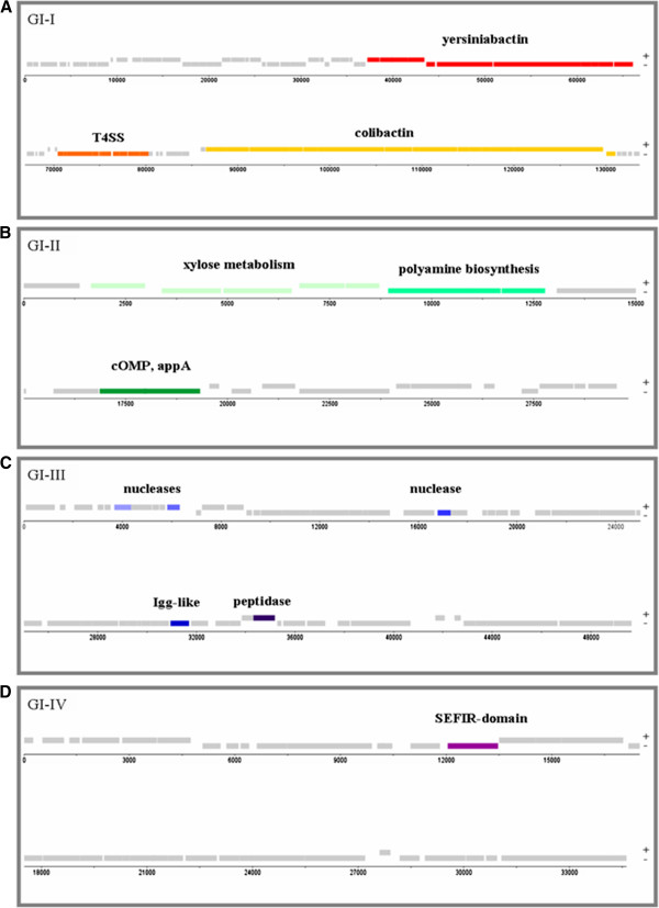
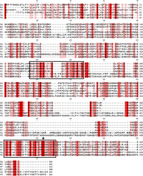
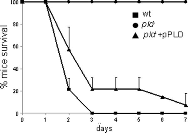
BACKGROUND: Klebsiella pneumoniae strains are pathogenic to animals and humans, in which they are both a frequent cause of nosocomial infections and a re-emerging cause of severe community-acquired infections. K. pneumoniae isolates of the capsular serotype K2 are among the most virulent. In order to identify novel putative virulence factors that may account for the severity of K2 infections, the genome sequence of the K2 reference strain Kp52.145 was determined and compared to two K1 and K2 strains of low virulence and to the reference strains MGH 78578 and NTUH-K2044. RESULTS: In addition to diverse functions related to host colonization and virulence encoded in genomic regions common to the four strains, four genomic islands specific for Kp52.145 were identified. These regions encoded genes for the synthesis of colibactin toxin, a putative cytotoxin outer membrane protein, secretion systems, nucleases and eukaryotic-like proteins. In addition, an insertion within a type VI secretion system locus included sel1 domain containing proteins and a phospholipase D family protein (PLD1). The pld1 mutant was avirulent in a pneumonia model in mouse. The pld1 mRNA was expressed in vivo and the pld1 gene was associated with K. pneumoniae isolates from severe infections. Analysis of lipid composition of a defective E. coli strain complemented with pld1 suggests an involvement of PLD1 in cardiolipin metabolism. CONCLUSIONS: Determination of the complete genome of the K2 reference strain identified several genomic islands comprising putative elements of pathogenicity. The role of PLD1 in pathogenesis was demonstrated for the first time and suggests that lipid metabolism is a novel virulence mechanism of K. pneumoniae.
Overview
The claims and statistics of this paper haven’t been checked yet.
Run the check — free, no account needed. It reads the open-access text and checks the sources it cites.
Integrity signals
Registry
Cites
Statistics
Figures
Verification
Claims the paper makes
The claims and statistics of this paper haven’t been checked yet.
Run the check — free, no account needed. It reads the open-access text and checks the sources it cites.
Claims other papers make about this one
Each is checked against this paper’s own text: does it say what the citing paper says it does?
The cited source says what the claim attributes to it.
In the citing paper
“From a genome-wide search of K. pneumoniae strain Kp52.145 (19), we found four active structural genes that were >50% identical to E. coli fepA: the plasmid pII locus 0027 and the chromosomal loci 1658, 2380, and 4984.”
From Siderophore-mediated iron acquisition by Klebsiella pneumoniae (2024) · cites this paper as [19]
Self-check
Percentages, sums and reported differences re-computed from the paper’s own numbers.
The self-check needs the paper’s full text.
Citations
Cites
Gapped BLAST and PSI-BLAST: a new generation of protein database search programs
Altschul, S. (1997) · Nucleic Acids Research · doi:10.1093/nar/25.17.3389 · Cited by 73,302
A RAPID METHOD OF TOTAL LIPID EXTRACTION AND PURIFICATION
Bligh, E. G., & Dyer, W. J. (1959) · Canadian Journal of Biochemistry and Physiology · doi:10.1139/o59-099 · Cited by 54,751
The RAST Server: Rapid Annotations using Subsystems Technology
Aziz, R. K., Bartels, D., Best, A. A., DeJongh, M., Disz, T. L., Edwards, R. A., Formsma, K., Gerdes, S., Glass, E. M., Kubal, M., et al. (2008) · BMC Genomics · doi:10.1186/1471-2164-9-75 · Cited by 11,893
Podschun, R., & Ullmann, U. (1998) · Clinical Microbiology Reviews · doi:10.1128/cmr.11.4.589 · Cited by 2,701
The lipocalin protein family: structure and function
FLOWER, D. R. (1996) · Biochemical Journal · doi:10.1042/bj3180001 · Cited by 1,679
Klebsiella pneumoniae liver abscess: a new invasive syndrome
Siu, L.-K. K., Yeh, K., Lin, J., Fung, C., & Chang, F. (2012) · The Lancet Infectious Diseases · doi:10.1016/s1473-3099(12)70205-0 · Cited by 907
An Endonuclease Allows Streptococcus pneumoniae to Escape from Neutrophil Extracellular Traps
Beiter, K., Wartha, F., Albiger, B., Normark, S. J., Zychlinsky, A., & Henriques‐Normark, B. (2006) · Current Biology · doi:10.1016/j.cub.2006.01.056 · Cited by 588
Boyer, F., Fichant, G. A., Berthod, J., Vandenbrouck, Y., & Attrée, I. (2009) · BMC Genomics · doi:10.1186/1471-2164-10-104 · Cited by 575
Capsule Polysaccharide Mediates Bacterial Resistance to Antimicrobial Peptides
Campos, M. A. L., Vargas, M. A., Regueiro, V., Llompart, C. M., Albertí, S., & Bengoechea, J. A. (2004) · Infection and Immunity · doi:10.1128/iai.72.12.7107-7114.2004 · Cited by 515
Self-citationBrisse, S., Fèvre, C., Passet, V., Issenhuth-Jeanjean, S., Tournebize, R., Diancourt, L., & Grimont, P. A. D. (2009) · PLoS ONE · doi:10.1371/journal.pone.0004982 · Cited by 493
Self-citation
Cited by
Sorted by citations
Klebsiella pneumoniae: Going on the Offense with a Strong Defense
Paczosa, M. K., & Mecsas, J. C. (2016) · Microbiology and Molecular Biology Reviews · doi:10.1128/mmbr.00078-15 · Cited by 2,090
Hypervirulent Klebsiella pneumoniae
Russo, T. A., & Marr, C. M. (2019) · Clinical Microbiology Reviews · doi:10.1128/cmr.00001-19 · Cited by 1,398
Population genomics of Klebsiella pneumoniae
Wyres, K. L., Lam, M. M. C., & Holt, K. E. (2020) · Nature Reviews Microbiology · doi:10.1038/s41579-019-0315-1 · Cited by 1,175
Colonization, Infection, and the Accessory Genome of Klebsiella pneumoniae
Martin, R. M., & Bachman, M. A. (2018) · Frontiers in Cellular and Infection Microbiology · doi:10.3389/fcimb.2018.00004 · Cited by 1,067
Hypervirulent Klebsiella pneumoniae – clinical and molecular perspectives
Choby, J. E., Howard-Anderson, J. R., & Weiss, D. S. (2019) · Journal of Internal Medicine · doi:10.1111/joim.13007 · Cited by 747
Metadata
- Authors
- Lery, L. M., Frangeul, L., Tomas, A., Passet, V., Almeida, A. S., Bialek-Davenet, S., Barbe, V., Bengoechea, J. A., Sansonetti, P., Brisse, S., & Tournebize, R.
- Affiliations
- Institut Pasteur (FR)
- Centre National de la Recherche Scientifique (FR)
- Inserm (FR)
- Commissariat à l'Énergie Atomique et aux Énergies Alternatives (FR)
- Queen's University Belfast (GB)
- Corresponding author
- Tournebize, Régis
- ORCID
- Venue
- BMC Biology
- Published
- 2014-05-29
- Volume
- 12
- Issue
- 1
- Pages
- 41-41
- ISSN
- 1741-7007 (ISSN-L)
- PMC
- PMC4068068
- OpenAlex
- W2129183301
- Cited by
- 181
- References
- 88
- Citations by year
- 2026: 8 · 2025: 8 · 2024: 12 · 2023: 15 · 2022: 37 · 2021: 11 · 2020: 21 · 2019: 14 · 2018: 15 · 2017: 10 · 2016: 9 · 2015: 15 · 2014: 5
- FWCI
- 9.16
- Type
- journal article
- Language
- en
- Open access
- gold
- doi.org
- Licence
- cc-by
- Locations
- BMC Biology (publishedVersion, cc-by)
- PubMed Central (publishedVersion, cc-by)
- PubMed (publishedVersion)
- Research Portal (Queen's University Belfast) (acceptedVersion, other-oa)
- HAL (submittedVersion, other-oa)
- Docusalut (Conselleria de Salut i Consum del Govern de les Illes Balears) (submittedVersion, cc-by)
- Hispana (submittedVersion, cc-by)
- Funders
- Fundação para a Ciência e a Tecnologia
- Topics
- Life Sciences › Immunology and Microbiology › Microbiology › Escherichia coli research studies
- Health Sciences › Medicine › Microbiology (medical) › Antibiotic Resistance in Bacteria
- Life Sciences › Immunology and Microbiology › Microbiology › Enterobacteriaceae and Cronobacter Research
- Keywords
- Klebsiella pneumoniae, virulence factors, phospholipase D, phospholipase D1, genomic islands, type VI secretion system, cardiolipin metabolism, lipid metabolism, bacterial pathogenesis, comparative genomics
- MeSH terms
- Amino Acid Sequence, Animals, Bacterial Proteins, Genes, Bacterial, Multigene Family, Klebsiella pneumoniae, Molecular Sequence Data, Phospholipase D, Plasmids, Virulence, Mutagenesis, Insertional, Sequence Alignment, Genome, Bacterial, Virulence Factors, Genomic Islands, Lipid Metabolism, Mice, Bacterial Secretion Systems, Molecular Sequence Annotation
About these checks
Registry, retraction, Cites and statistics results are computed, not judged, with one exception: a printed reference that no index lists is read once by a model before it counts as a citation error. Claims are extracted by a model and kept only when their quote appears verbatim in the text; each cited source is then checked, with a model’s help, for whether it says what the claim relies on. Those verdicts can be wrong — read the claim’s quote and the cited source before relying on one.
Statistics and arithmetic are recomputed, not judged.
Confidence bands are shown only for fields calibrated on our annotated test set. None is calibrated yet, so no band is shown.
What counts as a claim ›How we measure confidence ›How we check this ›
Are you an author, or do you see a problem with this page? Request a review
