

The DOI matches its Crossref record; 3 of the 3 references it cites were found in a registry or an index.
Brain CT registration using hybrid supervised convolutional neural network
Open accessPublished version of a preprint
Abstract
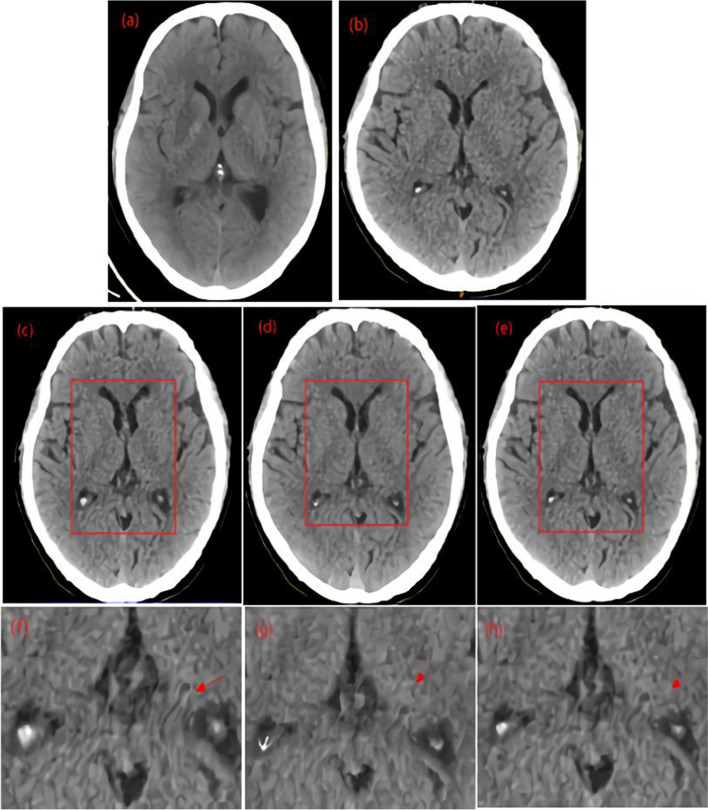
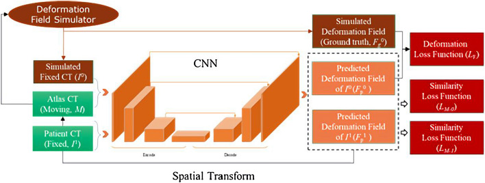
Background: Image registration is an essential step in the automated interpretation of the brain computed tomography (CT) images of patients with acute cerebrovascular disease (ACVD). However, performing brain CT registration accurately and rapidly remains greatly challenging due to the large intersubject anatomical variations, low resolution of soft tissues, and heavy computation costs. To this end, the HSCN-Net, a hybrid supervised convolutional neural network, was developed for precise and fast brain CT registration. Method: HSCN-Net generated synthetic deformation fields using a simulator as one supervision for one reference–moving image pair to address the problem of lack of gold standards. Furthermore, the simulator was designed to generate multiscale affine and elastic deformation fields to overcome the registration challenge posed by large intersubject anatomical deformation. Finally, HSCN-Net adopted a hybrid loss function constituted by deformation field and image similarity to improve registration accuracy and generalization capability. In this work, 101 CT images of patients were collected for model construction (57), evaluation (14), and testing (30). HSCN-Net was compared with the classical Demons and VoxelMorph models. Qualitative analysis through the visual evaluation of critical brain tissues and quantitative analysis by determining the endpoint error (EPE) between the predicted sparse deformation vectors and gold-standard sparse deformation vectors, image normalized mutual information (NMI), and the Dice coefficient of the middle cerebral artery (MCA) blood supply area were carried out to assess model performance comprehensively. Results: HSCN-Net and Demons had a better visual spatial matching performance than VoxelMorph, and HSCN-Net was more competent for smooth and large intersubject deformations than Demons. The mean EPE of HSCN-Net (3.29 mm) was less than that of Demons (3.47 mm) and VoxelMorph (5.12 mm); the mean Dice of HSCN-Net was 0.96, which was higher than that of Demons (0.90) and VoxelMorph (0.87); and the mean NMI of HSCN-Net (0.83) was slightly lower than that of Demons (0.84), but higher than that of VoxelMorph (0.81). Moreover, the mean registration time of HSCN-Net (17.86 s) was shorter than that of VoxelMorph (18.53 s) and Demons (147.21 s). Conclusion: The proposed HSCN-Net could achieve accurate and rapid intersubject brain CT registration.
Overview
The claims and statistics of this paper haven’t been checked yet.
Run the check — free, no account needed. It reads the open-access text and checks the sources it cites.
Integrity signals
Registry
Cites
Statistics
Figures
Verification
Claims the paper makes
The claims and statistics of this paper haven’t been checked yet.
Run the check — free, no account needed. It reads the open-access text and checks the sources it cites.
Self-check
Percentages, sums and reported differences re-computed from the paper’s own numbers.
The self-check needs the paper’s full text.
Citations
Cites
U-Net: Convolutional Networks for Biomedical Image Segmentation
Ronneberger, O., Fischer, P., & Brox, T. (2015) · Lecture notes in computer science · doi:10.1007/978-3-319-24574-4_28 · Cited by 99,404
Deep learning in medical image registration: a survey
Haskins, G., Kruger, U., & Yan, P. (2020) · Machine Vision and Applications · doi:10.1007/s00138-020-01060-x · Cited by 704
Quicksilver: Fast predictive image registration – A deep learning approach
Yang, X., Kwitt, R., Styner, M., & Niethammer, M. (2017) · NeuroImage · doi:10.1016/j.neuroimage.2017.07.008 · Cited by 630
Research works printed but not linked: 25
- Lee SH. Stroke revisited: diagnosis and treatment of ischemic stroke. In: Critical and medical management in acute stage of ischemic stroke (Chapter 14). 2017. p. 157–69. https://doi.org/10.1007/978-981-10-1424-6. · DOI
- Jung SM, Whangbo TK. Estimating aspect score for stroke from brain CT images based-on deep-learning. Adv Sci Technol. 2018;150:342–6. · DOI
- Mayer A, Zholkover A, Portnoy O, et al. Deformable registration of trans-rectal ultrasound (TRUS) and magnetic resonance imaging (MRI) for focal prostate brachytherapy. Int J Comput Assist Radiol Surg. 2016;11(6):1015–23. · DOI
- Broggi S, Scalco E, Belli ML, et al. A comparative evaluation of 3 different free-form deformable image registration and contour propagation methods for head and neck MRI: the case of parotid changes during radiotherapy. Technol Cancer Res Treat. 2017;16(3):373–81. · DOI
- Reaungamornrat S, Silva TD, Uneri A, et al. MIND demons: symmetric diffeomorphic deformable registration of MR and CT for image-guided spine surgery. IEEE Trans Med Imaging. 2016;35(11):2413–24. · DOI
- Krivov E, Pisov M, Belyaev M. MRI augmentation via elastic registration for brain lesions segmentation. In: International MICCAI brainlesion workshop. Cham: Springer; 2017. · DOI
- Liu X, Xing F, Yang C, et al. Symmetric-constrained irregular structure inpainting for brain MRI registration with tumor pathology. 2021. · DOI
- Uus A, Zhang T, Jackson LH, et al. Deformable slice-to-volume registration for motion correction of fetal body and placenta MRI. IEEE Trans Med Imaging. 2020;39(9):2750–9. · DOI
- Yu W, Tannast M, et al. Non-rigid free-form 2D–3D registration using a B-spline-based statistical deformation model. Pattern Recogn. 2017;63:689–99. · DOI
- Liu C, Zhang L, Wang L, et al. Multi-scale B-spline medical image registration based on pixel reconstruction. Intell Comput Appl. 2019;9(1):4.
- Wodzinski M, Skalski A, Ciepiela I, et al. Application of demons image registration algorithms in resected breast cancer lodge localization. In: 2017 signal processing: algorithms, architectures, arrangements, and applications (SPA). IEEE; 2017. · DOI
- Eppenhof K, Pluim J. Pulmonary CT registration through supervised learning with convolutional neural networks. IEEE Trans Med Imaging. 2019;38(5):1097–105. · DOI
- Dalca AV, Balakrishnan G, Guttag J, et al. Unsupervised learning for fast probabilistic diffeomorphic registration. 2018. · DOI
- Fan J, Cao X, Yap PT, et al. BIRNet: Brain image registration using dual-supervised fully convolutional networks. Med Image Anal. 2019;54:193–206. · DOI · Paper page
- Han R, Jones CK, Ketcha MD, et al. Deformable MR-CT image registration using an unsupervised end-to-end synthesis and registration network for endoscopic neurosurgery. In: Image-guided procedures, robotic interventions, and modeling. 2021. · DOI
- Kim B, Kim J, Lee J G, et al. Unsupervised deformable image registration using cycle-consistent CNN. 2019. · DOI
- Zhu W, Myronenko A, Xu Z, et al. NeurReg: neural registration and its application to image segmentation. In: 2020 IEEE winter conference on applications of computer vision (WACV). IEEE; 2020. · DOI
- Haskins G, Kruger U, Yan P. Deep learning in medical image registration: a survey. Mach Vis Appl. 2020;31(1–2):1–18.
- Mok T, Chung A. Large deformation diffeomorphic image registration with laplacian pyramid networks. 2020. · DOI
- Abualigah L, Diabat A, Mirjalili S, et al. The Arithmetic Optimization Algorithm. Comput Methods Appl Mech Eng. 2021;376:113609. · DOI
- Abualigah L, Yousri D, Elaziz MA, et al. Aquila optimizer: a novel meta-heuristic optimization algorithm. Comput Ind Eng. 2021;157:107250. · DOI
- Kingma D, Ba J. Adam: a method for stochastic optimization. 2014;6(12). arXiv:1412.6980. arXiv preprint · arXiv
- Abualigah L, Diabat A, Sumari P, Gandomi AH. A novel evolutionary arithmetic optimization algorithm for multilevel thresholding segmentation of covid-19 ct images. Processes. 2021;9(7):1155. https://doi.org/10.3390/pr9071155. · DOI
- Studholme C, Hill D, Hawkes DJ. An overlap invariant entropy measure of 3D medical image alignment. Pattern Recogn. 1999;32(1):71–86. · DOI
- Sudre C H, Li W, Vercauteren T, et al. Generalised Dice overlap as a deep learning loss function for highly unbalanced segmentations. In: International workshop on deep learning in medical image analysis international workshop on multimodal learning for clinical decision support. 2017. · DOI · Paper page
Metadata
- Authors
- Yuan, H., Yang, M., Qian, S., Wang, W., Jia, X., & Huang, F.
- Affiliations
- Neusoft (China) (CN)
- Neusoft Medical Systems Co., Ltd. (China) (CN)
- Corresponding author
- Yang, Minglei
- Venue
- BioMedical Engineering OnLine
- Published
- 2021-12
- Volume
- 20
- Issue
- 1
- Pages
- 131-131
- ISSN
- 1475-925X (ISSN-L)
- PMC
- PMC8715595
- OpenAlex
- W4225781432
- Cited by
- 4
- References
- 3
- Citations by year
- 2026: 1 · 2024: 1 · 2023: 1 · 2022: 1
- FWCI
- 0.27
- Type
- journal article
- Language
- en
- Open access
- gold
- doi.org
- Licence
- Locations
- BioMedical Engineering OnLine (publishedVersion, cc-by)
- Research Square (acceptedVersion, cc-by)
- PubMed Central (publishedVersion, cc-by)
- PubMed (publishedVersion)
- DOAJ (DOAJ: Directory of Open Access Journals) (submittedVersion, cc-by-sa)
- Funders
- shenyang science and technology plan fund (20-201-4-10)
- Topics
- Physical Sciences › Computer Science › Computer Vision and Pattern Recognition › Medical Image Segmentation Techniques
- Physical Sciences › Computer Science › Computer Vision and Pattern Recognition › Brain Tumor Detection and Classification
- Health Sciences › Medicine › Health Informatics › Artificial Intelligence in Healthcare and Education
- Keywords
- image registration, hybrid loss function, acute cerebrovascular disease, demons, VoxelMorph, endpoint error, registration speed, brain CT images
- MeSH terms
- Algorithms, Brain, Humans, Image Processing, Computer-Assisted, Tomography, X-Ray Computed, Neural Networks, Computer
About these checks
Registry, retraction, Cites and statistics results are computed, not judged, with one exception: a printed reference that no index lists is read once by a model before it counts as a citation error. Claims are extracted by a model and kept only when their quote appears verbatim in the text; each cited source is then checked, with a model’s help, for whether it says what the claim relies on. Those verdicts can be wrong — read the claim’s quote and the cited source before relying on one.
Statistics and arithmetic are recomputed, not judged.
Confidence bands are shown only for fields calibrated on our annotated test set. None is calibrated yet, so no band is shown.
What counts as a claim ›How we measure confidence ›How we check this ›
Are you an author, or do you see a problem with this page? Request a review
How to cite
Cite CoChat for the checks on this page, and the paper for its own findings.
- This page
- CoChat. (2026). Citation check: Brain CT registration using hybrid supervised convolutional neural network. https://app.cochat.ai/papers/10.1186/s12938-021-00971-8
- The paper
- Yuan, H., Yang, M., Qian, S., Wang, W., Jia, X., & Huang, F. (2021). Brain CT registration using hybrid supervised convolutional neural network. BioMedical Engineering OnLine. https://doi.org/10.1186/s12938-021-00971-8
