

The DOI matches its Crossref record.
Elevated microRNA-129-5p level ameliorates neuroinflammation and blood-spinal cord barrier damage after ischemia-reperfusion by inhibiting HMGB1 and the TLR3-cytokine pathway
Abstract
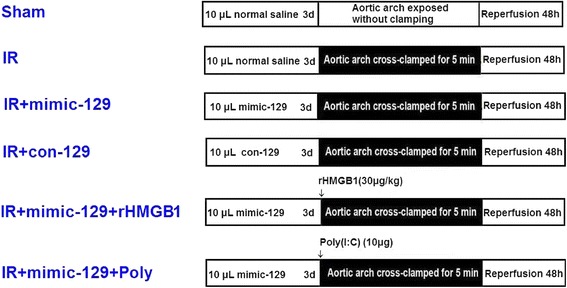
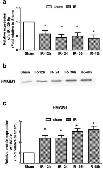
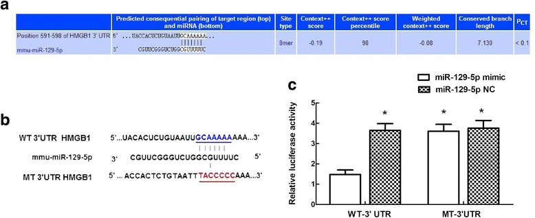
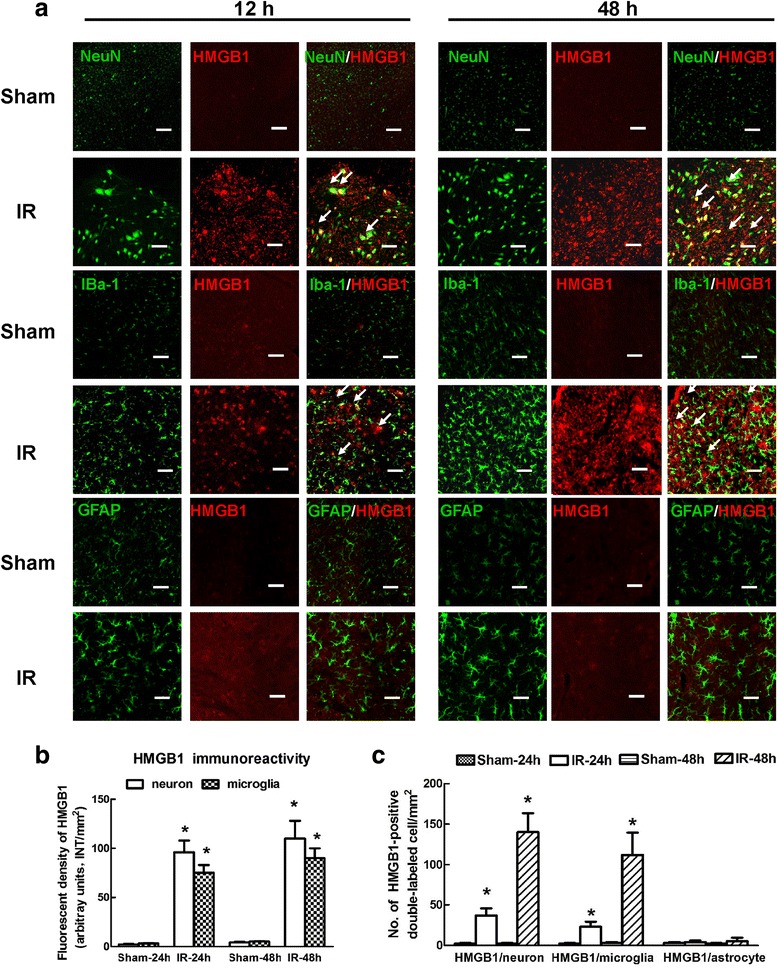
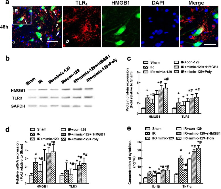
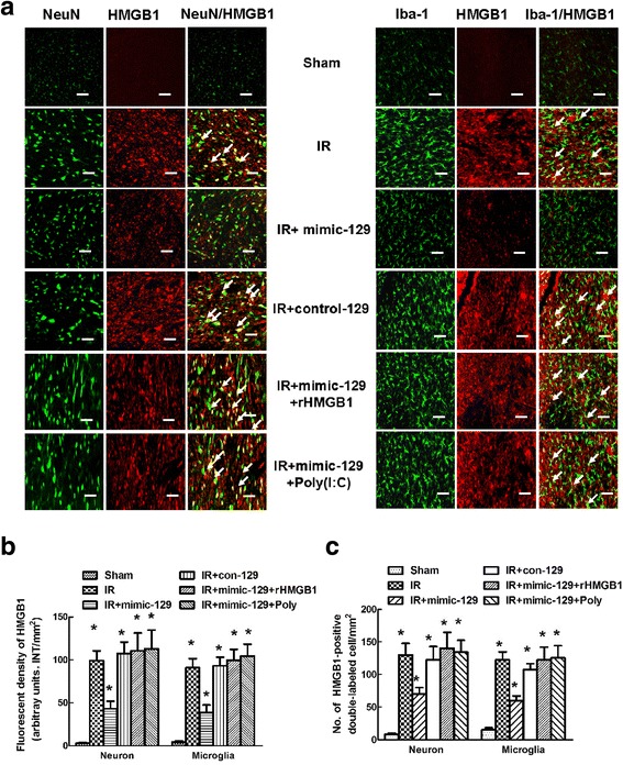
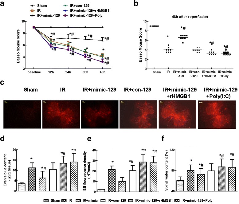
Background: Ischemia-reperfusion (IR) affects microRNA (miR) expression and causes substantial inflammation. Multiple roles of the tumor suppressor miR-129-5p in cerebral IR have recently been reported, but its functions in the spinal cord are unclear. Here, we investigated the role of miR-129-5p after spinal cord IR, particularly in regulating high-mobility group box-1 (HMGB1) and the Toll-like receptor (TLR)-3 pathway. Methods: Ischemia was induced via 5-min occlusion of the aortic arch. The relationship between miR-129-5p and HMGB1 was elucidated via RT-PCR, western blotting, and luciferase assays. The cellular distribution of HMGB1 was determined via double immunofluorescence. The effect of miR-129-5p on the expression of HMGB1, TLR3, and downstream cytokines was evaluated using synthetic miRs, rHMGB1, and the TLR3 agonist Poly(I:C). Blood-spinal cord barrier (BSCB) permeability was examined by measuring Evans blue (EB) dye extravasation and the water content. Results: The temporal miR-129-5p and HMGB1 expression profiles and luciferase assay results indicated that miR-129-5p targeted HMGB1. Compared with the Sham group, the IR group had higher HMGB1 immunoreactivity, which was primarily distributed in neurons and microglia. Intrathecal injection of the miR-129-5p mimic significantly decreased the HMGB1, TLR3, interleukin (IL)-1β and tumor necrosis factor (TNF)-α levels and the double-labeled cell count 48 h post-surgery, whereas rHMGB1 and Poly(I:C) reversed these effects. Injection of miR-129-5p mimic preserved motor function and prevented BSCB leakage based on increased Basso Mouse Scale scores and decreased EB extravasation and water content, whereas injection rHMGB1 and Poly(I:C) aggravated these injuries. Conclusions: Increasing miR-129-5p levels protect against IR by ameliorating inflammation-induced neuronal and BCSB damage by inhibiting HMGB1 and TLR3-associated cytokines.
Overview
The claims and statistics of this paper haven’t been checked yet.
Run the check — free, no account needed. It reads the open-access text and checks the sources it cites.
Integrity signals
Registry
Cites
Statistics
Figures
Verification
Claims the paper makes
The claims and statistics of this paper haven’t been checked yet.
Run the check — free, no account needed. It reads the open-access text and checks the sources it cites.
Claims other papers make about this one
Each is checked against this paper’s own text: does it say what the citing paper says it does?
The cited source says what the claim attributes to it.
In the citing paper
“increasing the levels of miR-129-5p in a mouse model for ischemia ameliorated the expression of proinflammatory cytokines and restored motor function [58].”
From A role for astrocytic miR-129-5p in frontotemporal dementia (2025) · cites this paper as [58]
In the citing paper
“increasing the levels of miR-129-5p in a mouse model for ischemia ameliorated the expression of proinflammatory cytokines and restored motor function [58].”
From A role for astrocytic miR-129-5p in frontotemporal dementia (2025) · cites this paper as [58]
Self-check
Percentages, sums and reported differences re-computed from the paper’s own numbers.
The self-check needs the paper’s full text.
Citations
Cites
No references could be resolved yet.
Research works printed but not linked: 33
- He F, Shi E, Yan L, Li J, Jiang X. Inhibition of micro-ribonucleic acid-320 attenuates neurologic injuries after spinal cord ischemia. J Thorac Cardiovasc Surg. 2015;150:398–406. doi: 10.1016/j.jtcvs.2015.03.066. · DOI
- Li XQ, Lv HW, Tan WF, Wang ZL, Fang B, Ma H. MiR-27a ameliorates inflammatory damage to the blood-spinal cord barrier after spinal cord ischemia-reperfusion injury in rats by down-regulating TICAM-2 of the TLR4 signaling pathway. J Neuroinflammation. 2015;12:25. doi: 10.1186/s12974-015-0246-3. · DOI
- Fang B, Li XQ, Bao NR, Tan WF, Chen FS, Pi XL, et al. Role of autophagy in the bimodal stage after spinal cord ischemia reperfusion injury in rats. Neuroscience. 2016;328:107–16. doi: 10.1016/j.neuroscience.2016.04.019. · DOI
- Bell MT, Puskas F, Agoston VA, Cleveland JC Jr, Freeman KA, Gamboni F, et al. Toll-like receptor 4-dependent microglial activation mediates spinal cord ischemia-reperfusion injury. Circulation. 2013;128(11 Suppl 1):S152–6. doi: 10.1161/CIRCULATIONAHA.112.000024. · DOI
- Li XQ, Wang J, Fang B, Tan WF, Ma H. Intrathecal antagonism of microglial TLR4 reduces inflammatory damage to blood-spinal cord barrier following ischemia/reperfusion injury in rats. Mol Brain. 2014;7(1):28. doi: 10.1186/1756-6606-7-28. · DOI
- Zhao G, Fu C, Wang L, Zhu L, Yan Y, Xiang Y, et al. Down-regulation of nuclear HMGB1 reduces ischemia-induced HMGB1 translocation and release and protects against liver ischemia-reperfusion injury. Sci Rep. 2017;7:46272. doi: 10.1038/srep46272. · DOI
- Chen KB, Uchida K, Nakajima H, Yayama T, Hirai T, Rodriguez Guerrero A, et al. High-mobility group box-1 and its receptors contribute to proinflammatory response in the acute phase of spinal cord injury in rats. Spine (Phila Pa 1976). 2011;36(25):2122–9. doi: 10.1097/BRS.0b013e318203941c. · DOI
- Gu JJ, Chen JB, Zhang JH, Zhang H, Wang SS. Recombinant human soluble thrombomodulin protects against brain injury in a CVST rat model, via downregulation of the HMGB1-RAGE axis. Mol Med Rep. 2016;14(6):5217–22. doi: 10.3892/mmr.2016.5891. · DOI
- Das N, Dewan V, Grace PM, Gunn RJ, Tamura R, Tzarum N, et al. HMGB1 activates proinflammatory signaling via TLR5 leading to allodynia. Cell Rep. 2016;17(4):1128–40. doi: 10.1016/j.celrep.2016.09.076. · DOI
- Liu AH, Wu YT, Wang YP. MicroRNA-129-5p inhibits the development of autoimmune encephalomyelitis-related epilepsy by targeting HMGB1 through the TLR4/NF-kB signaling pathway. Brain Res Bull. 2017;132:139–49. doi: 10.1016/j.brainresbull.2017.05.004. · DOI
- Zhai F, Zhang X, Guan Y, Yang X, Li Y, Song G, et al. Expression profiles of microRNAs after focal cerebral ischemia/reperfusion injury in rats. Neural Regen Res. 2015;7(12):917–23. doi: 10.1016/j.brainresbull.2017.05.004. · DOI
- Balsam LB. Spinal cordischemia-reperfusion injury: MicroRNAs and mitophagy at a crossroads. J Thorac Cardiovasc Surg. 2017; doi: 10.1016/j.jtcvs.2017.06.010. · DOI
- Kouhkan F, Mobarra N, Soufi-Zomorrod M, Keramati F, Hosseini Rad SM, Fathi-Roudsari M, et al. MicroRNA-129-1 acts as tumour suppressor and induces cell cycle arrest of GBM cancer cells through targeting IGF2BP3 and MAPK. J Med Genet. 2016;53(1):24–33. doi: 10.1136/jmedgenet-2015-103225. · DOI
- Ma XL, Li SY, Shang F. Effect of microRNA-129-5p targeting HMGB1-RAGE signaling pathway on revascularization in a collagenase-induced intracerebral hemorrhage rat model. Biomed Pharmacother. 2017;93:238–44. doi: 10.1016/j.biopha.2017.06.012. · DOI
- Li C, Peng S, Liu X, Han C, Wang X, Jin T, et al. Glycyrrhizin, a direct HMGB1 antagonist, ameliorates inflammatory infiltration in a model of autoimmune thyroiditis via inhibition of TLR2-HMGB1 signaling. Thyroid. 2017;27(5):722–31. doi: 10.1089/thy.2016.0432. · DOI
- Qin L, Crews FT. Chronic ethanol increases systemic TLR3 agonist-induced neuroinflammation and neurodegeneration. J Neuroinflammation. 2012;9:130. doi: 10.1186/1742-2094-9-130. · DOI
- Yanai H, Ban T, Wang Z, Choi MK, Kawamura T, Negishi H, et al. HMGB proteins function as universal sentinels for nucleic-acid-mediated innate immune responses. Nature. 2009;462:99–103. doi: 10.1186/1742-2094-9-130. · DOI
- Alexopoulou L, Holt AC, Medzhitov R, Flavell RA. Recognition of double-stranded RNA and activation of NF-kappaB by Toll-like receptor 3. Nature. 2001;413(6857):732–8. doi: 10.1038/35099560. · DOI · Paper page
- Zhang H, Verkman AS. Longitudinally extensive NMO spinal cord pathology produced by passive transfer of NMO-IgG in mice lacking complement inhibitor CD59. J Autoimmun. 2014;53:67–77. doi: 10.1016/j.jaut.2014.02.011. · DOI
- Luo J, Chen J, He L. mir-129-5p attenuates irradiation-induced autophagy and decreases radioresistance of breast cancer cells by targeting HMGB1. Med Sci Monit. 2015;21:4122–9. 10.12659/MSM.896661. · DOI
- Li XQ, Lv HW, Tan WF, Fang B, Wang H, Ma H. Role of the TLR4 pathway in blood-spinal cord barrier dysfunction during the bimodal stage after ischemia/reperfusion injury in rats. J Neuroinflammation. 2014;11:62. doi: 10.1186/1742-2094-11-62. · DOI
- Tsai CH, Liu SC, Wang YH, Su CM, Huang CC, Hsu CJ, et al. Osteopontin inhibition of miR-129-3p enhances IL-17 expression and monocyte migration in rheumatoid arthritis. Biochim Biophys Acta. 2017;1861(2):15–22. doi: 10.1016/j.bbagen.2016.11.015. · DOI
- Sepramaniam S, Tan JR, Tan KS, DeSilva DA, Tavintharan S, Woon FP, et al. Circulating microRNAs as biomarkers of acute stroke. Int J Mol Sci. 2014;15(1):1418–32. doi: 10.3390/ijms15011418. · DOI
- Li XQ, Fang B, Tan WF, Wang ZL, Sun XJ, Zhang ZZ, et al. miR-320a affects spinal cord edema through negatively regulating aquaporin-1 of blood–spinal cord barrier during bimodal stage after ischemia reperfusion injury in rats. BMC Neurosci. 2016;17:10. doi: 10.1186/s12868-016-0243-1. · DOI
- Stevens NE, Chapman MJ, Fraser CK, Kuchel TR, Hayball JD, Diener KR. Therapeutic targeting of HMGB1 during experimental sepsis modulates the inflammatory cytokine profile to one associated with improved clinical outcomes. Sci Rep. 2017;7(1):5850. doi: 10.1038/s41598-017-06205-z. · DOI
- Zheng C, Liu C, Wang W, Tang G, Dong L, Zhou J, et al. Ethanol extracts from Portulaca oleracea L. attenuated ischemia/reperfusion induced rat neural injury through inhibition of HMGB1 induced inflammation. Am J Transl Res. 2016;8(11):5016–24. PMCID: PMC5126344
- Jiang W, Sun R, Wei H, Tian Z. Toll-like receptor 3 ligand attenuates LPS-induced liver injury by down-regulation of toll-like receptor 4 expression on macrophages. Proc Natl Acad Sci U S A. 2005;102(47):17077–82. doi: 10.1073/pnas.0504570102. · DOI
- Lai R, Gu M, Jiang W, Lin W, Xu P, Liu Z, et al. Raf kinase inhibitor protein preferentially promotes TLR3-triggered signaling and inflammation. J Immunol. 2017;198(10):4086–95. doi: 10.4049/jimmunol.1601672. · DOI
- Garg AD, Nowis D, Golab J, Vandenabeele P, Krysko DV, Agostinis P. Immunogenic cell death, DAMPs and anticancer therapeutics: an emerging amalgamation. Biochim Biophys Acta. 1805;2010:53–71. doi: 10.1016/j.bbcan.2009.08.003. · DOI
- Li H, Li Q, Guo T, He W, Dong C, Wang Y. LncRNA CRNDE triggers inflammation through the TLR3-NF-κB-cytokine signaling pathway. Tumour Biol. 2017;39(6):1010428317703821. doi: 10.1177/1010428317703821. · DOI
- Suresh MV, Thomas B, Machado-Aranda D, Dolgachev VA, Kumar Ramakrishnan S, Talarico N, et al. Double-stranded RNA interacts with toll-like receptor 3 in driving the acute inflammatory response following lung contusion. Crit Care Med. 2016;44(11):e1054–66. doi: 10.1097/CCM.0000000000001879. · DOI
- Yamagata K, Sone N, Suguyama S, Nabika T. Different effects of arginine vasopressin on high-mobility group box 1 expression in astrocytes isolated from stroke-prone spontaneously hypertensive rats and congenic SHRpch1_18 rats. Int J Exp Pathol. 2016;97(2):97–106. doi: 10.1111/iep.12172. · DOI
- Ohnishi M, Monda A, Takemoto R, Fujimoto Y, Sugitani M, Iwamura T, et al. High-mobility group box 1 up-regulates aquaporin 4 expression via microglia-astrocyte interaction. Neurochem Int. 2014;5:32–8. doi: 10.1016/j.neuint.2014.05.007. · DOI
Cited by
Sorted by citations
A role for astrocytic miR-129-5p in frontotemporal dementia
Kaurani, L., Pradhan, R., Schröder, S., Burkhardt, S., Schuetz, A.-L., Krüger, D. M., Pena, T., Heutink, P., Sananbenesi, F., & Fischer, A. (2025) · Translational Psychiatry · doi:10.1038/s41398-025-03338-y · Cited by 10
Metadata
- Authors
- Li, X.-Q., Chen, F.-S., Tan, W.-F., Fang, B., Zhang, Z.-L., & Ma, H.
- ORCID
- Li, Xiao-Qian
- Venue
- Journal of Neuroinflammation
- Published
- 2017-10-23
- Volume
- 14
- Issue
- 1
- ISSN
- 1742-2094
- PMC
- PMC5654055
- Cited by
- 88
- References
- 0
- Language
- en
- Update notices
- correction (2021-12-28)
- Funders
- National Natural Science Foundation of China (81601053, 81401000)
- Scientific Research Project of Liaoning Province Department of Education (LK201636))
- Science and Technology Program of Liaoning (2012408002)
About these checks
Registry, retraction, Cites and statistics results are computed, not judged, with one exception: a printed reference that no index lists is read once by a model before it counts as a citation error. Claims are extracted by a model and kept only when their quote appears verbatim in the text; each cited source is then checked, with a model’s help, for whether it says what the claim relies on. Those verdicts can be wrong — read the claim’s quote and the cited source before relying on one.
Statistics and arithmetic are recomputed, not judged.
Confidence bands are shown only for fields calibrated on our annotated test set. None is calibrated yet, so no band is shown.
What counts as a claim ›How we measure confidence ›How we check this ›
Are you an author, or do you see a problem with this page? Request a review
