

The DOI matches its Crossref record; 32 of the 32 references it cites were found in a registry or an index.
Safety and clinical activity with an anti-PD-1 antibody JS001 in advanced melanoma or urologic cancer patients
Open accessAbstract
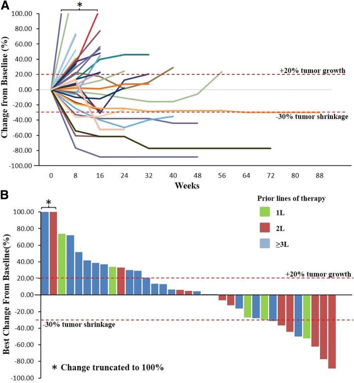
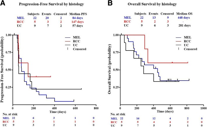
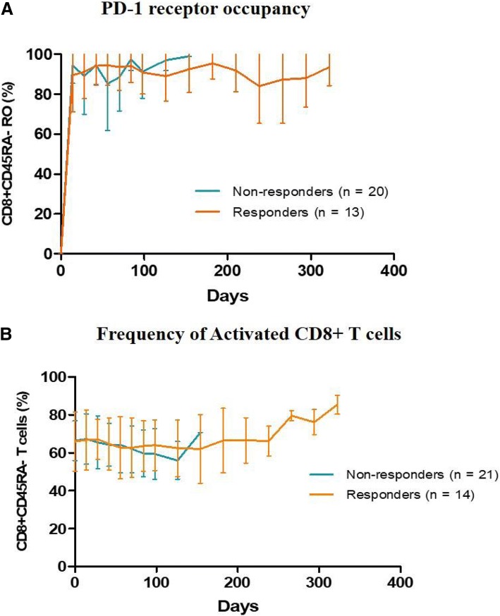
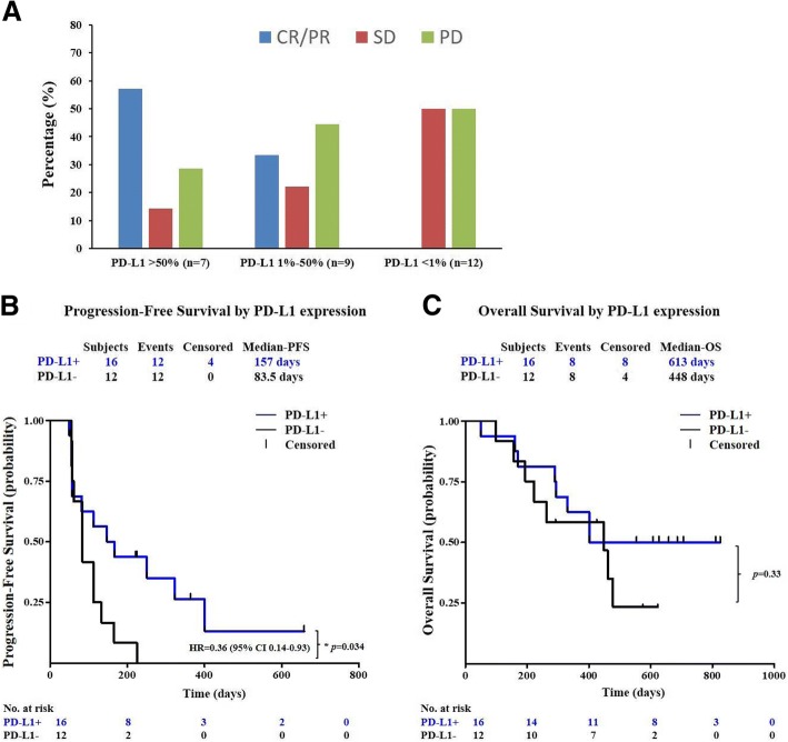
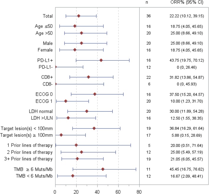
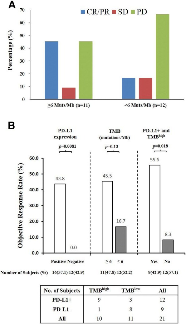
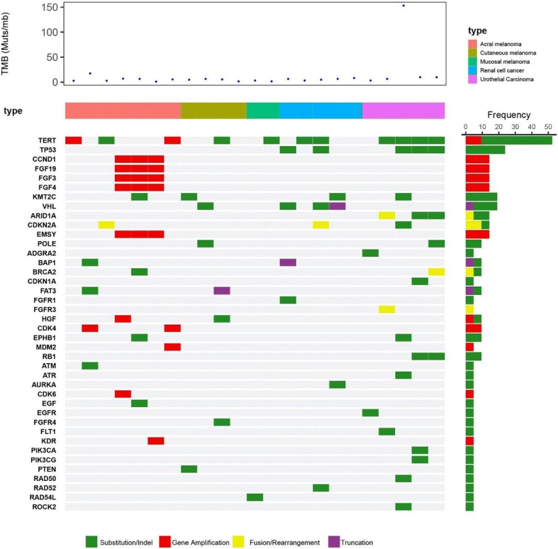
BACKGROUND: monoclonal antibody against the programmed death-1 (PD-1) receptor, blocks the interaction of PD-1 with its ligands and promotes T cell activation in preclinical studies. This phase I study is designed to evaluate the safety, tolerability, and clinical activity of JS001 in advanced melanoma or urologic cancer patients who are refractory to standard systemic therapy. PATIENTS AND METHODS: In the dose escalation cohorts, subjects initially received a single-dose, intravenous infusion of JS001, and were followed for 28 days followed by multi-dose infusions every 2 weeks. In the dose expansion cohorts, subjects received multi-dose infusions every 2 weeks. Clinical response was evaluated after each 8-week treatment cycle according to RECIST v1.1 criteria. RESULTS: Thirty-six subjects diagnosed with advanced melanoma (n = 22), urothelial cancer (UC) (n = 8), or renal cell cancer (RCC) (n = 6) were enrolled. Melanoma subjects included 14 acral and 4 mucosal subtypes. JS001 was well tolerated, and no dose-limiting toxicity was observed. By the safety data cutoff date, 100% of subjects had treatment-related adverse events (TRAE) with most adverse events being grade 1 or 2, and ≥ grade 3 TRAEs occurred in 36%. Among all 36 subjects, 1 confirmed complete response (acral melanoma), 7 confirmed partial responses (2 acral melanoma, 1 mucosal melanoma, 2 UC, and 2 RCC), and 10 stable disease were observed, for an objective response rate of 22.2% (95% CI, 10.1 to 39.2), and a disease control rate of 50.0% (95% CI, 32.9 to 67.1). Clinical responses were correlated with PD-L1 expression on tumor cells, the presence of tumor infiltrating lymphocytes (TIL), baseline tumor volume, ECOG performance status, serum LDH levels, high percentage of activated CD8+ T cells and CD3- CD16+ CD54+ NK cells in the peripheral blood as well as tumor mutational burden (TMB). CONCLUSION: JS001 was well tolerated and demonstrated promising anti-tumor activity in UC and RCC as well as in previously underexplored acral and mucosal melanoma subtypes. Subjects with an immune-active profile in the tumor microenvironment or in peripheral blood responded favorably to JS001 treatment. The completion of the current phase I study has led to the initiation of the first prospective anti-PD-1 registration trial in Asia focusing on acral and mucosal melanoma subtypes, representative of the regional disease epidemiology. TRIAL REGISTRATION: Clinical Trial ID: NCT02836795 , registered July 19, 2016, retrospectively registered.
Overview
The claims and statistics of this paper haven’t been checked yet.
Run the check — free, no account needed. It reads the open-access text and checks the sources it cites.
Integrity signals
Registry
Cites
Statistics
Figures
Verification
Claims the paper makes
The claims and statistics of this paper haven’t been checked yet.
Run the check — free, no account needed. It reads the open-access text and checks the sources it cites.
Claims other papers make about this one
Each is checked against this paper’s own text: does it say what the citing paper says it does?
In the citing paper
“At present, antibodies blocking PD-1 or its ligand PD-L1 have been approved to treat various solid and hematologic malignancies (Table 1) [7–12].”
From Combination strategies with PD-1/PD-L1 blockade: current advances and future directions (2022) · cites this paper as [12]
Self-check
Percentages, sums and reported differences re-computed from the paper’s own numbers.
The self-check needs the paper’s full text.
Citations
Cites
PD-1 and PD-L1 Immune Checkpoint Blockade to Treat Breast Cancer
Hartkopf, A. D., Taran, F., Wallwiener, M., Walter, C. B., Krämer, B., Grischke, E., & Brucker, S. Y. (2016) · Breast Care · doi:10.1159/000453569 · Cited by 30,989
Adverse Renal Effects of Immune Checkpoint Inhibitors: A Narrative Review
Wanchoo, R., Karam, S., Uppal, N. N., Barta, V. S., Deray, G., Devoe, C., Launay‐Vacher, V., Jhaveri, K. D., & on behalf of Cancer and Kidney International Network Workgroup on Immune Checkpoint Inhibitors (2017) · American Journal of Nephrology · doi:10.1159/000455014 · Cited by 15,320
Freeman, G. J., Long, A. J., Iwai, Y., Bourque, K., Chernova, T., Nishimura, H., Fitz, L. J., Malenkovich, N., Okazaki, T., Byrne, M. C., et al. (2000) · The Journal of Experimental Medicine · doi:10.1084/jem.192.7.1027 · Cited by 5,294
Tumor-associated B7-H1 promotes T-cell apoptosis: A potential mechanism of immune evasion
Dong, H., Strome, S. E., Salomao, D. R., Tamura, H., Hirano, F., Flies, D. B., Roche, P. C., Lu, J., Zhu, G., Tamada, K., et al. (2002) · Nature Medicine · doi:10.1038/nm730 · Cited by 5,025
Safety and Tumor Responses with Lambrolizumab (Anti–PD-1) in Melanoma
Hamid, O., Robert, C., DAUD, A. I., Hodi, F. S., Hwu, W., Kefford, R. F., Wolchok, J. D., Hersey, P., Joseph, R. W., Weber, J. S., et al. (2013) · New England Journal of Medicine · doi:10.1056/nejmoa1305133 · Cited by 3,427
Ishida, Y., Agata, Y., Shibahara, K., & Honjo, T. (1992) · The EMBO Journal · doi:10.1002/j.1460-2075.1992.tb05481.x · Cited by 3,259
Zou, W., Wolchok, J. D., & Chen, L. (2016) · Science Translational Medicine · doi:10.1126/scitranslmed.aad7118 · Cited by 2,400
Topalian, S. L., Sznol, M., McDermott, D. F., Kluger, H. M., Carvajal, R. D., Sharfman, W. H., Brahmer, J. R., Lawrence, D. P., Atkins, M. B., Powderly, J. D., et al. (2014) · Journal of Clinical Oncology · doi:10.1200/jco.2013.53.0105 · Cited by 2,222
Taube, J. M., Anders, R. A., Young, G. D., Xu, H., Sharma, R., McMiller, T. L., Chen, S., Klein, A. P., Pardoll, D. M., Topalian, S. L., et al. (2012) · Science Translational Medicine · doi:10.1126/scitranslmed.3003689 · Cited by 2,157
Regulatory T cells, tumour immunity and immunotherapy
Zou, W. (2006) · Nature reviews. Immunology · doi:10.1038/nri1806 · Cited by 2,135
Cited by
Sorted by citations
Combination strategies with PD-1/PD-L1 blockade: current advances and future directions
Yi, M., Zheng, X., Niu, M., Zhu, S., Ge, H., & Wu, K. (2022) · Molecular Cancer · doi:10.1186/s12943-021-01489-2 · Cited by 1,445
Perioperative Toripalimab Plus Chemotherapy for Patients With Resectable Non–Small Cell Lung Cancer
Lü, S., Zhang, W., Wu, L., Wang, W., Zhang, P., Neotorch Investigators, Fang, W., Xing, W., Chen, Q., Yang, L., et al. (2024) · JAMA · doi:10.1001/jama.2023.24735 · Cited by 405
Self-citationSignal pathways of melanoma and targeted therapy
Guo, W., Wang, H., & Li, C. (2021) · Signal Transduction and Targeted Therapy · doi:10.1038/s41392-021-00827-6 · Cited by 419
T Cell Dysfunction in Cancer Immunity and Immunotherapy
Xia, A., Zhang, Y., Jiang, X., Yin, T., & Lu, X. (2019) · Frontiers in Immunology · doi:10.3389/fimmu.2019.01719 · Cited by 344
Toripalimab Plus Chemotherapy for Recurrent or Metastatic Nasopharyngeal Carcinoma
Mai, H., Chen, Q., Chen, D., Hu, C., Yang, K., Wen, J., Li, J., Shi, Y., Jin, F., Xu, R., et al. (2023) · JAMA · doi:10.1001/jama.2023.20181 · Cited by 299
Self-citation
Metadata
- Authors
- Tang, B., Yan, X., Sheng, X., Si, L., Cui, C., Kong, Y., Mao, L., Lian, B., Bai, X., Wang, X., Li, S., Zhou, L., Yu, J., Dai, J., Wang, K., Hu, J., Dong, L., Song, H., Wu, H., Feng, H., Yao, S., Chi, Z., & Guo, J.
- Affiliations
- Peking University Cancer Hospital (CN)
- Beijing Radiation Center (CN)
- Junshi Biosciences (China) (CN)
- Corresponding author
- Chi, Zhihong
- Guo, Jun
- ORCID
- Venue
- Journal of Hematology & Oncology
- Published
- 2019-01-14
- Volume
- 12
- Issue
- 1
- Pages
- 7-7
- ISSN
- 1756-8722 (ISSN-L)
- PMC
- PMC6332582
- OpenAlex
- W2923110622
- Cited by
- 169
- References
- 32
- Citations by year
- 2026: 5 · 2025: 12 · 2024: 21 · 2023: 30 · 2022: 31 · 2021: 32 · 2020: 26 · 2019: 12
- FWCI
- 11.39
- Type
- journal article
- Language
- en
- Open access
- gold
- doi.org
- Licence
- Locations
- Journal of Hematology & Oncology (publishedVersion, cc-by)
- Europe PMC (PubMed Central) (publishedVersion, other-oa)
- PubMed Central (publishedVersion, cc-by)
- PubMed (publishedVersion)
- DOAJ (DOAJ: Directory of Open Access Journals) (submittedVersion, cc-by-sa)
- Funders
- Beijing Municipal Administration of Hospitals Clinical medicine Development of special funding support (ZYLX201603)
- National Science &Technology Major Project “Key New Drug Creation and Manufacturing Program” (2015ZX09102017)
- Topics
- Health Sciences › Medicine › Oncology › Cancer Immunotherapy and Biomarkers
- Health Sciences › Medicine › Oncology › Cutaneous Melanoma Detection and Management
- Life Sciences › Immunology and Microbiology › Immunology › Immunotherapy and Immune Responses
- Keywords
- anti-PD-1, advanced melanoma, urothelial carcinoma, renal cell carcinoma, immune checkpoint blockade, safety and tolerability, objective response rate, PD-L1 expression, tumor-infiltrating lymphocytes, tumor mutational burden, acral melanoma, mucosal melanoma, predictive biomarkers, dose escalation, dose expansion, natural killer cells
- MeSH terms
- Progression-Free Survival, Adult, Aged, Antineoplastic Agents, Biopsy, Carcinoma, Renal Cell, Dose-Response Relationship, Drug, Female, Follow-Up Studies, Humans, Immunoglobulin G, Infusions, Intravenous, Lymphocyte Activation, Male, Melanoma, Middle Aged, Skin Neoplasms, Biomarkers, Tumor, Urologic Neoplasms, CD8-Positive T-Lymphocytes, B7-H1 Antigen, Programmed Cell Death 1 Receptor, Antibodies, Monoclonal, Humanized
About these checks
Registry, retraction, Cites and statistics results are computed, not judged, with one exception: a printed reference that no index lists is read once by a model before it counts as a citation error. Claims are extracted by a model and kept only when their quote appears verbatim in the text; each cited source is then checked, with a model’s help, for whether it says what the claim relies on. Those verdicts can be wrong — read the claim’s quote and the cited source before relying on one.
Statistics and arithmetic are recomputed, not judged.
Confidence bands are shown only for fields calibrated on our annotated test set. None is calibrated yet, so no band is shown.
What counts as a claim ›How we measure confidence ›How we check this ›
Are you an author, or do you see a problem with this page? Request a review
