

The DOI matches its Crossref record; 2 of the 2 references it cites were found in a registry or an index.
Huc-MSCs-derived exosomes alleviate non-alcoholic steatohepatitis by regulating macrophages polarization through miR-24-3p/STING axis
Open accessAbstract
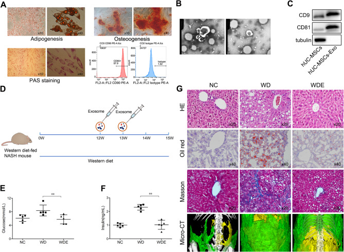
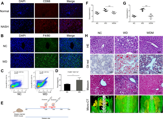
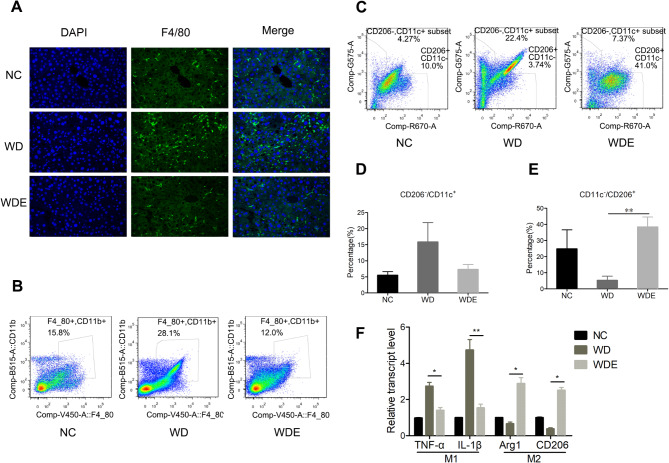
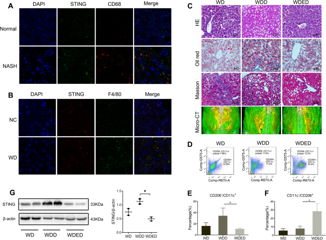
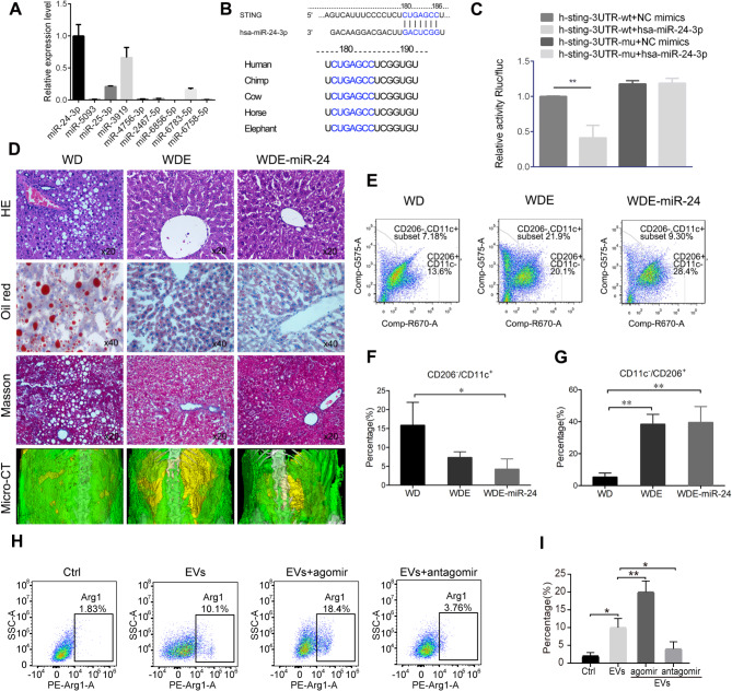
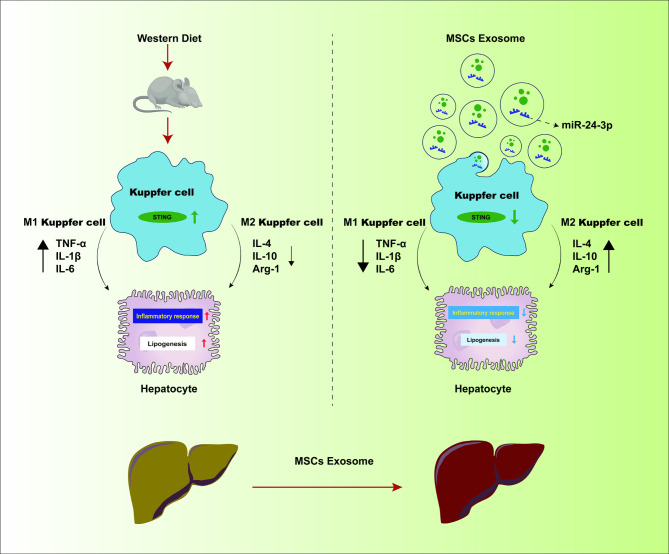
BACKGROUND: There's a scarcity of drugs effective against nonalcoholic steatohepatitis (NASH). Exosomes from Human umbilical cord mesenchymal stem cells (huc-MSCs) show potential in managing glycolipid metabolism and the immune response. Therefore, further investigations are required to explore their application in NASH and the underlying mechanisms. METHODS: C57BL/6J mice were fed with a western diet for 12 weeks to induce NASH, and huc-MSCs exosomes (MSCs-exo) were administered during the feeding period. The effect of MSCs-exo was evaluated by monitoring changes in body weight, fat distribution, blood glucose, and insulin levels, and analyzing pathological alterations in liver tissue. Mechanism investigations were carried out using flow cytometry, immunofluorescence staining, and other experimental techniques. RESULTS: MSCs-exo could reduce liver fat, inflammation, fibrosis, and improved metabolism to alleviate the progression of NASH. Besides, MSCs-exo could decrease macrophage accumulation in the liver, encouraging M2 over M1 macrophage polarization. Furthermore, our study found that MSCs-exo had a high expression of miR-24-3p, which may regulate macrophage polarization by targeting the interferon-stimulated genes (STING) gene in macrophages, with its overexpression amplifying MSCs-exo's NASH benefits. CONCLUSIONS: These findings suggest that the therapeutic effect of MSCs-exo on NASH may be attributed to the regulation of macrophage M2 polarization through miR-24-3p targeting STING. This provides a scientific basis for future clinical application.
Overview
The claims and statistics of this paper haven’t been checked yet.
Run the check — free, no account needed. It reads the open-access text and checks the sources it cites.
Integrity signals
Registry
Cites
Statistics
Figures
Verification
Claims the paper makes
The claims and statistics of this paper haven’t been checked yet.
Run the check — free, no account needed. It reads the open-access text and checks the sources it cites.
Claims other papers make about this one
Each is checked against this paper’s own text: does it say what the citing paper says it does?
The cited source says what the claim attributes to it.
In the citing paper
“Recently, MSC-Exos have been reported to alleviate NASH by regulating macrophage polarization through miR-24-3p/STING axis [209].”
From Mechanisms, efficacy, and future perspectives of cellular-based therapies for liver fibrosis/cirrhosis: focusing on mesenchymal stromal cells (2025) · cites this paper as [209]
Self-check
Percentages, sums and reported differences re-computed from the paper’s own numbers.
The self-check needs the paper’s full text.
Citations
Cites
Tsuchida, T., Lee, Y. A., Fujiwara, N., Ybanez, M., Allen, B., Martins, S., Fiel, M. I., Goossens, N., Chou, H.-I., Hoshida, Y., et al. (2018) · Journal of Hepatology · doi:10.1016/j.jhep.2018.03.011 · Cited by 583
The Role of Innate Immune Cells in Nonalcoholic Steatohepatitis
Cai, J., Zhang, X., & Li, H. (2019) · Hepatology · doi:10.1002/hep.30506 · Cited by 207
Research works printed but not linked: 39
- Powell EE, Wong VW, Rinella M. Non-alcoholic fatty liver disease. Lancet (London England). 2021;397:2212–24. · DOI · Paper page
- Chalasani N, Younossi Z, Lavine JE, Charlton M, Cusi K, Rinella M et al. The diagnosis and management of nonalcoholic fatty liver disease: Practice guidance from the American Association for the Study of Liver Diseases, Hepatology (Baltimore, Md.), 67 (2018) 328–357. · DOI · Paper page
- Schuster S, Cabrera D, Arrese M, Feldstein AE. Triggering and resolution of inflammation in NASH, Nature reviews. Gastroenterol Hepatol. 2018;15:349–64.
- Friedman SL, Neuschwander-Tetri BA, Rinella M, Sanyal AJ. Mechanisms of NAFLD development and therapeutic strategies. Nat Med. 2018;24:908–22. · DOI · Paper page
- EASL-EASD-EASO. Clinical practice guidelines for the management of non-alcoholic fatty liver disease. J Hepatol. 2016;64:1388–402. · DOI · Paper page
- Huang DQ, El-Serag HB, Loomba R. Global epidemiology of NAFLD-related HCC: trends, predictions, risk factors and prevention, Nature reviews. Gastroenterol Hepatol. 2021;18:223–38.
- Wang YH, Wu DB, Chen B, Chen EQ, Tang H. Progress in mesenchymal stem cell-based therapy for acute liver failure. Stem Cell Res Ther. 2018;9:227. · DOI
- Wu DB, Chen EQ, Tang H. Stem cell transplantation for the treatment of end-stage liver disease. World J Hepatol. 2018;10:907–10. · DOI
- Uccelli A, Moretta L, Pistoia V. Mesenchymal stem cells in health and disease. Nat Rev Immunol. 2008;8:726–36. · DOI
- Qi Y, Ma J, Li S, Liu W. Applicability of adipose-derived mesenchymal stem cells in treatment of patients with type 2 diabetes. Stem Cell Res Ther. 2019;10:274. · DOI
- Liu X, Zheng P, Wang X, Dai G, Cheng H, Zhang Z, et al. A preliminary evaluation of efficacy and safety of Wharton’s jelly mesenchymal stem cell transplantation in patients with type 2 diabetes mellitus. Stem Cell Res Ther. 2014;5:57. · DOI
- Wang M, Song L, Strange C, Dong X, Wang H. Therapeutic effects of adipose stem cells from Diabetic mice for the treatment of type 2 diabetes. Mol Therapy: J Am Soc Gene Therapy. 2018;26:1921–30. · DOI
- Winkler S, Borkham-Kamphorst E, Stock P, Brückner S, Dollinger M, Weiskirchen R, et al. Human mesenchymal stem cells towards non-alcoholic steatohepatitis in an immunodeficient mouse model. Exp Cell Res. 2014;326:230–9. · DOI
- Yang Z, Xia Q, Lu D, Yue H, Zhang J, Li Y, et al. Human mesenchymal stem cells treatment improved hepatic lesions and reversed gut microbiome disorder in non-alcoholic steatohepatitis. Aging. 2020;12:21660–73. · DOI
- Phinney DG, Pittenger MF, Review C. MSC-Derived exosomes for cell-free therapy, stem cells (Dayton. Ohio). 2017;35:851–8.
- Mead B, Tomarev S. Bone marrow-derived mesenchymal stem cells-derived exosomes promote survival of retinal ganglion cells through miRNA-Dependent mechanisms. Stem Cells Translational Med. 2017;6:1273–85. · DOI
- Su T, Xiao Y, Xiao Y, Guo Q, Li C, Huang Y, et al. Bone marrow mesenchymal stem cells-derived exosomal MiR-29b-3p regulates Aging-Associated insulin resistance. ACS Nano. 2019;13:2450–62.
- Ha DH, Kim HK, Lee J, Kwon HH, Park GH, Yang SH et al. Mesenchymal Stem/Stromal Cell-Derived Exosomes for Immunomodulatory Therapeutics and Skin Regeneration, Cells, 9 (2020). · DOI
- Yagi H, Soto-Gutierrez A, Navarro-Alvarez N, Nahmias Y, Goldwasser Y, Kitagawa Y, et al. Reactive bone marrow stromal cells attenuate systemic inflammation via sTNFR1. Mol Therapy: J Am Soc Gene Therapy. 2010;18:1857–64. · DOI
- Nowacki M, Nazarewski Ł, Pokrywczyńska M, Kloskowski T, Tyloch D, Pietkun K, et al. Long-term influence of bone marrow-derived mesenchymal stem cells on liver ischemia-reperfusion injury in a rat model. Ann Transplant. 2015;20:132–40. · DOI
- Devi A, Pahuja I, Singh SP, Verma A, Bhattacharya D, Bhaskar A, et al. Revisiting the role of mesenchymal stem cells in tuberculosis and other infectious diseases. Cell Mol Immunol. 2023;20:600–12. · DOI
- Prakash N, Kim J, Jeon J, Kim S, Arai Y, Bello AB, et al. Progress and emerging techniques for biomaterial-based derivation of mesenchymal stem cells (MSCs) from pluripotent stem cells (PSCs). Biomaterials Res. 2023;27:31. · DOI · Paper page
- Shi H, Zhou K, Wang M, Wang N, Song Y, Xiong W, et al. Integrating physicomechanical and biological strategies for BTE: biomaterials-induced osteogenic differentiation of MSCs. Theranostics. 2023;13:3245–75. · DOI
- Wang M, Zhao N, Wang C, Jin ZB, Zhang L. Immunomodulatory properties of mesenchymal stem cells: a potential therapeutic strategy for allergic rhinitis. Allergy. 2023;78:1425–40. · DOI
- Yang X, Li Q, Liu W, Zong C, Wei L, Shi Y, et al. Mesenchymal stromal cells in hepatic fibrosis/cirrhosis: from pathogenesis to treatment. Cell Mol Immunol. 2023;20:583–99. · DOI · Paper page
- Ala M. The beneficial effects of mesenchymal stem cells and their exosomes on myocardial infarction and critical considerations for enhancing their efficacy. Ageing Res Rev. 2023;89:101980. · DOI
- Giovannelli L, Bari E, Jommi C, Tartara F, Armocida D, Garbossa D, et al. Mesenchymal stem cell secretome and extracellular vesicles for neurodegenerative diseases: risk-benefit profile and next steps for the market access. Bioactive Mater. 2023;29:16–35. · DOI · Paper page
- Wang C, Xu M, Fan Q, Li C, Zhou X. Therapeutic potential of exosome-based personalized delivery platform in chronic inflammatory diseases. Asian J Pharm Sci. 2023;18:100772.
- Sun Y, Shi H, Yin S, Ji C, Zhang X, Zhang B, et al. Human mesenchymal stem cell derived exosomes alleviate type 2 diabetes Mellitus by reversing Peripheral insulin resistance and relieving β-Cell Destruction. ACS Nano. 2018;12:7613–28. · DOI
- Tilg H, Moschen AR, Roden M. NAFLD and Diabetes Mellitus, Nature reviews. Gastroenterol Hepatol. 2017;14:32–42.
- Cai J, Zhang XJ, Li H. Role of Innate Immune Signaling in non-alcoholic fatty liver disease, trends in endocrinology and metabolism. Volume 29. TEM; 2018. pp. 712–22. · DOI
- Kazankov K, Jørgensen SMD, Thomsen KL, Møller HJ, Vilstrup H, George J, et al. The role of macrophages in nonalcoholic fatty liver disease and nonalcoholic steatohepatitis, Nature reviews. Gastroenterol Hepatol. 2019;16:145–59.
- Qiao JT, Cui C, Qing L, Wang LS, He TY, Yan F, et al. Activation of the STING-IRF3 pathway promotes hepatocyte inflammation, apoptosis and induces metabolic disorders in nonalcoholic fatty liver disease. Metab Clin Exp. 2018;81:13–24. · DOI
- Petrasek J, Iracheta-Vellve A, Csak T, Satishchandran A, Kodys K, Kurt-Jones EA, et al. STING-IRF3 pathway links endoplasmic reticulum stress with hepatocyte apoptosis in early alcoholic liver disease. Proc Natl Acad Sci USA. 2013;110:16544–9. · DOI
- Luo X, Li H, Ma L, Zhou J, Guo X, Woo SL et al. Expression of STING Is Increased in Liver Tissues From Patients With NAFLD and Promotes Macrophage-Mediated Hepatic Inflammation and Fibrosis in Mice, Gastroenterology, 155 (2018) 1971–1984.e4. · DOI · Paper page
- Yu Y, Liu Y, An W, Song J, Zhang Y, Zhao X. STING-mediated inflammation in Kupffer cells contributes to progression of nonalcoholic steatohepatitis. J Clin Investig. 2019;129:546–55. · DOI
- Fabian MR, Sonenberg N. The mechanics of miRNA-mediated gene silencing: a look under the hood of miRISC. Nat Struct Mol Biol. 2012;19:586–93. · DOI
- Khan M, Harms JS, Liu Y, Eickhoff J, Tan JW, Hu T, et al. Brucella suppress STING expression via miR-24 to enhance infection. PLoS Pathog. 2020;16:e1009020. · DOI
- Xu T, Chu Q, Cui J. Rhabdovirus-Inducible MicroRNA-210 modulates antiviral innate Immune Response via Targeting STING/MITA in Fish. J Immunol (Baltimore Md: 1950). 2018;201:982–94. · DOI
Cited by
Sorted by citations
Pan, X., Gao, T., & Wang, B. (2025) · Cell & Bioscience · doi:10.1186/s13578-025-01520-9 · Cited by 2
“Recently, MSC-Exos have been reported to alleviate NASH by regulating macrophage polarization through miR-24-3p/STING axis [209].”
Metadata
- Authors
- Jiang, W., Zeng, Q., Liu, C.-H., Wang, Y., Wang, S., Chen, E., Wang, M., Zhou, T., Bai, L., Wu, D., & Tang, H.
- Affiliations
- Sichuan University (CN)
- Corresponding author
- Wu, Dongbo
- Tang, Hong
- Venue
- Stem Cell Research & Therapy
- Published
- 2025-02-21
- Volume
- 16
- Issue
- 1
- Pages
- 74-74
- ISSN
- 1757-6512 (ISSN-L)
- PMC
- PMC11846240
- OpenAlex
- W4407842915
- Cited by
- 26
- References
- 2
- Citations by year
- 2026: 15 · 2025: 11
- FWCI
- 7.82
- Type
- journal article
- Language
- en
- Open access
- gold
- doi.org
- Licence
- Locations
- Stem Cell Research & Therapy (publishedVersion, cc-by-nc-nd)
- PubMed Central (publishedVersion, cc-by-nc-nd)
- PubMed (publishedVersion)
- DOAJ (DOAJ: Directory of Open Access Journals) (submittedVersion)
- Funders
- National Key Research and Development Program of China (2022YFC2304800)
- 1.35 project for disciplines of excellence, West China Hospital, Sichuan University (ZYGD23030)
- National Natural Science Foundation of China (82172254, 81900512)
- the Science and Technological Supports Project of Sichuan Province, China (2024NSFSC1690)
- Topics
- Life Sciences › Biochemistry, Genetics and Molecular Biology › Molecular Biology › Extracellular vesicles in disease
- Health Sciences › Medicine › Hepatology › Liver Disease Diagnosis and Treatment
- Life Sciences › Biochemistry, Genetics and Molecular Biology › Cell Biology › Mesenchymal stem cell research
- Keywords
- nonalcoholic steatohepatitis, exosomes, human umbilical cord mesenchymal stem cells, macrophage polarization, M2 macrophages, miR-24-3p, STING, liver fibrosis, liver inflammation, lipid metabolism
- MeSH terms
- STING Protein, Animals, Humans, Liver, Macrophages, Male, Membrane Proteins, Mice, Inbred C57BL, Umbilical Cord, MicroRNAs, Mice, Exosomes, Mesenchymal Stem Cells, Non-alcoholic Fatty Liver Disease
About these checks
Registry, retraction, Cites and statistics results are computed, not judged, with one exception: a printed reference that no index lists is read once by a model before it counts as a citation error. Claims are extracted by a model and kept only when their quote appears verbatim in the text; each cited source is then checked, with a model’s help, for whether it says what the claim relies on. Those verdicts can be wrong — read the claim’s quote and the cited source before relying on one.
Statistics and arithmetic are recomputed, not judged.
Confidence bands are shown only for fields calibrated on our annotated test set. None is calibrated yet, so no band is shown.
What counts as a claim ›How we measure confidence ›How we check this ›
Are you an author, or do you see a problem with this page? Request a review
