

The DOI matches its Crossref record; 49 of the 49 references it cites were found in a registry or an index.
IGFBP7 inhibits cell proliferation by suppressing AKT activity and cell cycle progression in thyroid carcinoma
Open accessAbstract
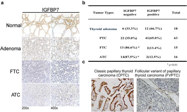
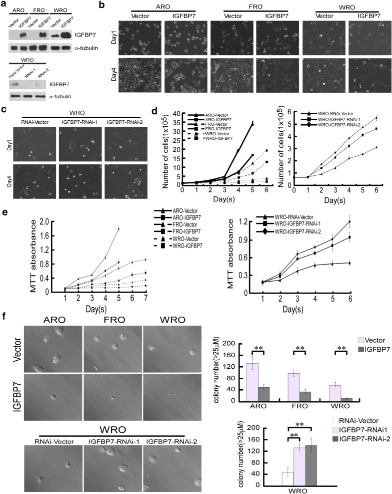
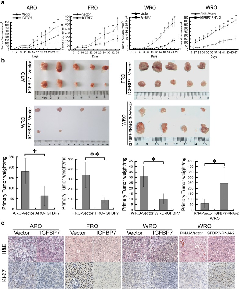
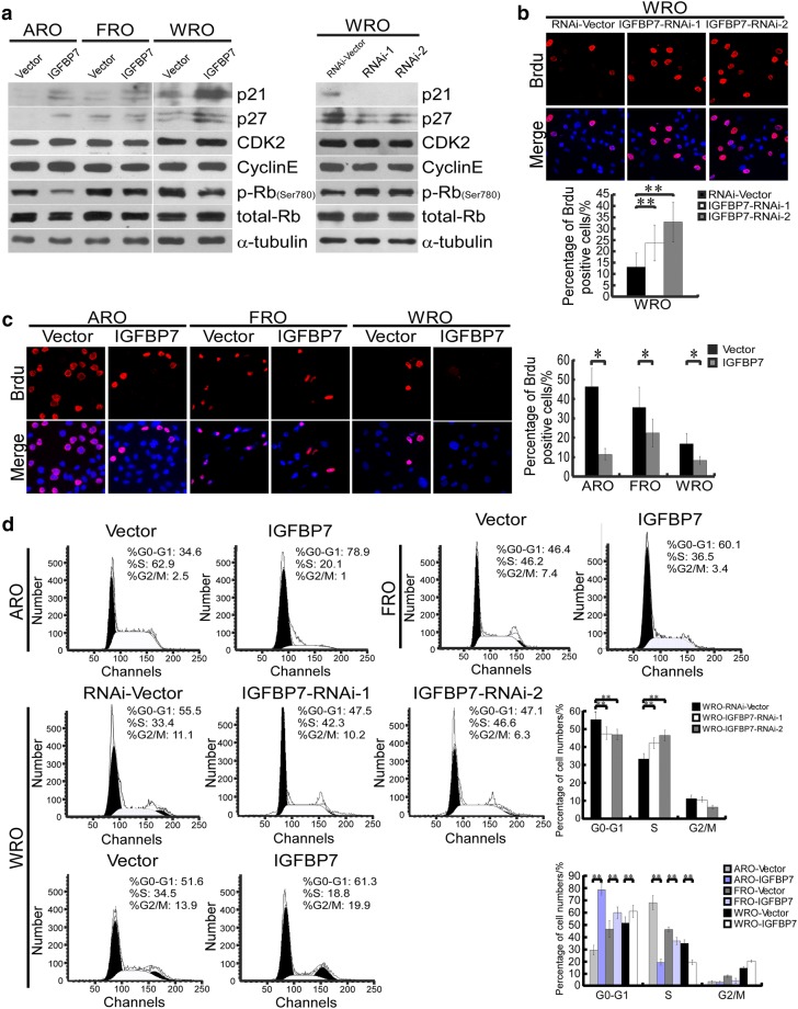
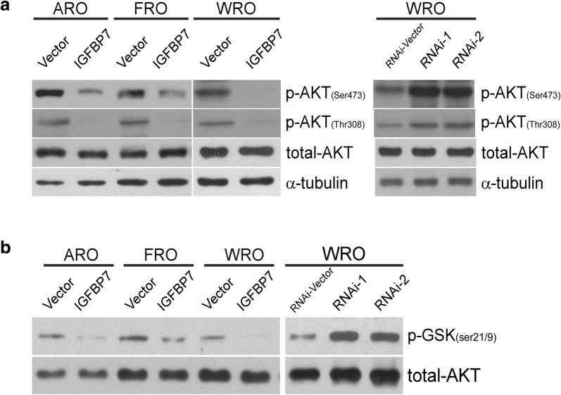
Thyroid cancer is the most common malignant endocrine tumor and is classified into papillary thyroid cancer (PTC), follicular thyroid cancer (FTC) and anaplastic thyroid cancer (ATC), which have substantially different characteristics. Insulin-like growth factor binding protein 7 (IGFBP7) has recently been recognized as a tumor suppressor in many cancer types. However, the expression pattern of IGFBP7 and its biological function in various types of thyroid carcinoma remain poorly understood. We found that the protein levels of IGFBP7 in FTC and ATC tissues were significantly lower or even absent compared with those in normal thyroid, benign thyroid adenoma and classical PTC tissues. Moreover, overexpression of IGFBP7 in two undifferentiated ATC cell lines, ARO and FRO, and one differentiated FTC cell line, WRO, significantly inhibited cell proliferation in vitro. In vivo experiments revealed that ectopic IGFBP7 expression markedly suppressed growth of tumor xenografts derived from these thyroid cancer cell lines, while IGFBP7 silencing accelerated tumor growth. At the mechanistic level, overexpression of IGFBP7 dramatically suppressed phosphorylation-mediated activation and kinase activity of AKT, causing an upregulation of cyclin-dependent kinase (CDK) inhibitors p27 Kip1 and p21 Cip1 and induction of G1/S cell cycle arrest, while silencing IGFBP7 exerted the opposite effects. IGFBP7 expression is decreased or even absent in FTC and ATC. Acting as a cell cycle repressor, IGFBP7 plays an important tumor-suppressive role in human thyroid cancer, especially in FTC and ATC subtypes and may represent a promising biomarker and therapeutic target for human thyroid cancer treatment.
Overview
The claims and statistics of this paper haven’t been checked yet.
Run the check — free, no account needed. It reads the open-access text and checks the sources it cites.
Integrity signals
Registry
Cites
Statistics
Figures
Verification
Claims the paper makes
The claims and statistics of this paper haven’t been checked yet.
Run the check — free, no account needed. It reads the open-access text and checks the sources it cites.
Claims other papers make about this one
Each is checked against this paper’s own text: does it say what the citing paper says it does?
The cited source says what the claim attributes to it.
In the citing paper
“A study revealed an association between IGFBP7 and the gene expression profiles related to cell cycle inhibitors, such as p21 and p27, in the context of thyroid carcinoma [23].”
From Ensemble of Time-Evolving SASP Gene Sets Identifies IGFBP7 and CDKN1A as a Potential Marker Pair for Senescent Fibroblast Subpopulations Across Tissues (2026) · cites this paper as [23]
Self-check
Percentages, sums and reported differences re-computed from the paper’s own numbers.
The self-check needs the paper’s full text.
Citations
Cites
Bray, F., Ferlay, J., Soerjomataram, I., Siegel, R. L., Torre, L. A., & Jemal, A. (2018) · CA: A Cancer Journal for Clinicians · doi:10.3322/caac.21492 · Cited by 84,109
Polyak, K., Lee, M.-H., Erdjument-Bromage, H., Koff, A., Roberts, J. M., Tempst, P., & Massagué, J. (1994) · Cell · doi:10.1016/0092-8674(94)90572-x · Cited by 2,118
Welch, H. G., & Black, W. C. (2010) · JNCI Journal of the National Cancer Institute · doi:10.1093/jnci/djq099 · Cited by 1,669
Cabanillas, M. E., McFadden, D. G., & Durante, C. (2016) · The Lancet · doi:10.1016/s0140-6736(16)30172-6 · Cited by 1,430
BRAF mutation in thyroid cancer
Xing, M. (2005) · Endocrine Related Cancer · doi:10.1677/erc.1.0978 · Cited by 1,300
Seoane, J., Le, H.-V., Shen, L., Anderson, S. A., & Massagué, J. (2004) · Cell · doi:10.1016/s0092-8674(04)00298-3 · Cited by 1,056
Preventing overdiagnosis: how to stop harming the healthy
Moynihan, R., Doust, J., & Henry, D. (2012) · BMJ · doi:10.1136/bmj.e3502 · Cited by 675
BRAF mutations and RET/PTC rearrangements are alternative events in the etiopathogenesis of PTC
Soares, P., Trovisco, V., Rocha, A. S., Lima, J. P., Castro, P., Preto, A., Máximo, V., Botelho, T. O., Seruca, R., & Sobrinho-Simões, M. A. (2003) · Oncogene · doi:10.1038/sj.onc.1206706 · Cited by 669
Dijkers, P., Medema, R. H., Pals, C., Banerji, L., Thomas, N., Lam, E., Burgering, B., Raaijmakers, J., Lammers, J.-W. J., Koenderman, L., et al. (2000) · Molecular and Cellular Biology · doi:10.1128/mcb.20.24.9138-9148.2000 · Cited by 621
Nakamura, N., Ramaswamy, S., Vázquez, F., Signoretti, S., Loda, M. F., & Sellers, W. R. (2000) · Molecular and Cellular Biology · doi:10.1128/mcb.20.23.8969-8982.2000 · Cited by 567
Ricarte‐Filho, J. C., Ryder, M. M., Chitale, D. A., Rivera, M., Heguy, A., Ladanyi, M., Janakiraman, M., Solit, D. B., Knauf, J. A., Tuttle, R. M., et al. (2009) · Cancer Research · doi:10.1158/0008-5472.can-09-0727 · Cited by 532
Anaplastic thyroid carcinoma: from clinicopathology to genetics and advanced therapies
Molinaro, E., Romei, C., Biagini, A., Sabini, E., Agate, L., Mazzeo, S., Materazzi, G., Sellari‐Franceschini, S., Ribechini, A., Torregrossa, L., et al. (2017) · Nature Reviews Endocrinology · doi:10.1038/nrendo.2017.76 · Cited by 494
Hou, P., Liu, D., Shan, Y., Hu, S., Studeman, K. D., Condouris, S., Wang, Y., Trink, A., El‐Naggar, A. K., Tallini, G., et al. (2007) · Clinical Cancer Research · doi:10.1158/1078-0432.ccr-06-1125 · Cited by 416
Pestell, R. G., Albanese, C., Reutens, A. T., Segall, J. E., Lee, R. J., & Arnold, A. (1999) · Endocrine Reviews · doi:10.1210/edrv.20.4.0373 · Cited by 379
MicroRNA-374a activates Wnt/β-catenin signaling to promote breast cancer metastasis
Cai, J., Guan, H., Fang, L., Yang, Y., Zhu, X., Yuan, J., Wu, J., & Li, M. (2013) · Journal of Clinical Investigation · doi:10.1172/jci65871 · Cited by 339
Self-citationMutation of the PIK3CA Gene in Anaplastic Thyroid Cancer
García‐Rostán, G., Costa, Â. M., Pereira‐Castro, I., Salvatore, G., Hernández, R., Hermsem, M. J. A., Herrero, A., Fusco, A., Cameselle‐Teijeiro, J., & Santoro, M. M. (2005) · Cancer Research · doi:10.1158/0008-5472.can-04-4259 · Cited by 336
New developments in the diagnosis and treatment of thyroid cancer
Schneider, D. F., & Chen, H. (2013) · CA A Cancer Journal for Clinicians · doi:10.3322/caac.21195 · Cited by 319
β-Catenin Dysregulation in Thyroid Neoplasms
Garcia-Rostan, G., Camp, R. L., Herrero, A., Carcangiu, M. L., Rimm, D. L., & Tallini, G. (2001) · The American Journal of Pathology · doi:10.1016/s0002-9440(10)64045-x · Cited by 277
Dinneen, S. F., Välimäki, M. J., Bergstralh, E. J., Goellner, J. R., Gorman, C. A., & HAY, I. D. (1995) · The Journal of Clinical Endocrinology & Metabolism · doi:10.1210/jcem.80.7.7608252 · Cited by 273
Frattini, M., Ferrario, C., Bressan, P., Balestra, D., Cecco, L. D., Mondellini, P., Bongarzone, I., Collini, P., Gariboldi, M., Pilotti, S., et al. (2004) · Oncogene · doi:10.1038/sj.onc.1207980 · Cited by 264
Aoki, M., Jiang, H., & Vogt, P. K. (2004) · Proceedings of the National Academy of Sciences · doi:10.1073/pnas.0405454101 · Cited by 240
Swisshelm, K. L., Ryan, K., Tsuchiya, K., & Sager, R. (1995) · Proceedings of the National Academy of Sciences · doi:10.1073/pnas.92.10.4472 · Cited by 203
The increasing incidence of small thyroid cancers: Where are the cases coming from?
Davies, L., Ouellette, M. C., Hunter, M. A., et al. (2010) · The Laryngoscope · doi:10.1002/lary.21076 · Cited by 189
Protein kinase DYRK2 is a scaffold that facilitates assembly of an E3 ligase
Maddika, S., & Chen, J. (2009) · Nature Cell Biology · doi:10.1038/ncb1848 · Cited by 179
Identification and characterization of genes differentially expressed in meningiomas.
Murphy, M. E., Pykett, M. J., Harnish, P. R., Zang, K. D., & George, D. L. (1993) · PubMed · Cited by 160
Adachi, M., Osawa, Y., Uchinami, H., Kitamura, T., Accili, D., & Brenner, D. A. (2007) · Gastroenterology · doi:10.1053/j.gastro.2007.01.033 · Cited by 159
Jiang, L., Lin, C., Song, L., Wu, J., Chen, B., Ying, Z., Fang, L., Xiao, Y., He, M., Li, J., et al. (2011) · Journal of Clinical Investigation · doi:10.1172/jci58849 · Cited by 146
Self-citationBurger, A. M., Zhang, X., Li, H., Ostrowski, J. L., Beatty, B., Venanzoni, M. C., Papas, T. S., & Seth, A. K. (1998) · Oncogene · doi:10.1038/sj.onc.1201772 · Cited by 133
IGFBP7 plays a potential tumor suppressor role in colorectal carcinogenesis
Ruan, W., Xu, E., Xu, F., Ma, Y., Deng, H., Huang, Q., Lv, B., Hu, H., Lin, J., Cui, J., et al. (2007) · Cancer Biology & Therapy · doi:10.4161/cbt.6.3.3702 · Cited by 106
Motti, M. L., Califano, D., Troncone, G., Marco, C. D., Migliaccio, I., Palmieri, E. A., Pezzullo, L., Palombini, L., Fusco, A., & Viglietto, G. (2005) · American Journal Of Pathology · doi:10.1016/s0002-9440(10)62295-x · Cited by 100
Increasing diagnosis of subclinical thyroid cancers leads to spurious improvements in survival rates
Ho, A. S., Davies, L., Nixon, I. J., Palmer, F. L., Wang, L. Y., Patel, S. G., Ganly, I., Wong, R. J., Tuttle, R. M., & Morris, L. G. T. (2015) · Cancer · doi:10.1002/cncr.29289 · Cited by 89
Chen, Y., Pacyna-Gengelbach, M., Ye, F., Knösel, T., Lund, P., Deutschmann, N., Schlüns, K., Kotb, W., Sers, C., Yasumoto, H., et al. (2007) · The Journal of Pathology · doi:10.1002/path.2132 · Cited by 78
Landberg, G., Östlund, H., Nielsen, N. H., Roos, G., Emdin, S. O., Burger, A. M., & Seth, A. K. (2001) · Oncogene · doi:10.1038/sj.onc.1204471 · Cited by 74
Characterization of Insulin-Like Growth Factor-Binding Protein-Related Protein-1 in Prostate Cells1
Hwa, V., Sprenger, C., Bermejo, A. L., Rosenfeld, R. G., & Plymate, S. R. (1998) · The Journal of Clinical Endocrinology & Metabolism · doi:10.1210/jcem.83.12.5341 · Cited by 69
Screening and overdiagnosis: public health implications
Bulliard, J., & Chioléro, A. (2015) · Public health reviews · doi:10.1186/s40985-015-0012-1 · Cited by 68
Yang, Y., Liu, L., Cai, J., Wu, J., Guan, H., Zhu, X., Yuan, J. X., & Li, M. (2014) · Biochemical and Biophysical Research Communications · doi:10.1016/j.bbrc.2014.06.076 · Cited by 63
Self-citationDrivdahl, R. H., Haugk, K. H., Sprenger, C. C. T., Nelson, P. S., Tennant, M. K., & Plymate, S. R. (2004) · Oncogene · doi:10.1038/sj.onc.1207603 · Cited by 62
Chioléro, A., Paccaud, F. M., Aujesky, D., Santschi, V., & Rodondi, N. (2015) · Swiss Medical Weekly · doi:10.4414/smw.2015.14060 · Cited by 47
Characterization of Insulin-Like Growth Factor-Binding Protein-Related Protein-1 in Prostate Cells
Hwa, V. (1998) · The Journal of Clinical Endocrinology & Metabolism · doi:10.1210/jc.83.12.4355 · Cited by 43
Ruan, W., Lin, J., Xu, E., Xu, F., Ma, Y., Deng, H., Huang, Q., Lv, B., Hu, H., Cui, J., et al. (2006) · Journal of Zhejiang University SCIENCE B · doi:10.1631/jzus.2006.b0929 · Cited by 41
Ito, Y., Higashiyama, T., Takamura, Y., Kobayashi, K., Miya, A., & Miyauchi, A. (2010) · World Journal of Surgery · doi:10.1007/s00268-010-0712-0 · Cited by 38
Miyake, N., Maeta, H., Horie, S., Kitamura, Y., Nanba, E., Kobayashi, K., & Terada, T. (2001) · Pathology International · doi:10.1046/j.1440-1827.2001.01269.x · Cited by 33
SOSTDC1 is down-regulated in non-small cell lung cancer and contributes to cancer cell proliferation
Liu, L., Wu, S., Yang, Y., Cai, J., Zhu, X., Wu, J., Li, M., & Guan, H. (2016) · Cell & Bioscience · doi:10.1186/s13578-016-0091-9 · Cited by 27
Self-citationLin, H.-C., Liou, M., Hsu, H., Hsieh, C., Chen, Y., Tseng, C., & Lin, J. (2015) · Oncotarget · doi:10.18632/oncotarget.6587 · Cited by 24
Tu (2010) · Oncology Reports · doi:10.3892/or_00000723 · Cited by 19
Burger, A. M., Zhang, X., & Seth, A. (1998) · European Journal of Cancer Prevention · doi:10.1097/00008469-199802001-00007 · Cited by 15
Tennant, M. K., Vessella, R. L., Sprenger, C. C. T., Sikes, R. A., Hwa, V., Baker, L. D., & Plymate, S. R. (2003) · The Prostate · doi:10.1002/pros.10223 · Cited by 12
[Undifferentiated carcinoma of the thyroid].
Amodeo, C., Caglià, P., Gandolfo, L., Veroux, M., Donati, M., & Immè, A. (2003) · PubMed · Cited by 6
Rôle majeur pour p27 dans l’activité anti-tumorale d’un inhibiteur du protéasome
Serres, M. P., & Besson, A. (2009) · médecine/sciences · doi:10.1051/medsci/2009253213 · Cited by 1
Research works printed but not linked: 1
- Serres MP, Besson A. Critical role for p27 in mediating the antitumoral activity of a proteasome inhibitor. Med Sci. 2009;25(3):213–4.
Cited by
Kim, H., Kummerfeld, E., Niedernhofer, L. J., Aliferis, C., Robbins, P. D., & Wang, J. (2026) · International Journal of Molecular Sciences · doi:10.3390/ijms27073012 · Cited by 1
“A study revealed an association between IGFBP7 and the gene expression profiles related to cell cycle inhibitors, such as p21 and p27, in the context of thyroid carcinoma [23].”
Metadata
- Authors
- Zhang, L., Lian, R., Zhao, J., Feng, X., Ye, R., Pan, L., Wu, J., Li, M., Huang, Y., & Cai, J.
- Affiliations
- Sun Yat-sen University (CN)
- First Affiliated Hospital of Guangzhou Medical University (CN)
- Corresponding author
- Huang, Yongbo
- Cai, Junchao
- ORCID
- Venue
- Cell & Bioscience
- Published
- 2019-06-06
- Volume
- 9
- Issue
- 1
- Pages
- 44-44
- ISSN
- 2045-3701 (ISSN-L)
- PMC
- PMC6555742
- OpenAlex
- W2948798348
- Cited by
- 36
- References
- 49
- Citations by year
- 2026: 3 · 2025: 2 · 2024: 6 · 2023: 5 · 2022: 2 · 2021: 8 · 2020: 7 · 2019: 3
- FWCI
- 4.06
- Type
- journal article
- Language
- en
- Update notices
- correction (2019-07-15)
- Open access
- gold
- doi.org
- Licence
- cc-by
- creativecommons.org/licenses/by/4.0/ (tdm)
- creativecommons.org/licenses/by/4.0/ (vor)
- Locations
- Cell & Bioscience (publishedVersion, cc-by)
- PubMed Central (publishedVersion, cc-by)
- PubMed (publishedVersion)
- DOAJ (DOAJ: Directory of Open Access Journals) (submittedVersion, cc-by-sa)
- Funders
- National Key Research and Development Program of China (2017YFA0106300)
- the Natural Science Foundation of China (81870069, 81500279, 81772473, 81472351)
- the Natural Science Foundation of Guangdong Province (2016A030306026)
- Pearl River S&T Nova Program of Guangzhou (201610010177)
- Topics
- Health Sciences › Medicine › Endocrinology, Diabetes and Metabolism › Thyroid Cancer Diagnosis and Treatment
- Health Sciences › Medicine › Endocrinology, Diabetes and Metabolism › Thyroid Disorders and Treatments
- Health Sciences › Medicine › Hepatology › Hepatocellular Carcinoma Treatment and Prognosis
- Keywords
- IGFBP7, thyroid cancer, follicular thyroid carcinoma, anaplastic thyroid carcinoma, papillary thyroid carcinoma, Akt signaling, cell cycle arrest, G1/S cell cycle arrest, p27, p21, cyclin-dependent kinase inhibitors, tumor suppressor, cell proliferation, tumor xenografts, biomarkers, therapeutic target
About these checks
Registry, retraction, Cites and statistics results are computed, not judged, with one exception: a printed reference that no index lists is read once by a model before it counts as a citation error. Claims are extracted by a model and kept only when their quote appears verbatim in the text; each cited source is then checked, with a model’s help, for whether it says what the claim relies on. Those verdicts can be wrong — read the claim’s quote and the cited source before relying on one.
Statistics and arithmetic are recomputed, not judged.
Confidence bands are shown only for fields calibrated on our annotated test set. None is calibrated yet, so no band is shown.
What counts as a claim ›How we measure confidence ›How we check this ›
Are you an author, or do you see a problem with this page? Request a review
Data sources
Bibliographic data: Semantic Scholar, OpenAlex, Crossref.
Contains information from the Semantic Scholar Academic Graph Datasets, made available under the ODC Attribution License (ODC-BY 1.0).
Semantic Scholar’s data is described in: Kinney, R., et al. (2023). The Semantic Scholar Open Data Platform. arXiv:2301.10140.
