Cited by 16RNA modifications and cancerRNA and protein synthesis mechanismsPI3K/AKT/mTOR signaling in cancer

The DOI matches its Crossref record; 1 of the 1 references it cites were found in a registry or an index.

METTL3 drives NSCLC metastasis by enhancing CYP19A1 translation and oestrogen synthesis

Open access

Published version of a preprint

Abstract

Background: METTL3 plays a significant role as a catalytic enzyme in mediating N6-methyladenosine (m 6 A) modification, and its importance in tumour progression has been extensively studied in recent years. However, the precise involvement of METTL3 in the regulation of translation in non-small cell lung cancer (NSCLC) remains unclear. Results: Here we discovered by clinical investigation that METTL3 expression is correlated with NSCLC metastasis. Ablation of METTL3 in NSCLC cells inhibits invasion and metastasis in vitro and in vivo. Subsequently, through translatomics data mining and experimental validation, we demonstrated that METTL3 enhances the translation of aromatase (CYP19A1), a key enzyme in oestrogen synthesis, thereby promoting oestrogen production and mediating the invasion and metastasis of NSCLC. Mechanistically, METTL3 interacts with translation initiation factors and binds to CYP19A1 mRNA, thus enhancing the translation efficiency of CYP19A1 in an m 6 A-dependent manner. Pharmacological inhibition of METTL3 enzymatic activity or translation initiation factor eIF4E abolishes CYP19A1 protein synthesis. Conclusions: Our findings indicate the crucial role of METTL3-mediated translation regulation in NSCLC and reveal the significance of METTL3/eIF4E/CYP19A1 signaling as a promising therapeutic target for anti-metastatic strategies against NSCLC. Graphical abstract:

Overview

The claims and statistics of this paper haven’t been checked yet.

Run the check — free, no account needed. It reads the open-access text and checks the sources it cites.

A few free checks a day · papers already verified open instantly · results are public

Integrity signals

Registry

Crossref · 10.1186/s13578-024-01194-9

Cites

1 of 1 references verified0 citation errors · 0 not checkable

Statistics

Figures

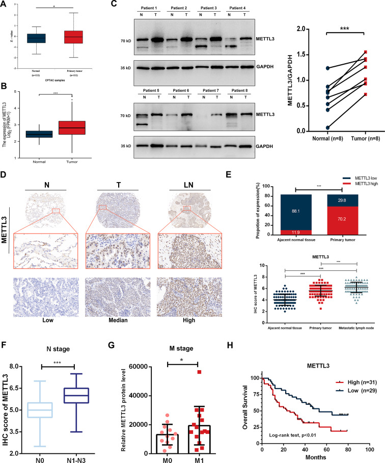
Fig. 1 METTL3 is highly expressed in primary lesions and metastatic lymph nodes of NSCLC, indicating a poor prognosis. (A) The CPTAC database indicates high expression of the METTL3 protein in lung adenocarcinoma tissues. (B) The TCGA database indicates high expression of METTL3 mRNA in NSCLC. (C) Western blot analysis indicates high expression of METTL3 in tumour tissues from 8 NSCLC patients. The right panel displays the quantification results of the left panel. (D) IHC staining of a tissue microarray from 96 patients indicates high expression of METTL3 in primary lesions and metastatic lymph nodes of NSCLC. (E) Quantification results of IHC staining in tissue microarrays. (F) METTL3 expression of primary tumours in N0 stage patients is significantly lower than that in N1-N3 stage patients. (G) DIA-MS results from late-stage NSCLC revealed that METTL3 expression of primary tumours in M0 stage patients is significantly lower than that in M1 stage patients. (H) Survival analysis demonstrates the relationship between METTL3 expression in tissue microarrays and the prognosis of NSCLC patients, with high expression of METTL3 suggesting poor prognosis
Fig. 1 METTL3 is highly expressed in primary lesions and metastatic lymph nodes of NSCLC, indicating a poor prognosis. (A) The CPTAC database indicates high expression of the METTL3 protein in lung adenocarcinoma tissues. (B) The TCGA database indicates high expression of METTL3 mRNA in NSCLC. (C) Western blot analysis indicates high expression of METTL3 in tumour tissues from 8 NSCLC patients. The right panel displays the quantification results of the left panel. (D) IHC staining of a tissue microarray from 96 patients indicates high expression of METTL3 in primary lesions and metastatic lymph nodes of NSCLC. (E) Quantification results of IHC staining in tissue microarrays. (F) METTL3 expression of primary tumours in N0 stage patients is significantly lower than that in N1-N3 stage patients. (G) DIA-MS results from late-stage NSCLC revealed that METTL3 expression of primary tumours in M0 stage patients is significantly lower than that in M1 stage patients. (H) Survival analysis demonstrates the relationship between METTL3 expression in tissue microarrays and the prognosis of NSCLC patients, with high expression of METTL3 suggesting poor prognosisCC BY · PubMed Central
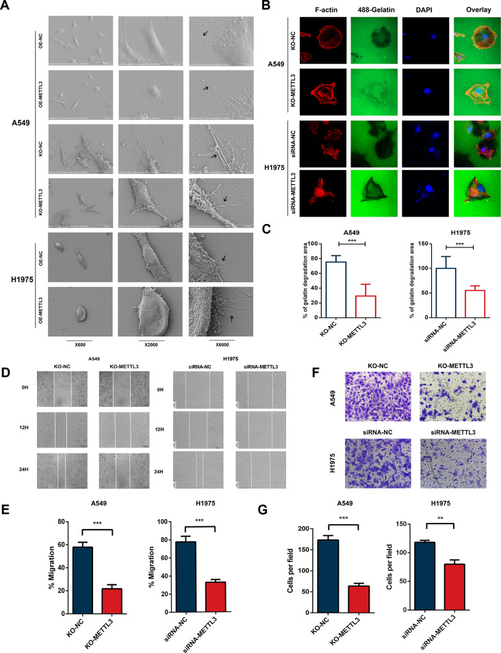
Fig. 2 METTL3 promotes migration, invasion and invadopodia formation in NSCLC cells. (A) Scanning electron microscopy showed that overexpression of METTL3 in A549 and H1975 cells resulted in increased and elongated pseudopodia, while knockout of METTL3 led to decreased and shortened pseudopodia in the cells. (B-C) In the fluorescence matrix degradation experiment, the depth and extent of extracellular matrix degradation were significantly reduced when METTL3 was knocked out in A549 cells using CRISPR‒Cas9 technology. Furthermore, the depth and extent of extracellular matrix degradation were reduced in H1975 cells after METTL3 knockdown. (D-E) The wound healing experiment after METTL3 knockout demonstrated a significant decrease in cell migration ability in A549 cells. After transfection with siRNA targeting METTL3, there is a significant decrease in the migration ability of H1975 cells. (F-G) Transwell invasion experiments after METTL3 knockout showed a significant decrease in cell invasion ability of A549 cells. After transfection with siRNA targeting METTL3, there is a significant decrease in the invasion ability of H1975 cells
Fig. 2 METTL3 promotes migration, invasion and invadopodia formation in NSCLC cells. (A) Scanning electron microscopy showed that overexpression of METTL3 in A549 and H1975 cells resulted in increased and elongated pseudopodia, while knockout of METTL3 led to decreased and shortened pseudopodia in the cells. (B-C) In the fluorescence matrix degradation experiment, the depth and extent of extracellular matrix degradation were significantly reduced when METTL3 was knocked out in A549 cells using CRISPR‒Cas9 technology. Furthermore, the depth and extent of extracellular matrix degradation were reduced in H1975 cells after METTL3 knockdown. (D-E) The wound healing experiment after METTL3 knockout demonstrated a significant decrease in cell migration ability in A549 cells. After transfection with siRNA targeting METTL3, there is a significant decrease in the migration ability of H1975 cells. (F-G) Transwell invasion experiments after METTL3 knockout showed a significant decrease in cell invasion ability of A549 cells. After transfection with siRNA targeting METTL3, there is a significant decrease in the invasion ability of H1975 cellsCC BY · PubMed Central
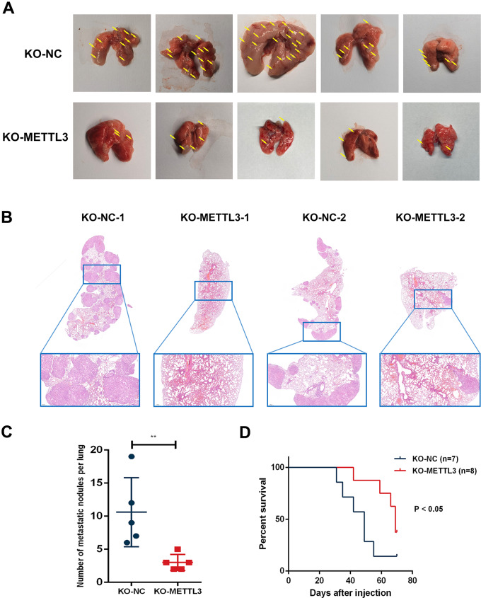
Fig. 3 METTL3 depletion inhibits metastatic potential of lung cancer cells in vivo. (A) Macroscopic image of mouse lungs injected with NC-METTL3 cells and KO-METTL3 cells via the tail vein. The NC-METTL3 group exhibited a significantly higher number of metastatic tumours than the KO-METTL3 group. (B) Histological sections of mouse lungs with HE staining. The NC-METTL3 group showed a significantly higher number of metastatic noduless than the KO-METTL3 group. (C) Quantification of lung metastatic nodules in mice from the NC-METTL3 group and the KO-METTL3 group. The NC-METTL3 group demonstrated significantly more lung metastatic tumours than the KO-METTL3 group. (D) Kaplan‒Meier survival curves for the KO-NC group and KO-METTL3 group
Fig. 3 METTL3 depletion inhibits metastatic potential of lung cancer cells in vivo. (A) Macroscopic image of mouse lungs injected with NC-METTL3 cells and KO-METTL3 cells via the tail vein. The NC-METTL3 group exhibited a significantly higher number of metastatic tumours than the KO-METTL3 group. (B) Histological sections of mouse lungs with HE staining. The NC-METTL3 group showed a significantly higher number of metastatic noduless than the KO-METTL3 group. (C) Quantification of lung metastatic nodules in mice from the NC-METTL3 group and the KO-METTL3 group. The NC-METTL3 group demonstrated significantly more lung metastatic tumours than the KO-METTL3 group. (D) Kaplan‒Meier survival curves for the KO-NC group and KO-METTL3 groupCC BY · PubMed Central
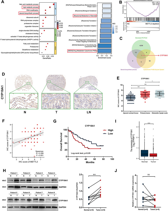
Fig. 4 CYP19A1 is the potential target of METTL3-mediated translation regulation. (A-B) GO-KEGG analysis and GSEA of genes with decreased translation efficiency after METTL3 knockout indicated the association between METTL3 and the steroid signalling pathway. (C) Genes downregulated in translation efficiency after METTL3 depletion were crossed with genes involved in steroid biosynthetic processes and the cytochrome P450 enzyme system to obtain CYP19A1. (D-E) IHC staining analysis of patient tissue microarray showed that compared to adjacent tissues, CYP19A1 protein was highly expressed in both the primary lesions and metastatic lymph nodes of NSCLC. (F) The results of IHC analysis indicated that CYP19A1 expression was positively correlated with METTL3 expression in NSCLC. (G) Survival analysis of the tissue microarray indicated that high expression of CYP19A1 suggests poor prognosis of NSCLC patients. (H) Western blotting analysis validated that the CYP19A1 protein is highly expressed in NSCLC tissues compared with paired normal lung tissues. (I) Data from the TCGA database indicated that CYP19A1 mRNA expression in NSCLC is lower than that in normal lung tissue. (J) RT‒qPCR indicated that CYP19A1 mRNA expression in NSCLC is not higher than that in paired normal lung tissue
Fig. 4 CYP19A1 is the potential target of METTL3-mediated translation regulation. (A-B) GO-KEGG analysis and GSEA of genes with decreased translation efficiency after METTL3 knockout indicated the association between METTL3 and the steroid signalling pathway. (C) Genes downregulated in translation efficiency after METTL3 depletion were crossed with genes involved in steroid biosynthetic processes and the cytochrome P450 enzyme system to obtain CYP19A1. (D-E) IHC staining analysis of patient tissue microarray showed that compared to adjacent tissues, CYP19A1 protein was highly expressed in both the primary lesions and metastatic lymph nodes of NSCLC. (F) The results of IHC analysis indicated that CYP19A1 expression was positively correlated with METTL3 expression in NSCLC. (G) Survival analysis of the tissue microarray indicated that high expression of CYP19A1 suggests poor prognosis of NSCLC patients. (H) Western blotting analysis validated that the CYP19A1 protein is highly expressed in NSCLC tissues compared with paired normal lung tissues. (I) Data from the TCGA database indicated that CYP19A1 mRNA expression in NSCLC is lower than that in normal lung tissue. (J) RT‒qPCR indicated that CYP19A1 mRNA expression in NSCLC is not higher than that in paired normal lung tissueCC BY · PubMed Central
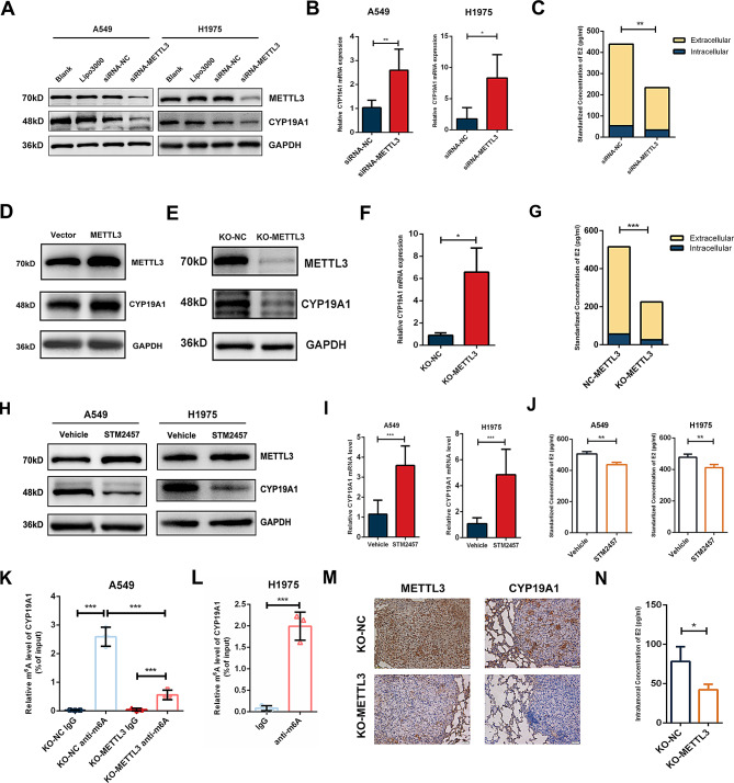
Fig. 5 METTL3 mediates m6A modification of CYP19A1 and promotes the synthesis of E2. (A) WB experiments demonstrated that knockdown of METTL3 using siRNA significantly reduced the protein expression of CYP19A1. (B) PCR experiments showed an upwards trend in the mRNA expression of CYP19A1 following siRNA knockdown of METTL3. (C) ELISA revealed a decrease in intracellular and extracellular levels of E2 after siRNA knockdown of METTL3. (D) CYP19A1 is upregulated after transfection of plasmids for METTL3 overexpression. (E) Protein expression of CYP19A1 is significantly downregulated after CRISPR‒Cas9 knockout of METTL3. (F) PCR indicated an upwards trend in the mRNA expression of CYP19A1 following CRISPR‒Cas9 knockout of METTL3. (G) ELISA demonstrated a significant decrease in intracellular and extracellular levels of E2 after CRISPR‒Cas9 knockout of METTL3. (H) Treatment with 5 µM STM2457 for 3 days led to a significant decrease in the protein level of CYP19A1. (I) Treatment with 5 µM STM2457 for 3 days led to a significant increase in the mRNA level of CYP19A1. (J) Treatment with 5 µM STM2457 for 3 days led to a significant reduction in E2 levels. (K) MeRIP-qPCR revealed that CYP19A1 mRNA was enriched in m6A modification and the relative levels of m6A whin CYP19A1 were significantly downregulated after METTL3 knockout in A549 cells. (L) MeRIP-qPCR revealed that CYP19A1 mRNA was enriched in m6A modification in H1975 cells. (M-N) CYP19A1 protein expression and intratumoural E2 concentration in lung metastasis of KO-METTL3 tumours are significantly downregulated
Fig. 5 METTL3 mediates m6A modification of CYP19A1 and promotes the synthesis of E2. (A) WB experiments demonstrated that knockdown of METTL3 using siRNA significantly reduced the protein expression of CYP19A1. (B) PCR experiments showed an upwards trend in the mRNA expression of CYP19A1 following siRNA knockdown of METTL3. (C) ELISA revealed a decrease in intracellular and extracellular levels of E2 after siRNA knockdown of METTL3. (D) CYP19A1 is upregulated after transfection of plasmids for METTL3 overexpression. (E) Protein expression of CYP19A1 is significantly downregulated after CRISPR‒Cas9 knockout of METTL3. (F) PCR indicated an upwards trend in the mRNA expression of CYP19A1 following CRISPR‒Cas9 knockout of METTL3. (G) ELISA demonstrated a significant decrease in intracellular and extracellular levels of E2 after CRISPR‒Cas9 knockout of METTL3. (H) Treatment with 5 µM STM2457 for 3 days led to a significant decrease in the protein level of CYP19A1. (I) Treatment with 5 µM STM2457 for 3 days led to a significant increase in the mRNA level of CYP19A1. (J) Treatment with 5 µM STM2457 for 3 days led to a significant reduction in E2 levels. (K) MeRIP-qPCR revealed that CYP19A1 mRNA was enriched in m6A modification and the relative levels of m6A whin CYP19A1 were significantly downregulated after METTL3 knockout in A549 cells. (L) MeRIP-qPCR revealed that CYP19A1 mRNA was enriched in m6A modification in H1975 cells. (M-N) CYP19A1 protein expression and intratumoural E2 concentration in lung metastasis of KO-METTL3 tumours are significantly downregulatedCC BY · PubMed Central
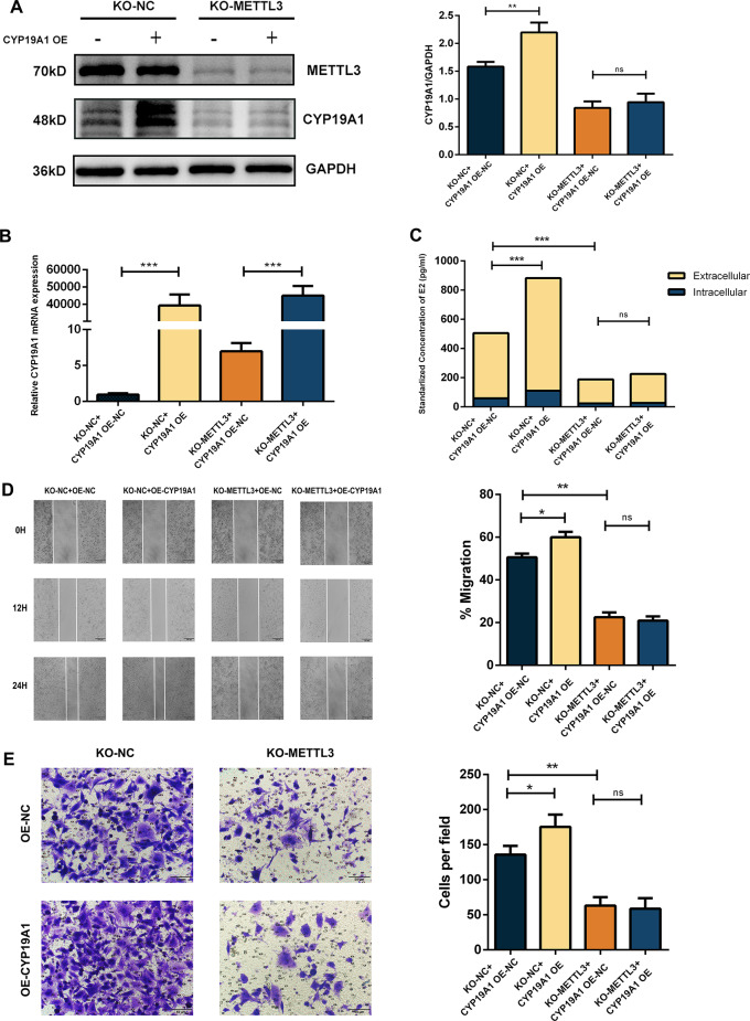
Fig. 6 Translation of CYP19A1 mRNA is dependent on the regulation of METTL3. (A) In KO-METTL3 cells, the transient transfection of plasmids was used to upregulate the mRNA levels of CYP19A1, while METTL3 deficiency abolished its effect on CYP19A1 protein expression. However, in the KO-NC group, transient overexpression of CYP19A1 mRNA successfully led to overexpressed CYP19A1 protein. (B) PCR results showed that both the KO-METTL3 and KO-NC groups transfected with plasmids transiently overexpressed CYP19A1 mRNA. (C) In the KO-NC group, transient overexpression of CYP19A1 mRNA led to upregulation of E2 levels, while in the KO-METTL3 group, METTL3 deficiency decreased intracellular and extracellular E2 secretion. (D) Wound healing experiments showed that in the KO-NC group, transient upregulation of CYP19A1 mRNA significantly enhanced cell migration ability. However, transient upregulation of CYP19A1 mRNA did not significantly alter the cell migration ability under METTL3 deficiency. (E) Transwell invasion assay showed that in the KO-NC group, transient upregulation of CYP19A1 mRNA significantly enhanced cell invasion ability. However, transient upregulation of CYP19A1 mRNA did not significantly alter the cell invasion ability under METTL3 deficiency
Fig. 6 Translation of CYP19A1 mRNA is dependent on the regulation of METTL3. (A) In KO-METTL3 cells, the transient transfection of plasmids was used to upregulate the mRNA levels of CYP19A1, while METTL3 deficiency abolished its effect on CYP19A1 protein expression. However, in the KO-NC group, transient overexpression of CYP19A1 mRNA successfully led to overexpressed CYP19A1 protein. (B) PCR results showed that both the KO-METTL3 and KO-NC groups transfected with plasmids transiently overexpressed CYP19A1 mRNA. (C) In the KO-NC group, transient overexpression of CYP19A1 mRNA led to upregulation of E2 levels, while in the KO-METTL3 group, METTL3 deficiency decreased intracellular and extracellular E2 secretion. (D) Wound healing experiments showed that in the KO-NC group, transient upregulation of CYP19A1 mRNA significantly enhanced cell migration ability. However, transient upregulation of CYP19A1 mRNA did not significantly alter the cell migration ability under METTL3 deficiency. (E) Transwell invasion assay showed that in the KO-NC group, transient upregulation of CYP19A1 mRNA significantly enhanced cell invasion ability. However, transient upregulation of CYP19A1 mRNA did not significantly alter the cell invasion ability under METTL3 deficiencyCC BY · PubMed Central
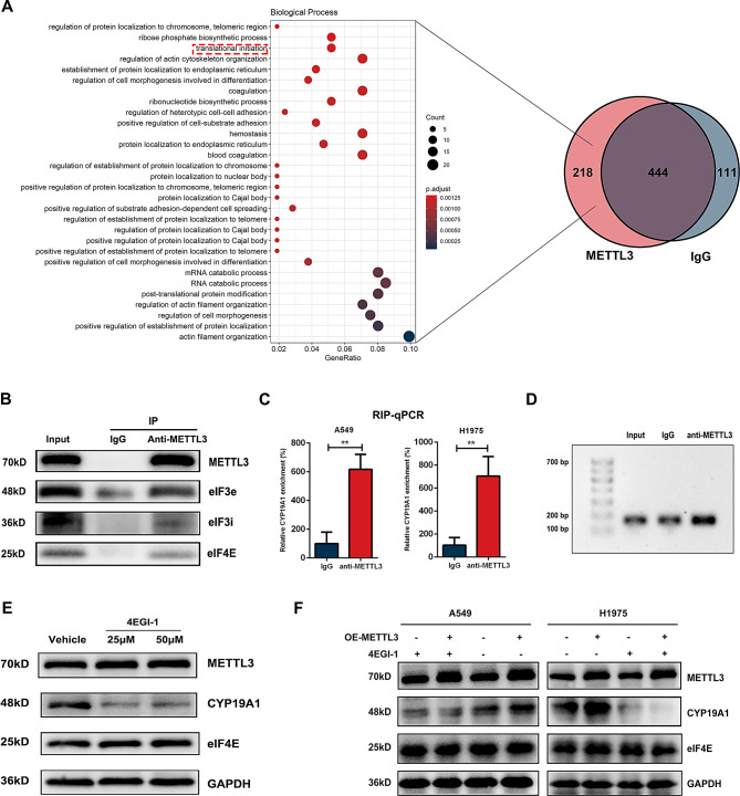
Fig. 7 METTL3 binds to CYP19A1 mRNA and interact with eIFs to regulates translation efficiency of CYP19A1. (A) Immunoprecipitation using anti-METTL3 followed by mass spectrometry analysis was performed to identify proteins that interact with METTL3. A total of 218 proteins were identified as specific binding proteins of METTL3, which are presented in a Venn diagram. GO analysis of the 218 proteins revealed enrichment in the translation initiation process. (B) Co-IP to confirm the interaction between METTL3 and translation initiation molecules identified by mass spectrometry, including eIF3e, eIF3i, and eIF4E. (C-D) RNA immunoprecipitation using anti-METTL3 antibody followed by qPCR validation showing direct binding of METTL3 to CYP19A1 mRNA in A549 and H1975 cells. Agarose gel electrophoresis of qPCR samples confirmed the enrichment of CYP19A1 mRNA in the anti-METTL3 group, confirming the binding between METTL3 and CYP19A1 mRNA. (E) Treatment with the eIF4E inhibitor 4EGI-1 significantly downregulated the protein expression of CYP19A1. (F) Administration of 4EGI-1 abolished the effect of METTL3 overexpression in promoting CYP19A1 expression
Fig. 7 METTL3 binds to CYP19A1 mRNA and interact with eIFs to regulates translation efficiency of CYP19A1. (A) Immunoprecipitation using anti-METTL3 followed by mass spectrometry analysis was performed to identify proteins that interact with METTL3. A total of 218 proteins were identified as specific binding proteins of METTL3, which are presented in a Venn diagram. GO analysis of the 218 proteins revealed enrichment in the translation initiation process. (B) Co-IP to confirm the interaction between METTL3 and translation initiation molecules identified by mass spectrometry, including eIF3e, eIF3i, and eIF4E. (C-D) RNA immunoprecipitation using anti-METTL3 antibody followed by qPCR validation showing direct binding of METTL3 to CYP19A1 mRNA in A549 and H1975 cells. Agarose gel electrophoresis of qPCR samples confirmed the enrichment of CYP19A1 mRNA in the anti-METTL3 group, confirming the binding between METTL3 and CYP19A1 mRNA. (E) Treatment with the eIF4E inhibitor 4EGI-1 significantly downregulated the protein expression of CYP19A1. (F) Administration of 4EGI-1 abolished the effect of METTL3 overexpression in promoting CYP19A1 expressionCC BY · PubMed Central

Verification

Claims the paper makes

The claims and statistics of this paper haven’t been checked yet.

Run the check — free, no account needed. It reads the open-access text and checks the sources it cites.

A few free checks a day · papers already verified open instantly · results are public

Self-check

Percentages, sums and reported differences re-computed from the paper’s own numbers.

The self-check needs the paper’s full text.

Citations

Cites

  • Global quantification of mammalian gene expression control

    Schwanhäusser, B., Busse, D., Li, N., Dittmar, G., Schuchhardt, J., Wolf, J., Chen, W., & Selbach, M. (2011) · Nature · doi:10.1038/nature10098 · Cited by 6,566 · cited 1×

    “N6-methyladenosine (m 6 A) is the most prevalent chemical modification found in eukaryotic RNA, and it plays crucial regulatory roles in RNA stability, splicing, and translation [7, 8].”
    Background

Show all 1 references

Research works printed but not linked: 44

  1. Bakir B, Chiarella AM, Pitarresi JR, Rustgi AK. EMT, MET, plasticity, and Tumor Metastasis. Trends Cell Biol. 2020;30:764–76. https://doi.org/10.1016/j.tcb.2020.07.003. · DOI
  2. Nürnberg A, Kitzing T, Grosse R. Nucleating actin for invasion. Nat Rev Cancer. 2011;11:177–87. https://doi.org/10.1038/nrc3003. · DOI
  3. Artym VV, Zhang Y, Seillier-Moiseiwitsch F, Yamada KM, Mueller SC. Dynamic interactions of cortactin and membrane type 1 matrix metalloproteinase at invadopodia: defining the stages of invadopodia formation and function. Cancer Res. 2006;66:3034–43. https://doi.org/10.1158/0008-5472.can-05-2177. · DOI
  4. Linder S, Cervero P. Mechanisms and roles of podosomes and invadopodia. Nat Rev Mol Cell Biol. 2023;24:86–106. https://doi.org/10.1038/s41580-022-00530-6. · DOI
  5. Eckert MA, et al. Twist1-induced invadopodia formation promotes tumor metastasis. Cancer Cell. 2011;19:372–86. https://doi.org/10.1016/j.ccr.2011.01.036. · DOI
  6. Yilmaz M, Christofori G. EMT, the cytoskeleton, and cancer cell invasion. Cancer Metastasis Rev. 2009;28:15–33. https://doi.org/10.1007/s10555-008-9169-0. · DOI
  7. Sendinc E, Shi Y. RNA m6A methylation across the transcriptome. Mol Cell. 2023;83:428–41. https://doi.org/10.1016/j.molcel.2023.01.006. · DOI
  8. Lin X, et al. RNA m(6)a methylation regulates the epithelial mesenchymal transition of cancer cells and translation of snail. Nat Commun. 2019;10(2065). https://doi.org/10.1038/s41467-019-09865-9. · DOI
  9. Wang Q, et al. Emerging role of RNA methyltransferase METTL3 in gastrointestinal cancer. J Hematol Oncol. 2020;13. https://doi.org/10.1186/s13045-020-00895-1. · DOI
  10. Choe J, et al. mRNA circularization by METTL3-eIF3h enhances translation and promotes oncogenesis. Nature. 2018;561:556–60. https://doi.org/10.1038/s41586-018-0538-8. · DOI
  11. Wei X, et al. METTL3 preferentially enhances non-m(6)a translation of epigenetic factors and promotes tumourigenesis. Nat Cell Biol. 2022;24:1278–90. https://doi.org/10.1038/s41556-022-00968-y. · DOI
  12. Su R, et al. METTL16 exerts an m(6)A-independent function to facilitate translation and tumorigenesis. Nat Cell Biol. 2022;24:205–16. https://doi.org/10.1038/s41556-021-00835-2. · DOI
  13. Huang Q, et al. 17β-estradiol upregulates IL6 expression through the ERβ pathway to promote lung adenocarcinoma progression. J Experimental Clin cancer Research: CR. 2018;37:133. https://doi.org/10.1186/s13046-018-0804-5. · DOI
  14. Meng W, et al. Upregulation of estrogen receptor beta protein but not mRNA predicts poor prognosis and may be associated with enhanced translation in non-small cell lung cancer: a systematic review and meta-analysis. J Thorac Disease. 2021;13:4281–300. https://doi.org/10.21037/jtd-21-658. · DOI
  15. Tang H, et al. Estrogen and insulin-like growth factor 1 synergistically promote the development of lung adenocarcinoma in mice. Int J Cancer. 2013;133:2473–82. https://doi.org/10.1002/ijc.28262. · DOI
  16. Wang Y, et al. ERβ promoted invadopodia formation-mediated non-small cell lung cancer metastasis via the ICAM1/p-Src/p-Cortactin signaling pathway. Int J Cancer. 2023;153:1287–99. https://doi.org/10.1002/ijc.34563. · DOI
  17. Fan S, et al. Estrogen promotes the metastasis of non–small cell lung cancer via estrogen receptor β by upregulation of toll–like receptor 4 and activation of the myd88/NF–κB/MMP2 pathway. Oncol Rep. 2020. https://doi.org/10.3892/or.2020.7574. · DOI
  18. Chen J, et al. Stearoyl-CoA Desaturases1 accelerates Non-small Cell Lung Cancer Metastasis by promoting aromatase expression to improve Estrogen Synthesis. Int J Mol Sci. 2023;24. https://doi.org/10.3390/ijms24076826. · DOI
  19. Stabile LP, et al. Preclinical evidence for combined use of aromatase inhibitors and NSAIDs as preventive agents of Tobacco-Induced Lung Cancer. J Thorac Oncology: Official Publication Int Association Study Lung Cancer. 2018;13:399–412. https://doi.org/10.1016/j.jtho.2017.11.126. · DOI
  20. Vu LP, et al. The N(6)-methyladenosine (m(6)A)-forming enzyme METTL3 controls myeloid differentiation of normal hematopoietic and leukemia cells. Nat Med. 2017;23:1369–76. https://doi.org/10.1038/nm.4416. · DOI
  21. Kim DS, et al. Smac mimetics and oncolytic viruses synergize in driving anticancer T-cell responses through complementary mechanisms. Nat Commun. 2017;8:344. https://doi.org/10.1038/s41467-017-00324-x. · DOI
  22. Zhou Y, Zeng P, Li YH, Zhang Z, Cui Q. SRAMP: prediction of mammalian N6-methyladenosine (m6A) sites based on sequence-derived features. Nucleic Acids Res. 2016;44:e91. https://doi.org/10.1093/nar/gkw104. · DOI
  23. Lehtiö J, Arslan T. Proteogenomics of non-small cell lung cancer reveals molecular subtypes associated with specific therapeutic targets and immune evasion mechanisms. 2, 1224–42, https://doi.org/10.1038/s43018-021-00259-9 (2021). · DOI
  24. Eddy RJ, Weidmann MD, Sharma VP, Condeelis JS. Tumor Cell Invadopodia: invasive protrusions that orchestrate metastasis. Trends Cell Biol. 2017;27:595–607. https://doi.org/10.1016/j.tcb.2017.03.003. · DOI
  25. Liu Y, Chudgar N, Mastrogiacomo B. A germline SNP in BRMS1 predisposes patients with lung adenocarcinoma to metastasis and can be ameliorated by targeting c-fos. 14, eabo1050, https://doi.org/10.1126/scitranslmed.abo1050 (2022). · DOI
  26. Chrysostomou S, Roy R. Repurposed floxacins targeting RSK4 prevent chemoresistance and metastasis in lung and bladder cancer. 13, https://doi.org/10.1126/scitranslmed.aba4627 (2021). · DOI
  27. Deng X, Qing Y. The roles and implications of RNA m(6)a modification in cancer. 20, 507–26, https://doi.org/10.1038/s41571-023-00774-x (2023). · DOI
  28. Mah V, et al. Expression levels of estrogen receptor beta in conjunction with aromatase predict survival in non-small cell lung cancer. Lung Cancer. 2011;74:318–25. https://doi.org/10.1016/j.lungcan.2011.03.009. · DOI
  29. Mah V, et al. Aromatase expression predicts survival in women with early-stage non small cell lung cancer. Cancer Res. 2007;67:10484–90. https://doi.org/10.1158/0008-5472.can-07-2607. · DOI
  30. Yankova E, et al. Small-molecule inhibition of METTL3 as a strategy against myeloid leukaemia. Nature. 2021;593:597–601. https://doi.org/10.1038/s41586-021-03536-w. · DOI
  31. Moerke NJ, et al. Small-molecule inhibition of the interaction between the translation initiation factors eIF4E and eIF4G. Cell. 2007;128:257–67. https://doi.org/10.1016/j.cell.2006.11.046. · DOI
  32. Ma L, et al. The essential roles of m(6)a RNA modification to stimulate ENO1-dependent glycolysis and tumorigenesis in lung adenocarcinoma. J Experimental Clin cancer Research: CR. 2022;41:36. https://doi.org/10.1186/s13046-021-02200-5. · DOI
  33. Xu X, Zhang P. METTL3-mediated m6A mRNA contributes to the resistance of carbon-ion radiotherapy in non-small-cell lung cancer. Cancer Sci. 2023;114:105–14. https://doi.org/10.1111/cas.15590. · DOI
  34. Yang Y, et al. Cigarette smoking, by accelerating the cell cycle, promotes the progression of non-small cell lung cancer through an HIF-1α-METTL3-m(6)A/CDK2AP2 axis. J Hazard Mater. 2023;455:131556. https://doi.org/10.1016/j.jhazmat.2023.131556. · DOI
  35. Yue B, et al. METTL3-mediated N6-methyladenosine modification is critical for epithelial-mesenchymal transition and metastasis of gastric cancer. Mol Cancer. 2019;18. https://doi.org/10.1186/s12943-019-1065-4. · DOI
  36. Li T, et al. METTL3 facilitates tumor progression via an m(6)A-IGF2BP2-dependent mechanism in colorectal carcinoma. Mol Cancer. 2019;18. https://doi.org/10.1186/s12943-019-1038-7. · DOI
  37. Bhat M, et al. Targeting the translation machinery in cancer. Nat Rev Drug Discov. 2015;14:261–78. https://doi.org/10.1038/nrd4505. · DOI · Paper page
  38. Knight JRP, Alexandrou C, Skalka GL, Vlahov N, Pennel K. MNK Inhibition sensitizes KRAS-Mutant Colorectal Cancer to mTORC1 inhibition by reducing eIF4E phosphorylation and c-MYC expression. Cancer Discov. 2021;11:1228–47. https://doi.org/10.1158/2159-8290.cd-20-0652. · DOI · Paper page
  39. Walsh D, Mohr I. Coupling 40S ribosome recruitment to modification of a cap-binding initiation factor by eIF3 subunit e. Genes Dev. 2014;28:835–40. https://doi.org/10.1101/gad.236752.113. · DOI
  40. Hara K, et al. Amino acid sufficiency and mTOR regulate p70 S6 kinase and eIF-4E BP1 through a common effector mechanism. J Biol Chem. 1998;273:14484–94. https://doi.org/10.1074/jbc.273.23.14484. · DOI
  41. Raught B, et al. Phosphorylation of eucaryotic translation initiation factor 4B Ser422 is modulated by S6 kinases. Embo j. 2004;23:1761–9. https://doi.org/10.1038/sj.emboj.7600193. · DOI
  42. Meng W, Han Y, Li B, Li H. The diverse role of RNA methylation in esophageal cancer. Acta Biochim Biophys Sin. 2023;55:988–1000. https://doi.org/10.3724/abbs.2023057. · DOI
  43. Xiao H, Zhao R, Meng W, Liao Y. Effects and translatomics characteristics of a small-molecule inhibitor of METTL3 against non-small cell lung cancer. J Pharm Anal. 2023;13:625–39. https://doi.org/10.1016/j.jpha.2023.04.009. · DOI
  44. Sun Y, et al. METTL3 promotes chemoresistance in small cell lung cancer by inducing mitophagy. J Experimental Clin cancer Research: CR. 2023;42:65. https://doi.org/10.1186/s13046-023-02638-9. · DOI

Metadata

Authors
Meng, W., Xiao, H., Zhao, R., Chen, J., Wang, Y., Mei, P., Li, H., & Liao, Y.
Affiliations
  • Shanghai Jiao Tong University (CN)
  • Huazhong University of Science and Technology (CN)
  • Kunming Medical University (CN)
  • Wuhan Union Hospital (CN)
Corresponding author
  • Li, Hecheng
  • Liao, Yongde
Venue
Cell & Bioscience
Published
2024-01-18
Volume
14
Issue
1
Pages
10-10
ISSN
2045-3701 (ISSN-L)
OpenAlex
W4391011947
Cited by
16
References
1
Citations by year
2026: 4 · 2025: 8 · 2024: 4
FWCI
2.17
Type
journal article
Language
en
Open access
Locations
  • Cell & Bioscience (publishedVersion, cc-by)
  • Research Square (acceptedVersion, cc-by)
  • PubMed Central (publishedVersion, cc-by)
  • PubMed (publishedVersion)
  • DOAJ (DOAJ: Directory of Open Access Journals) (submittedVersion, cc-by-sa)
Funders
  • National Natural Science Foundation of China (82072593)
  • Department of Science and Technology, Hubei Provincial People’s Government (2020BCB027)
Topics
  • Life Sciences › Biochemistry, Genetics and Molecular Biology › Molecular Biology › RNA modifications and cancer
  • Life Sciences › Biochemistry, Genetics and Molecular Biology › Molecular Biology › RNA and protein synthesis mechanisms
  • Life Sciences › Biochemistry, Genetics and Molecular Biology › Molecular Biology › PI3K/AKT/mTOR signaling in cancer
Keywords
METTL3, non-small cell lung cancer, metastasis, CYP19A1, aromatase, estrogen synthesis, N6-methyladenosine, mRNA methylation, translational regulation, eIF4E, translation initiation factors, tumor invasion, therapeutic target

About these checks

Registry, retraction, Cites and statistics results are computed, not judged, with one exception: a printed reference that no index lists is read once by a model before it counts as a citation error. Claims are extracted by a model and kept only when their quote appears verbatim in the text; each cited source is then checked, with a model’s help, for whether it says what the claim relies on. Those verdicts can be wrong — read the claim’s quote and the cited source before relying on one.

Statistics and arithmetic are recomputed, not judged.

Confidence bands are shown only for fields calibrated on our annotated test set. None is calibrated yet, so no band is shown.

What counts as a claim ›How we measure confidence ›How we check this ›

Are you an author, or do you see a problem with this page? Request a review

How to cite

Cite CoChat for the checks on this page, and the paper for its own findings.

This page
CoChat. (2026). Citation check: METTL3 drives NSCLC metastasis by enhancing CYP19A1 translation and oestrogen synthesis. https://app.cochat.ai/papers/10.1186/s13578-024-01194-9
The paper
Meng, W., Xiao, H., Zhao, R., Chen, J., Wang, Y., Mei, P., Li, H., & Liao, Y. (2024). METTL3 drives NSCLC metastasis by enhancing CYP19A1 translation and oestrogen synthesis. Cell & Bioscience. https://doi.org/10.1186/s13578-024-01194-9