

The DOI matches its Crossref record; 22 of the 22 references it cites were found in a registry or an index.
Neurocognitive benchmarks following transplant for emerging cerebral adrenoleukodystrophy
Open accessAbstract
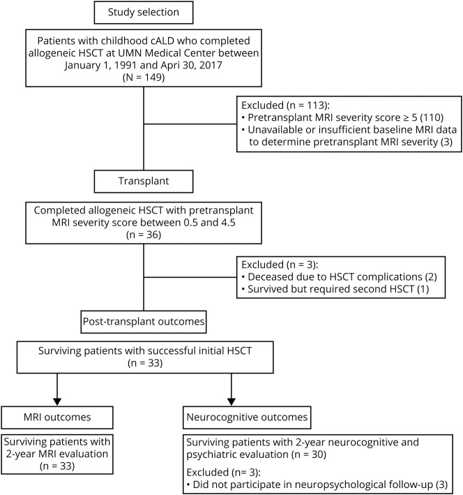
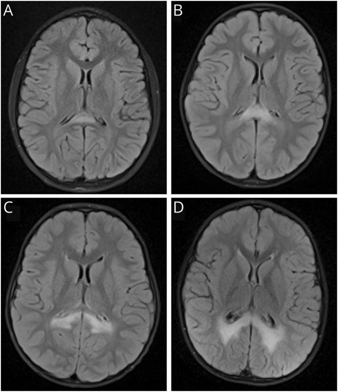
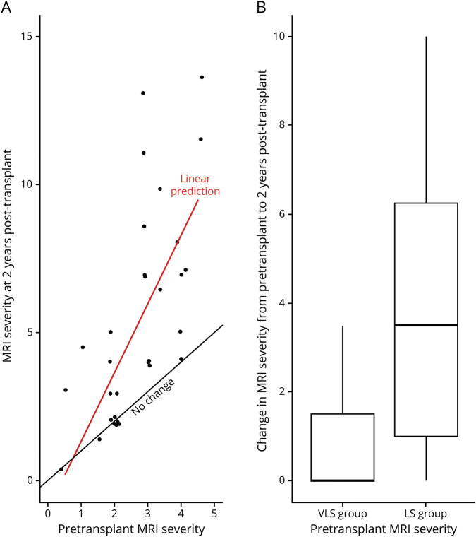
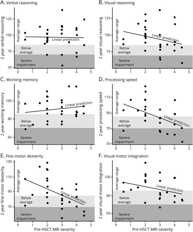
OBJECTIVE: To quantify benchmark treatment outcomes that may be enabled by newborn screening surveillance for X-linked adrenoleukodystrophy (ALD), we report neurocognitive, neuropsychiatric, and MRI change for boys who underwent hematopoietic stem cell transplant (HSCT) at initial stages of demyelination, prior to neurocognitive signs of disease. METHODS: Retrospective chart review identified 36 patients whose cerebral ALD was detected and treated early, with lesion severity less than 5 on the ALD-specific MRI scoring system. Median age at transplant was 7.3 years (range, 4.0-16.1). Progression of radiologic disease on MRI in the 2 years following HSCT was examined relative to the severity of the initial lesion for 33 patients, and longitudinal neurocognitive and neuropsychiatric outcomes were studied for 30 patients. RESULTS: Patients whose pretransplant lesion extended beyond the splenium of the corpus callosum and adjacent periventricular white matter (MRI severity score >2) demonstrated lower posttransplant neurocognitive scores, more neuropsychiatric symptoms, and more disease progression on MRI than patients with a less severe lesion. Changes from baseline neurocognitive functioning were greater at 2 years posttransplant as compared to 1 year. There was greater variance and risk of lesion progression as pretransplant MRI severity increased. CONCLUSION: To realize the full benefits of newborn screening, clinicians must detect very small demyelinating lesions during surveillance and intervene quickly. Novel interventions that reduce risks inherent in allogeneic transplantation are needed. Trial endpoints should include direct neurocognitive assessment and extend at least 2 years posttreatment to provide the greatest sensitivity to detect neurocognitive morbidity.
Overview
The claims and statistics of this paper haven’t been checked yet.
Run the check — free, no account needed. It reads the open-access text and checks the sources it cites.
Integrity signals
Registry
Cites
Statistics
Figures
Verification
Claims the paper makes
The claims and statistics of this paper haven’t been checked yet.
Run the check — free, no account needed. It reads the open-access text and checks the sources it cites.
Claims other papers make about this one
Each is checked against this paper’s own text: does it say what the citing paper says it does?
The source backs part of the claim, not all of it — or backs it for a different population, condition or comparison.
In the citing paper
“However, they are useful only as the first signs of abnormalities are noted on magnetic resonance imaging (MRI) of the brain, before the onset of any neurological symptoms.”
From Identification of a Novel Non-Canonical Splice-Site Variant in ABCD1 (2023) · cites this paper as [4]
Experimental — accuracy not yet measuredThe cited source says what the claim attributes to it.
In the citing paper
“Thus, prompt diagnosis can improve the prognosis of CALD in boys [2,3,4,5,6].”
From Identification of a Novel Non-Canonical Splice-Site Variant in ABCD1 (2023) · cites this paper as [4]
Self-check
Percentages, sums and reported differences re-computed from the paper’s own numbers.
The self-check needs the paper’s full text.
Citations
Cites
Fitting Linear Mixed-Effects Models Using lme4
Bates, D., Mächler, M., Bolker, B., & Walker, S. (2015) · Journal of Statistical Software · doi:10.18637/jss.v067.i01 · Cited by 86,835
Norman, G. R., Sloan, J. A., & Wyrwich, K. W. (2003) · Medical Care · doi:10.1097/01.mlr.0000062554.74615.4c · Cited by 4,700
Moser, H. W., Mahmood, A., & Raymond, G. V. (2007) · Nature Clinical Practice Neurology · doi:10.1038/ncpneuro0421 · Cited by 663
Engelen, M., Kemp, S., de Visser, M., van Geel, B. M., Wanders, R. J., Aubourg, P., & Poll-The, B. (2012) · Orphanet Journal of Rare Diseases · doi:10.1186/1750-1172-7-51 · Cited by 562
Peters, C., Charnas, L. R., Tan, Y., Ziegler, R. S., Shapiro, E. G., DeFor, T., Grewal, S. S., Orchard, P. J., Abel, S. L., Goldman, A. I., et al. (2004) · Blood · doi:10.1182/blood-2003-10-3402 · Cited by 374
Self-citationAdrenoleukodystrophy: a scoring method for brain MR observations.
(1994) · PubMed Central · Cited by 321
Miller, W. P., Rothman, S. M., Nascene, D., Kivisto, T., DeFor, T. E., Ziegler, R. S., Eisengart, J., Leiser, K., Raymond, G., Lund, T. C., et al. (2011) · Blood · doi:10.1182/blood-2011-01-329235 · Cited by 290
Self-citationAnalysis of MRI patterns aids prediction of progression in X-linked adrenoleukodystrophy
Loes, D. J., Fatemi, A., Melhem, E. R., Gupte, N., Bezman, L., Moser, H. W., & Raymond, G. V. (2003) · Neurology · doi:10.1212/01.wnl.0000079050.91337.83 · Cited by 268
Raymond, G. V., Aubourg, P., Paker, A., Escolar, M., Fischer, A., Blanche, S., Baruchel, A., Dalle, J.-H., Michel, G., Prasad, V., et al. (2019) · Biology of Blood and Marrow Transplantation · doi:10.1016/j.bbmt.2018.09.036 · Cited by 142
Self-citation(2000) · PubMed Central · Cited by 114
Cited by
Sorted by citations
Mallack, E. J., Turk, B. R., Yan, H., Price, C., Demetres, M., Moser, A. B., Becker, C., Hollandsworth, K., Adang, L., Vanderver, A., et al. (2021) · Journal of Inherited Metabolic Disease · doi:10.1002/jimd.12356 · Cited by 79
Self-citationTreatment of cerebral adrenoleukodystrophy: allogeneic transplantation and lentiviral gene therapy
Gupta, A. O., Raymond, G., Pierpont, E. I., Kemp, S., McIvor, R. S., Rayannavar, A., Miller, B., Lund, T. C., & Orchard, P. J. (2022) · Expert Opinion on Biological Therapy · doi:10.1080/14712598.2022.2124857 · Cited by 67
“who were identified to have ALD early in life and therefore able to receive treatment when cerebral lesions were quite small (MRI severity score < 5) [38].”
BackgroundSelf-citationMallack, E. J., Gao, K., Engelen, M., & Kemp, S. (2022) · Cells · doi:10.3390/cells11020283 · Cited by 60
Chiesa, R., Boelens, J. J., Duncan, C., Kühl, J., Sevin, C., Kapoor, N., Prasad, V. K., Lindemans, C. A., Jones, S. A., Amartino, H. M., et al. (2021) · Blood Advances · doi:10.1182/bloodadvances.2021005294 · Cited by 41
Self-citationPresymptomatic Lesion in Childhood Cerebral Adrenoleukodystrophy
Mallack, E. J., van Haren, K. P., Torrey, A. C., van de Stadt, S. I. W., Engelen, M., Raymond, G. V., Fatemi, A. S., & Eichler, F. (2022) · Neurology · doi:10.1212/wnl.0000000000200571 · Cited by 17
Gupta, A. O., Nascene, D. R., Shanley, R., Kenney‐Jung, D. L., Eisengart, J. B., Lund, T. C., Orchard, P. J., & Pierpont, E. I. (2021) · Journal of Inherited Metabolic Disease · doi:10.1002/jimd.12435 · Cited by 14
Self-citationMallack, E. J., Askin, G., van de Stadt, S. I. W., Caruso, P. A., Musolino, P. L., Engelen, M., Niogi, S. N., & Eichler, F. (2021) · American Journal of Neuroradiology · doi:10.3174/ajnr.a7250 · Cited by 13
Pierpont, E. I., Isaia, A., McCoy, E., Brown, S. J., Gupta, A. O., & Eisengart, J. B. (2022) · Journal of Inherited Metabolic Disease · doi:10.1002/jimd.12581 · Cited by 12
Self-citationNeurocognitive outcomes in pediatric hematological cancer survivors post‐HSCT: A systematic review
Ansari, S., Garg, A., & Khan, M. A. (2023) · Clinical Transplantation · doi:10.1111/ctr.15193 · Cited by 7
Hematopoietic stem cell therapy and ex vivo gene therapy for X-linked adrenoleukodystrophy
Eichler, F., & Kuehl, J.-S. (2024) · Handbook of clinical neurology · doi:10.1016/b978-0-323-90242-7.00018-3 · Cited by 7
Diffusion Tensor Imaging in Boys With Adrenoleukodystrophy
Pierpont, E. I., Labounek, R., Gupta, A. O., Lund, T. C., Orchard, P. J., Dobyns, W. B., Bondy, M. T., Paulson, A. L., Metz, A., Shanley, R., et al. (2024) · Neurology · doi:10.1212/wnl.0000000000209764 · Cited by 5
Self-citationCharacterizing visual processing deficits in cerebral adrenoleukodystrophy
Corre, C. S., Bambery, M., Bennett, C. R., Nagy, A. M., Manley, C. E., Winter, E., Peregoy, C., Kelly, D. C., Andonian, H., Maciel, S., et al. (2024) · Brain and Development · doi:10.1016/j.braindev.2024.09.008 · Cited by 4
Wang, Y., Tian, G., Ji, W., Wang, S., & Zhang, X. (2021) · Journal of Zhejiang University (Medical Sciences) · doi:10.3724/zdxbyxb-2021-0254 · Cited by 3
Wang, Y., Liu, D., Gao, H., Liu, W., & Mao, Y. (2025) · Journal of Pediatric Hematology/Oncology · doi:10.1097/mph.0000000000003069 · Cited by 2
Identification of a Novel Non-Canonical Splice-Site Variant in ABCD1
Zheng, F., Lin, Z., Hu, Y., Shi, X., Zhao, Q., & Lin, Z. (2023) · Journal of Clinical Medicine · doi:10.3390/jcm12020473 · Cited by 1
Hou, Y.-Q., Yan, W., Deng, Z., Lei, Z., Feng, M., & Song, X.-Q. (2026) · Research in Developmental Disabilities · doi:10.1016/j.ridd.2026.105316 · Cited by 1
Vapljanin, M., Milosavljević, M. N., & Јаnkovic, S. (2024) · Biotechnology & Biotechnological Equipment · doi:10.1080/13102818.2024.2342888 · Cited by 1
Adrenoleukodystrophy: Symptoms, Treatment and Newborn Screening. Literature Review and Clinical Case
Regelskytė, L., & Praninskienė, R. (2024) · Neurologijos seminarai · doi:10.15388/ns.2024.28.99.1
Recognizing Cerebral Pathology in Adrenoleukodystrophy Amid the Noise
Pierpont, E. I. (2025) · Neurology · doi:10.1212/wnl.0000000000214394
Self-citationLate-Onset X-linked Adrenoleukodystrophy: A Rare Cause of Progressive Spastic Paraparesis
Sequeira, S., Costa, A., Vargas, M., & Velon, A. G. (2025) · Cureus · doi:10.7759/cureus.99429
Rinaldi, S., Rinaldi, A., & Fontani, V. (2026) · Neurology International · doi:10.3390/neurolint18040063
Metadata
- Authors
- Pierpont, E. I., Nascene, D. R., Shanley, R., Kenney-Jung, D. L., Ziegler, R. S., Miller, W. P., Gupta, A. O., Lund, T. C., Orchard, P. J., & Eisengart, J. B.
- Affiliations
- University of Minnesota (US)
- From the Departments of Pediatrics (E.I.P., R.S.Z., W.P.M., A.O.G., T.C.L., P.J.O., J.B.E.), Radiology (D.R.N.), Bioinformatics and Biostatistics Core (R.S.), and Neurology (D.L.K.-J.), University of Minnesota, Minneapolis; and Sangamo Therapeutics (W.P.M.), Richmond, CA.
- Corresponding author
- Pierpont, Elizabeth I.
- ORCID
- Venue
- Neurology
- Published
- 2020-08-04
- Volume
- 95
- Issue
- 5
- Pages
- e591-e600
- ISSN
- 0028-3878 (ISSN-L)
- 1526-632X
- PMC
- PMC7455349
- OpenAlex
- W3039456116
- Cited by
- 21
- References
- 22
- Citations by year
- 2026: 2 · 2025: 3 · 2024: 5 · 2023: 2 · 2022: 4 · 2021: 4 · 2020: 1
- FWCI
- 1.82
- Type
- journal article
- Language
- en
- Open access
- hybrid
- doi.org
- Licence
- cc-by-nc-nd
- Locations
- Neurology (publishedVersion, cc-by-nc-nd)
- PubMed Central (publishedVersion, cc-by-nc-nd)
- PubMed (publishedVersion)
- Funders
- National Institutes of Health (UL1TR002494, KL2TR002492)
- National Center for Advancing Translational Sciences
- Topics
- Life Sciences › Biochemistry, Genetics and Molecular Biology › Clinical Biochemistry › Metabolism and Genetic Disorders
- Health Sciences › Medicine › Genetics (clinical) › Lysosomal Storage Disorders Research
- Health Sciences › Medicine › Clinical Neurology › Multiple Sclerosis Research Studies
- Keywords
- cerebral adrenoleukodystrophy, X-linked adrenoleukodystrophy, hematopoietic stem cell transplantation, newborn screening, neuropsychological outcomes, neuropsychiatric symptoms, demyelination, splenium of corpus callosum, periventricular white matter, disease progression, clinical trial endpoints
- MeSH terms
- Adolescent, Adrenoleukodystrophy, Child, Child, Preschool, Humans, Infant, Newborn, Magnetic Resonance Imaging, Male, Neonatal Screening, Treatment Outcome, Hematopoietic Stem Cell Transplantation, Benchmarking, Early Diagnosis
About these checks
Registry, retraction, Cites and statistics results are computed, not judged, with one exception: a printed reference that no index lists is read once by a model before it counts as a citation error. Claims are extracted by a model and kept only when their quote appears verbatim in the text; each cited source is then checked, with a model’s help, for whether it says what the claim relies on. Those verdicts can be wrong — read the claim’s quote and the cited source before relying on one.
Statistics and arithmetic are recomputed, not judged.
Confidence bands are shown only for fields calibrated on our annotated test set. None is calibrated yet, so no band is shown.
What counts as a claim ›How we measure confidence ›How we check this ›
Are you an author, or do you see a problem with this page? Request a review
