

The DOI matches its Crossref record; 33 of the 35 references it cites were found in a registry or an index.
Lymphatic pathology in asymptomatic and symptomatic children with Wuchereria bancrofti infection in children from Odisha, India and its reversal with DEC and albendazole treatment
Open accessAbstract
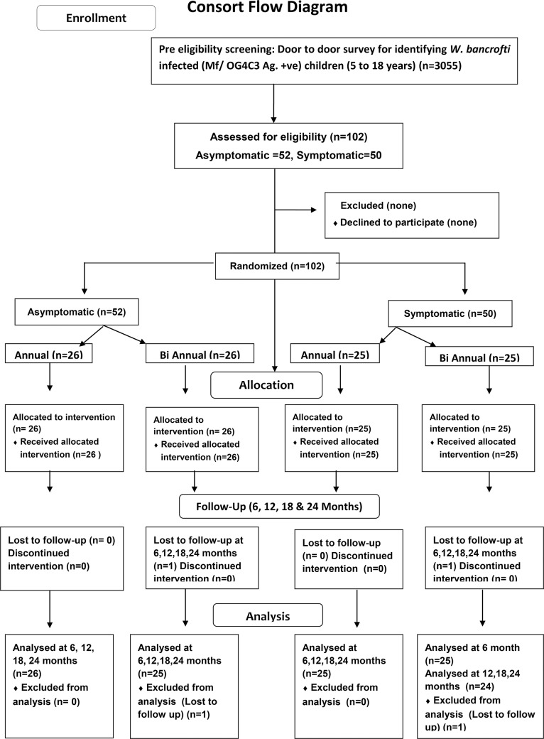
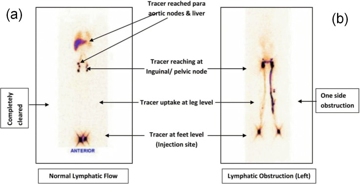
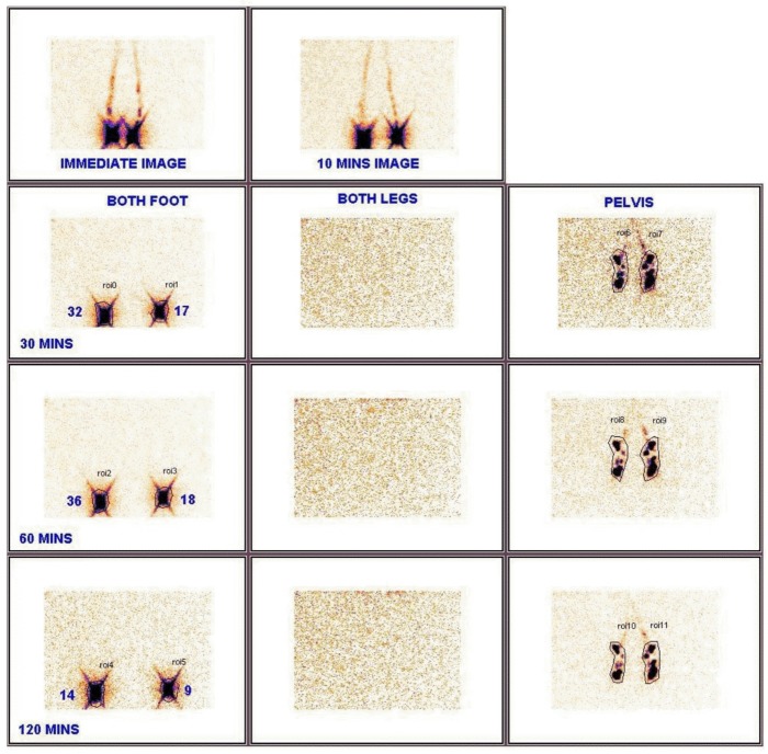
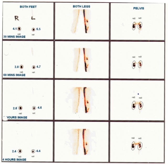
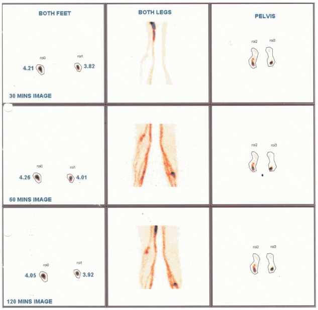
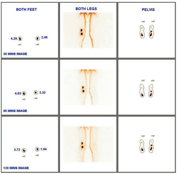
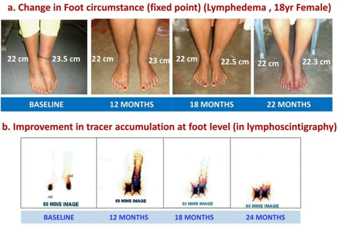
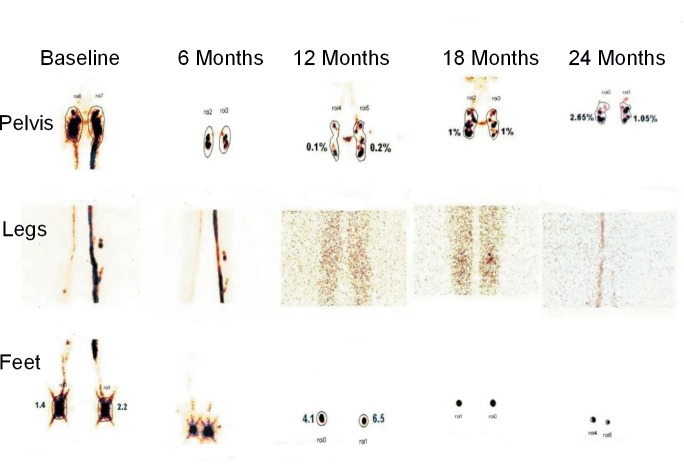
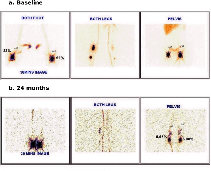
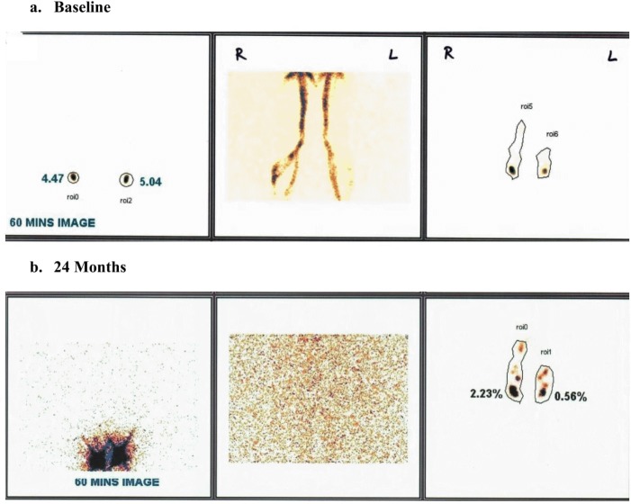
BACKGROUND: Once interruption of transmission of lymphatic filariasis is achieved, morbidity prevention and management becomes more important. A study in Brugia malayi filariasis from India has shown sub-clinical lymphatic pathology with potential reversibility. We studied a Wuchereria bancrofti infected population, the major contributor to LF globally. METHODS: Children aged 5-18 years from Odisha, India were screened for W. bancrofti infection and disease. 102 infected children, 50 with filarial disease and 52 without symptoms were investigated by lymphoscintigraphy and then randomized to receive a supervised single oral dose of DEC and albendazole which was repeated either annually or semi-annually. The lymphatic pathology was evaluated six monthly for two years. FINDINGS: Baseline lymphoscintigraphy showed abnormality in lower limb lymphatics in 80% of symptomatic (40/50) and 63·5% (33/52) of asymptomatic children. Progressive improvement in baseline pathology was seen in 70·8, 87·3, 98·6, and 98·6% of cases at 6, 12, 18, and 24 months follow up, while in 4·2, 22·5, 47·9 and 64·8%, pathology reverted to normal. This was independent of age (p = 0·27), symptomatic status (p = 0·57) and semi-annual/bi-annual dosing (p = 0·46). Six of eleven cases showed clinical reduction in lymphedema of legs. INTERPRETATION: A significant proportion of a young W. bancrofti infected population exhibited lymphatic pathology which was reversible with annual dosage of DEC and albendazole. This provides evidence for morbidity prevention & treatment of early lymphedema. It can also be used as a tool to improve community compliance during mass drug administration. TRIAL REGISTRATION: ClinicalTrials.gov No CTRI/2013/10/004121.
Overview
The claims and statistics of this paper haven’t been checked yet.
Run the check — free, no account needed. It reads the open-access text and checks the sources it cites.
Integrity signals
Registry
Cites
Statistics
Figures
Verification
Claims the paper makes
The claims and statistics of this paper haven’t been checked yet.
Run the check — free, no account needed. It reads the open-access text and checks the sources it cites.
Claims other papers make about this one
Each is checked against this paper’s own text: does it say what the citing paper says it does?
The cited source says what the claim attributes to it.
In the citing paper
“a significant proportion of children with W. bancrofti infection in India had their lymphatic pathology reversed when given the combination of albendazole and DEC annually or semiannually for 2 years [206]”
From Albendazole and Mebendazole as Anti-Parasitic and Anti-Cancer Agents: an Update (2021) · cites this paper as [206]
Self-check
Percentages, sums and reported differences re-computed from the paper’s own numbers.
The self-check needs the paper’s full text.
Citations
Cites
Strategies and tools for the control/elimination of lymphatic filariasis.
(1997) · Europe PMC (PubMed Central) · Cited by 621
Editorial: The Global Programme to Eliminate Lymphatic Filariasis
Ottesen, E. A. (2000) · Tropical Medicine & International Health · doi:10.1046/j.1365-3156.2000.00620.x · Cited by 574
Re-assessing the global prevalence and distribution of lymphatic filariasis
Michael, E., Bundy, D. A. P., & Grenfell, B. T. (1996) · Parasitology · doi:10.1017/s0031182000066646 · Cited by 516
Lymph-node concentration of radioactive colloidal gold following interstitial injection
Sherman, A. I., Ter‐Pogossian, M. M., & Tocus, W. T. T. A. O. E. C. (1953) · Cancer · doi:10.1002/1097-0142(195311)6:6 · Cited by 250
Lymphatic filariasis infection and disease: Control strategies
Ottesen, E. A., & Ramachandran, C. P. (1995) · Parasitology Today · doi:10.1016/0169-4758(95)80128-6 · Cited by 201
Self-citationDreyer, G., de Medeiros, Z. M., Netto, M. J., Leal, N. C., de Castro, L. G., & Piessens, W. F. (1999) · Transactions of the Royal Society of Tropical Medicine and Hygiene · doi:10.1016/s0035-9203(99)90140-2 · Cited by 150
Mand, S., Debrah, A. Y., Klarmann‐Schulz, U., Debrah, L. B., Marfo‐Debrekyei, Y., Kwarteng, A., Specht, S., Belda-Domene, A., Fimmers, R., Taylor, M. J., et al. (2012) · Clinical Infectious Diseases · doi:10.1093/cid/cis486 · Cited by 122
Freedman, D. O., Filho, P. J. D. A., Besh, S., Silva, M. C. M. E., Braga, C., & Maciel, A. (1994) · The Journal of Infectious Diseases · doi:10.1093/infdis/170.4.927 · Cited by 112
Filariasis in Travelers Presenting to the GeoSentinel Surveillance Network
Lipner, E. M., Law, M. A., Barnett, E. D., Keystone, J. S., von Sonnenburg, F., Loutan, L., Prevots, D. R., Klion, A. D., Nutman, T. B., & for the GeoSentinel Surveillance Network (2007) · PLoS neglected tropical diseases · doi:10.1371/journal.pntd.0000088 · Cited by 109
SHENOY, R. K., Kumaraswami, V., Suma, T., Rajan, K. V., & Radhakuttyamma, G. (1999) · Annals of Tropical Medicine and Parasitology · doi:10.1080/00034983.1999.11813433 · Cited by 100
Research works printed but not linked: 1
- 2002, Prospects of eliminating Lymphatic Filariasis in India, ICMR Bulletin, 32, 1
Cited by
Sorted by citations
Albendazole and Mebendazole as Anti-Parasitic and Anti-Cancer Agents: an Update
Chai, J.-Y., Jung, B.-K., & Hong, S.-J. (2021) · The Korean Journal of Parasitology · doi:10.3347/kjp.2021.59.3.189 · Cited by 286
Albendazole alone or in combination with microfilaricidal drugs for lymphatic filariasis
Macfarlane, C. L., Budhathoki, S. S., Johnson, S., Richardson, M., & Garner, P. (2019) · Cochrane Database of Systematic Reviews · doi:10.1002/14651858.cd003753.pub4 · Cited by 48
Furlong-Silva, J., Cross, S. D., Marriott, A. E., Pionnier, N. P., Archer, J., Steven, A., Schulte‐Merker, S., Mack, M., Hong, Y., Taylor, M. J., et al. (2021) · Journal of Clinical Investigation · doi:10.1172/jci140853 · Cited by 39
Advances in Antiwolbachial Drug Discovery for Treatment of Parasitic Filarial Worm Infections
Bakowski, M. A., & McNamara, C. W. (2019) · Tropical Medicine and Infectious Disease · doi:10.3390/tropicalmed4030108 · Cited by 38
Elimination of lymphatic filariasis in South East Asia
Specht, S., Suma, T. K., Pedrique, B., & Hoerauf, A. M. (2019) · BMJ · doi:10.1136/bmj.k5198 · Cited by 35
Metadata
- Authors
- Kar, S. K., Dwibedi, B., Das, B. K., Agrawala, B. K., Ramachandran, C. P., & Horton, J.
- Affiliations
- Siksha O Anusandhan University (IN)
- Regional Medical Research Center (IN)
- Utkal University (IN)
- Universiti Sains Malaysia (MY)
- Corresponding author
- Horton, John
- Venue
- PLOS Neglected Tropical Diseases
- Publisher
- Public Library of Science (PLoS)
- Published
- 2017-10-23
- Volume
- 11
- Issue
- 10
- Pages
- e0005631
- ISSN
- 1935-2727 (ISSN-L)
- 1935-2735
- PMC
- PMC5667936
- OpenAlex
- W2765419938
- Cited by
- 25
- References
- 35
- Citations by year
- 2026: 2 · 2025: 2 · 2024: 1 · 2022: 2 · 2021: 3 · 2020: 5 · 2019: 6 · 2018: 3 · 2017: 1
- FWCI
- 3.16
- Type
- journal article
- Language
- en
- Update notices
- new_version (2017-11-02)
- Open access
- gold
- doi.org
- Licence
- Locations
- PLoS neglected tropical diseases (publishedVersion, cc-by)
- Europe PMC (PubMed Central) (publishedVersion, cc-by)
- PubMed Central (publishedVersion, cc-by)
- PubMed (publishedVersion)
- DOAJ (DOAJ: Directory of Open Access Journals) (submittedVersion, cc-by-sa)
- Figshare (submittedVersion, cc-by)
- Greater South Information System
- Funders
- Bill and Melinda Gates Foundation (43922)
- Topics
- Health Sciences › Medicine › Infectious Diseases › Parasitic Diseases Research and Treatment
- Life Sciences › Immunology and Microbiology › Parasitology › Parasites and Host Interactions
- Health Sciences › Medicine › Public Health, Environmental and Occupational Health › Research on Leishmaniasis Studies
- Keywords
- lymphatic filariasis, Wuchereria bancrofti, lymphatic pathology, lymphoscintigraphy, diethylcarbamazine, albendazole, mass drug administration, lymphedema, children, Odisha
- MeSH terms
- Administration, Oral, Adolescent, Animals, Child, Child, Preschool, Diethylcarbamazine, Elephantiasis, Filarial, Female, Filaricides, Humans, India, Lymphatic System, Lymphedema, Male, Wuchereria bancrofti, Albendazole, Treatment Outcome, Asymptomatic Infections, Lymphoscintigraphy
About these checks
Registry, retraction, Cites and statistics results are computed, not judged, with one exception: a printed reference that no index lists is read once by a model before it counts as a citation error. Claims are extracted by a model and kept only when their quote appears verbatim in the text; each cited source is then checked, with a model’s help, for whether it says what the claim relies on. Those verdicts can be wrong — read the claim’s quote and the cited source before relying on one.
Statistics and arithmetic are recomputed, not judged.
Confidence bands are shown only for fields calibrated on our annotated test set. None is calibrated yet, so no band is shown.
What counts as a claim ›How we measure confidence ›How we check this ›
Are you an author, or do you see a problem with this page? Request a review
How to cite
Cite CoChat for the checks on this page, and the paper for its own findings.
- This page
- CoChat. (2026). Citation check: Lymphatic pathology in asymptomatic and symptomatic children with Wuchereria bancrofti infection in children from Odisha, India and its reversal with DEC and albendazole treatment. https://app.cochat.ai/papers/10.1371/journal.pntd.0005631
- The paper
- Kar, S. K., Dwibedi, B., Das, B. K., Agrawala, B. K., Ramachandran, C. P., & Horton, J. (2017). Lymphatic pathology in asymptomatic and symptomatic children with Wuchereria bancrofti infection in children from Odisha, India and its reversal with DEC and albendazole treatment. PLOS Neglected Tropical Diseases. https://doi.org/10.1371/journal.pntd.0005631
