

The DOI matches its Crossref record.
Respiratory Evolution Facilitated the Origin of Pterosaur Flight and Aerial Gigantism
Open accessAbstract
Pterosaurs, enigmatic extinct Mesozoic reptiles, were the first vertebrates to achieve true flapping flight. Various lines of evidence provide strong support for highly efficient wing design, control, and flight capabilities. However, little is known of the pulmonary system that powered flight in pterosaurs. We investigated the structure and function of the pterosaurian breathing apparatus through a broad scale comparative study of respiratory structure and function in living and extinct archosaurs, using computer-assisted tomographic (CT) scanning of pterosaur and bird skeletal remains, cineradiographic (X-ray film) studies of the skeletal breathing pump in extant birds and alligators, and study of skeletal structure in historic fossil specimens. In this report we present various lines of skeletal evidence that indicate that pterosaurs had a highly effective flow-through respiratory system, capable of sustaining powered flight, predating the appearance of an analogous breathing system in birds by approximately seventy million years. Convergent evolution of gigantism in several Cretaceous pterosaur lineages was made possible through body density reduction by expansion of the pulmonary air sac system throughout the trunk and the distal limb girdle skeleton, highlighting the importance of respiratory adaptations in pterosaur evolution, and the dramatic effect of the release of physical constraints on morphological diversification and evolutionary radiation.
Overview
The claims and statistics of this paper haven’t been checked yet.
Run the check — free, no account needed. It reads the open-access text and checks the sources it cites.
Integrity signals
Registry
Cites
Statistics
Figures
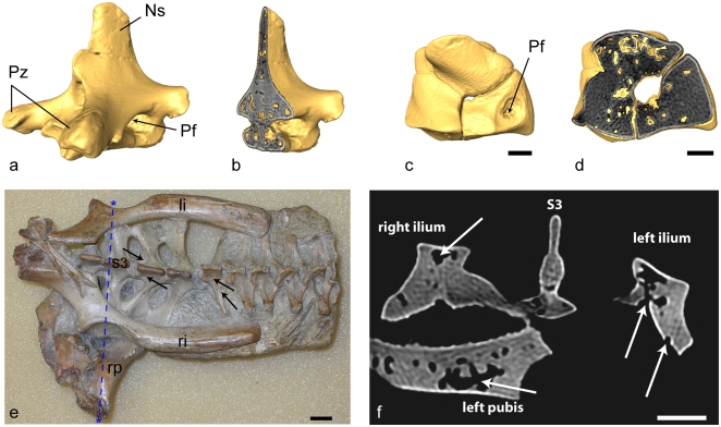
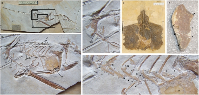
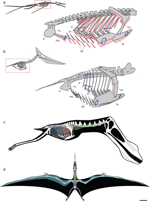
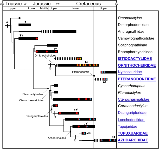
Verification
Claims the paper makes
The claims and statistics of this paper haven’t been checked yet.
Run the check — free, no account needed. It reads the open-access text and checks the sources it cites.
Claims other papers make about this one
Each is checked against this paper’s own text: does it say what the citing paper says it does?
The check wasn’t confident either way, so it gives no verdict — never disputed.
In the citing paper
“Bats had arboreal, gliding antecedents [124] and pterosaurs, whose origins are unknown [125], are suspected to have also been arboreal gliders [8], [126].”
From Assessing Arboreal Adaptations of Bird Antecedents: Testing the Ecological Setting of the Origin of the Avian Flight Stroke (2011) · cites this paper as [125]
Experimental — accuracy not yet measured
Self-check
Percentages, sums and reported differences re-computed from the paper’s own numbers.
The self-check needs the paper’s full text.
Citations
Cites
Still being checked.
Research works printed but not linked: 40
- K Padian, 1983, A functional analysis of flying and walking in pterosaurs., Paleobiology, 9, 218, 10.1017/S009483730000765X · DOI
- K Padian, 1992, The wings of pterosaurs., American Journal of Science, 293, 91, 10.2475/ajs.293.A.91 · DOI
- MT Wilkinson, 2005, High lift function of the pteroid bone and forewing of pterosaurs., Proceedings of the Royal Society of London B: Biological Sciences, 273, 119, 10.1098/rspb.2005.3278 · DOI
- DR Carrier, 2000, The evolution of pelvic aspiration in archosaurs., Paleobiology, 26, 271, 10.1666/0094-8373(2000)026<0271:TEOPAI>2.0.CO;2 · DOI
- JA Ruben, 2003, Respiratory and reproductive paleophysiology of dinosaurs and early birds., Physiological and Biochemical Zoology, 76, 141, 10.1086/375425 · DOI
- D Carrier, 1987, The evolution of locomotor stamina in tetrapods: circumventing a mechanical constraint., Paleobiology, 13(3), 326, 10.1017/S0094837300008903 · DOI
- CG Farmer, 2000, Pelvic aspiration in the American alligator (Alligator mississippiensis)., Journal of Experimental Biology, 203, 1679, 10.1242/jeb.203.11.1679 · DOI
- JW Hicks, 1999, Gas exchange potential in reptilian lungs: implications for the dinosaur- avian connection., Respiration Physiology, 117, 73, 10.1016/S0034-5687(99)00060-2 · DOI
- T Owerkowicz, 1999, Contribution of gular pumping to lung ventilation in monitor lizards., Science, 284, 1661, 10.1126/science.284.5420.1661 · DOI
- MT Wilkinson, 2008, Three-dimensional geometry of a pterosaur wing skeleton, and its implications for aerial and terrestrial locomotion., Zoological Journal of the Linnean Society, 154, 27, 10.1111/j.1096-3642.2008.00409.x · DOI
- PM O'Connor, 2006, Postcranial pneumaticity: an evaluation of soft-tissue influences on the postcranial skeleton and the reconstruction of pulmonary anatomy in archosaurs., Journal of Morphology, 267, 1199, 10.1002/jmor.10470 · DOI
- PM O'Connor, 2005, Basic avian pulmonary design and flow-through ventilation in nonavian theropod dinosaurs., Nature, 436, 253, 10.1038/nature03716 · DOI
- PM O'Connor, 2004, Pulmonary pneumaticity in the postcranial skeleton of extant Aves: a case study examining Anseriformes., Journal of Morphology, 261, 141, 10.1002/jmor.10190 · DOI
- LPAM Claessens, 2005, The evolution of breathing mechanisms in the Archosauria [Ph.D. thesis]., 258
- LPAM Claessens, 2004, Archosaurian respiration and the pelvic girdle aspiration breathing of crocodyliforms., Proceedings of the Royal Society of London B: Biological Sciences, 271, 1461, 10.1098/rspb.2004.2743 · DOI
- LPAM Claessens, 2004, Dinosaur gastralia; origin, morphology, and function., Journal of Vertebrate Paleontology, 24, 89, 10.1671/A1116-8 · DOI
- P Sereno, 2008, Evidence for avian intrathoracic air sacs in a new predatory dinosaur from Argentina., PLoS ONE, 3, e3303, 10.1371/journal.pone.0003303 · DOI
- CG Farmer, 2006, On the origin of avian air sacs., Respiratory Physiology & Neurobiology, 154, 89, 10.1016/j.resp.2006.04.014 · DOI
- LPAM Claessens, A cineradiographic study of lung ventilation in Alligator mississippiensis., Journal of Experimental Zoology, Part A
- LPAM Claessens, The skeletal kinematics of lung ventilation in three basal bird taxa (emu, tinamou, and guinea fowl)., Journal of Experimental Zoology, Part A
- R Wild, 1978, Die Flugsaurier (Reptilia, Pterosauria) aus der Oberen Trias von Cene bei Bergamo, Italien., Bollettino della società paleontologica Italiana, 17, 176
- SC Bennett, 2001, The osteology and functional morphology of the Late Cretaceous pterosaur Pteranodon., Palaeontographica A, 260, 1, 10.1127/pala/260/2001/1 · DOI
- K Zimmer, 1935, Beiträge zur Mechanik der Atmung bei den Vögeln in Stand und Flug auf Grund anatomisch-physiologischer und experimenteller Studien., Zoologica (Stuttgart), 33, 1
- JR Codd, 2005, Activity of three muscles associated with the uncinate processes of the giant Canada goose Branta canadensis maximus., Journal of Experimental Biology, 208, 849, 10.1242/jeb.01489 · DOI
- JR Codd, 2008, Avian-like breathing mechanics in maniraptoran dinosaurs., Proceedings of the Royal Society of London B: Biological Sciences, 275, 157, 10.1098/rspb.2007.1233 · DOI
- PG Tickle, 2007, Functional significance of uncinate processes in birds., Journal of Experimental Biology, 210, 3955, 10.1242/jeb.008953 · DOI
- SF Perry, 1983, Reptilian lungs. Functional anatomy and evolution., Advances in Anatomy, Embryology, and Cell Biology, 79, 1
- DO Kuethe, 1988, Fluid mechanical valving of air flow in bird lungs., Journal of Experimental Biology, 136, 1, 10.1242/jeb.136.1.1 · DOI
- R Owen, 1859, Monograph on the fossil Reptilia of the Cretaceous Formations. Supplement No. 1. Pterosauria (Pterodactylus)., Palaeontographical Society, 1
- MJ Wedel, 2003, The evolution of vertebral pneumaticity in sauropod dinosaurs., Journal of Vertebrate Paleontology, 23, 344, 10.1671/0272-4634(2003)023[0344:TEOVPI]2.0.CO;2 · DOI
- H-R Duncker, 1971, The lung air sac system of birds., Advances in Anatomy, Embryology, and Cell Biology, 45, 1
- KF Liem, 1989, Respiratory gas bladders in teleosts: functional conservatism and morphological diversity., American Zoologist, 29, 333, 10.1093/icb/29.1.333 · DOI
- DM Martill, 1989, Exceptionally well-preserved pterosaur wing membrane from the Cretaceous of Brazil., Nature, 340, 138, 10.1038/340138a0 · DOI
- F Bignon, 1889, Contribution a l'etude de la pneumaticite chez les oiseaux., Mémoires de la Société zoologique de France, 2, 260
- AR Akester, 1973, Subcutaneous air pouches in the Marabou stork., Journal of Zoology (London), 170, 493, 10.1111/j.1469-7998.1973.tb05063.x · DOI
- F Broili, 1927, Ein Rhamphorhynchus mit Spuren von Haarbedeckung., Sitzungsberichte der Bayerischen Akademie der Wissenschaften, mathemathisch-naturwissenschaftliche Abteilung, 1927, 49
- AG Sharov, 1971, [New flying reptiles from the Mesozoic of Kazakhstan and Kirghizia][In Russian]., Transactions of the Palaeontological Institute, 130, 104
- LM Witmer, 2003, Neuroanatomy of flying reptiles and implications for flight, posture and behaviour., Science, 425, 950
- P Wellnhofer, 1975, Die Rhamphorhynchoidea (Pterosauria) der Oberjura-Plattenkalke Süddeutschlands. Teil III., Palaeontographica A, 149, 1
- P Wellnhofer, 1991, Weitere Pterosaurierfunde aus der Santana-Formation (Apt) der Chapada do Araripe, Brasilien., Paleontographica, 215, 43
Cited by
Sorted by citations
Paleohistological estimation of bone growth rate in extinct archosaurs
Cubo, J., Le Roy, N., Martinez-Maza, C., & Montes, L. (2012) · Paleobiology · doi:10.1666/08093.1 · Cited by 123
A new Jurassic theropod from China documents a transitional step in the macrostructure of feathers
Lefèvre, U., Cau, A., Cincotta, A., Hu, D., Chinsamy, A., Escuillié, F., & Godefroit, P. (2017) · The Science of Nature · doi:10.1007/s00114-017-1496-y · Cited by 62
FOTH, C., BRUSATTE, S. L., & BUTLER, R. J. (2012) · Journal of Evolutionary Biology · doi:10.1111/j.1420-9101.2012.02479.x · Cited by 61
Dececchi, T. A., & Larsson, H. C. E. (2011) · PLoS ONE · doi:10.1371/journal.pone.0022292 · Cited by 56
“unknown [125], are suspected to have also been arboreal gliders [8,126].”
BackgroundHone, D., Henderson, D. M., Therrien, F., & Habib, M. B. (2015) · PeerJ · doi:10.7717/peerj.1191 · Cited by 34
Metadata
- Authors
- Claessens, L. P. A. M., O'Connor, P. M., & Unwin, D. M.
- Affiliations
- College of the Holy Cross (US)
- University of Leicester (GB)
- Corresponding author
- Claessens, Leon P. A. M.
- Venue
- PLoS ONE
- Publisher
- Public Library of Science (PLoS)
- Published
- 2009-02-18
- Volume
- 4
- Issue
- 2
- Pages
- e4497
- ISSN
- 1932-6203 (ISSN-L)
- PMC
- PMC2637988
- OpenAlex
- W1981670163
- Cited by
- 156
- References
- 0
- Citations by year
- 2026: 5 · 2025: 14 · 2024: 9 · 2023: 5 · 2022: 4 · 2021: 10 · 2020: 11 · 2019: 7 · 2018: 5 · 2017: 14 · 2016: 5 · 2015: 5 · 2014: 7 · 2013: 15 · 2012: 13
- FWCI
- 10.16
- Type
- journal article
- Language
- en
- Open access
- gold
- doi.org
- Locations
- PLoS ONE (publishedVersion, cc-by)
- PubMed Central (publishedVersion, cc-by)
- PubMed (publishedVersion)
- DOAJ (DOAJ: Directory of Open Access Journals) (submittedVersion, cc-by-sa)
- Figshare (submittedVersion, cc-by)
- Leicester Research Archive (University of Leicester) (submittedVersion, other-oa)
- Funders
- National Science Foundation (0206169)
- Harvard University
- University of Leicester
- Deutsche Forschungsgemeinschaft
- Topics
- Physical Sciences › Earth and Planetary Sciences › Paleontology › Paleontology and Evolutionary Biology
- Health Sciences › Medicine › Anatomy › Comparative Animal Anatomy Studies
- Physical Sciences › Earth and Planetary Sciences › Paleontology › Evolution and Paleontology Studies
- Keywords
- pterosaurs, respiratory system, flapping flight, convergent evolution, archosaurs, birds, alligators, Cretaceous, evolutionary radiation
- MeSH terms
- Alligators and Crocodiles, Animals, Birds, Biological Evolution, Flight, Animal, Fossils, Reptiles, Respiratory System, Skeleton
About these checks
Registry, retraction, Cites and statistics results are computed, not judged, with one exception: a printed reference that no index lists is read once by a model before it counts as a citation error. Claims are extracted by a model and kept only when their quote appears verbatim in the text; each cited source is then checked, with a model’s help, for whether it says what the claim relies on. Those verdicts can be wrong — read the claim’s quote and the cited source before relying on one.
Statistics and arithmetic are recomputed, not judged.
Confidence bands are shown only for fields calibrated on our annotated test set. None is calibrated yet, so no band is shown.
What counts as a claim ›How we measure confidence ›How we check this ›
Are you an author, or do you see a problem with this page? Request a review
