

The DOI matches its Crossref record; 49 of the 49 references it cites were found in a registry or an index.
A Partial Structural and Functional Rescue of a Retinitis Pigmentosa Model with Compacted DNA Nanoparticles
Open accessAbstract
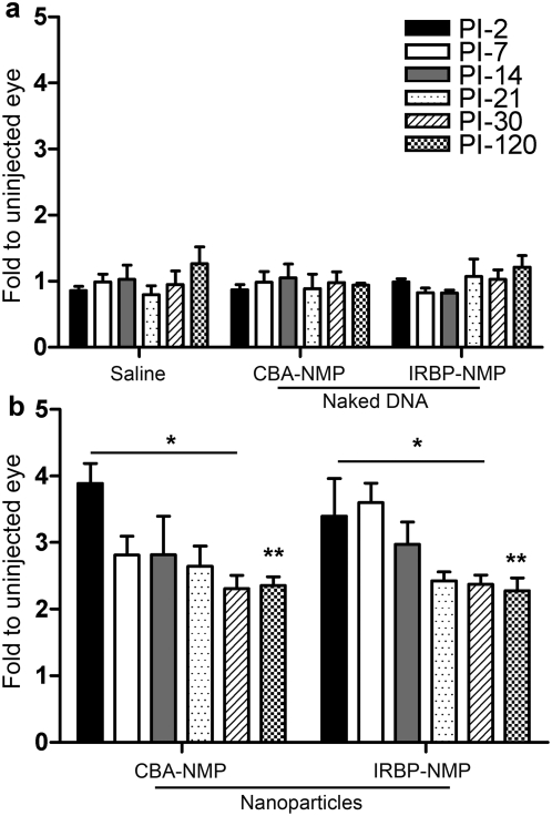
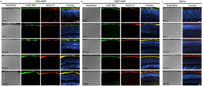
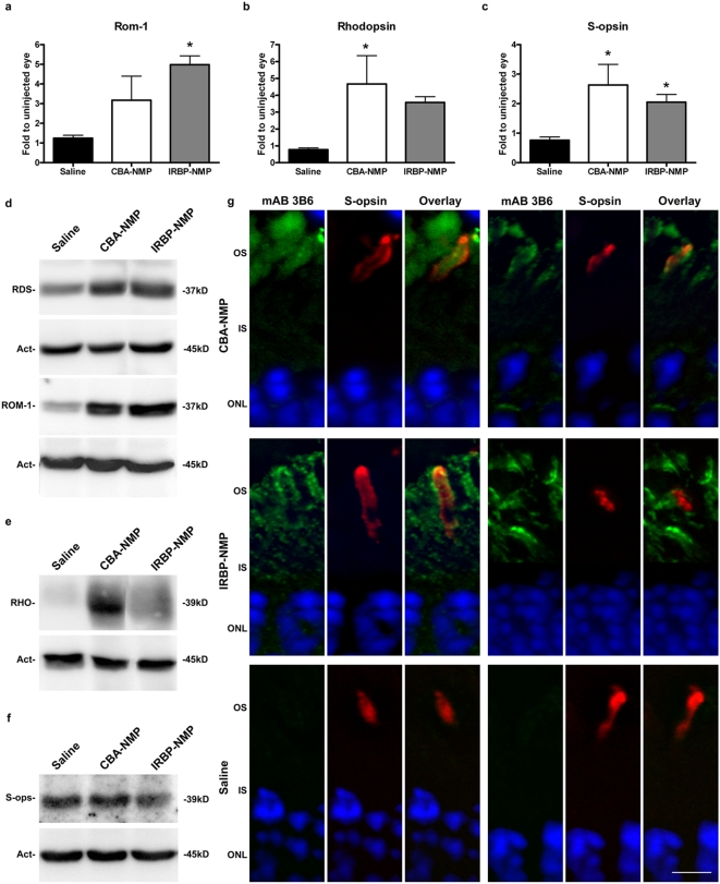
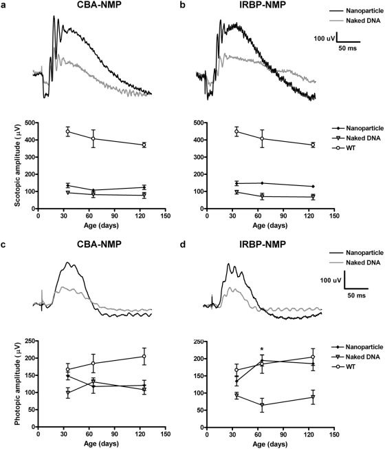
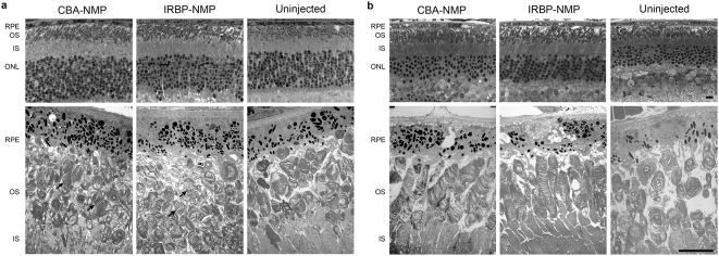
Previously we have shown that compacted DNA nanoparticles can drive high levels of transgene expression after subretinal injection in the mouse eye. Here we delivered compacted DNA nanoparticles containing a therapeutic gene to the retinas of a mouse model of retinitis pigmentosa. Nanoparticles containing the wild-type retinal degeneration slow (Rds) gene were injected into the subretinal space of rds(+/-) mice on postnatal day 5. Gene expression was sustained for up to four months at levels up to four times higher than in controls injected with saline or naked DNA. The nanoparticles were taken up into virtually all photoreceptors and mediated significant structural and biochemical rescue of the disease without histological or functional evidence of toxicity. Electroretinogram recordings showed that nanoparticle-mediated gene transfer restored cone function to a near-normal level in contrast to transfer of naked plasmid DNA. Rod function was also improved. These findings demonstrate that compacted DNA nanoparticles represent a viable option for development of gene-based interventions for ocular diseases and obviate major barriers commonly encountered with non-viral based therapies.
Overview
The claims and statistics of this paper haven’t been checked yet.
Run the check — free, no account needed. It reads the open-access text and checks the sources it cites.
Integrity signals
Registry
Cites
Statistics
Figures
Verification
Claims the paper makes
The claims and statistics of this paper haven’t been checked yet.
Run the check — free, no account needed. It reads the open-access text and checks the sources it cites.
Claims other papers make about this one
Each is checked against this paper’s own text: does it say what the citing paper says it does?
In the citing paper
“Single-molecule DNA nanoparticles compacted by polyethylene glycol (PEG)-substituted lysine peptides (CK30-PEG) have been used to carry payloads up to 20 kb in size,82,83 with several groups demonstrating their safety in BALB/c mice and rds+/− Rho-/- or Abca4-/- mouse models of retinal degeneration.82–86”
From Gene Therapy for Retinal Degenerative Diseases: Progress, Challenges, and Future Directions (2023) · cites this paper as [82]
Self-check
Percentages, sums and reported differences re-computed from the paper’s own numbers.
The self-check needs the paper’s full text.
Citations
Cites
A new mathematical model for relative quantification in real-time RT-PCR
Pfaffl, M. W. (2001) · Nucleic Acids Research · doi:10.1093/nar/29.9.e45 · Cited by 34,610
Safety and Efficacy of Gene Transfer for Leber's Congenital Amaurosis
Maguire, A. M., Simonelli, F., Pierce, E. A., Pugh, E. N., Mingozzi, F., Bennicelli, J., Banfi, S., Marshall, K. A., Testa, F., Surace, E. M., et al. (2008) · New England Journal of Medicine · doi:10.1056/nejmoa0802315 · Cited by 2,145
Effect of gene therapy on visual function in Leber’s congenital amaurosis
Bainbridge, J. W. B., Smith, A. J., Barker, S. S., Robbie, S., Henderson, R., Balaggan, K., Viswanathan, A., Holder, G. E., Stockman, A., Tyler, N., et al. (2008) · New England Journal of Medicine · doi:10.1056/nejmoa0802268 · Cited by 1,924
Gene therapy restores vision in a canine model of childhood blindness
Acland, G. M., Aguirre, G. D., Ray, J., Zhang, Q., Aleman, T. S., Cideciyan, A. V., Pearce-Kelling, S. E., Anand, V., Zeng, Y., Maguire, A. M., et al. (2001) · Nature Genetics · doi:10.1038/ng0501-92 · Cited by 1,202 · cited 1×
“Viral gene therapy has been remarkably successful in treating some types of ocular diseases, the most notable example of which to date being the AAV-mediated long-term rescue of vision in Briard dogs harboring a mutation in RPE65 [1,2].”
BackgroundElectroporation and RNA interference in the rodent retina in vivo and in vitro
Matsuda, T., & Cepko, C. L. (2003) · Proceedings of the National Academy of Sciences · doi:10.1073/pnas.2235688100 · Cited by 1,032
Cell fate determination in the vertebrate retina.
Cepko, C. L., Austin, C. P., Yang, X.-J., Alexiades, M. R., & Ezzeddine, D. (1996) · Proceedings of the National Academy of Sciences · doi:10.1073/pnas.93.2.589 · Cited by 989
Rods and cones in the mouse retina. I. Structural analysis using light and electron microscopy
Carter‐Dawson, L. D., & LaVail, M. M. (1979) · The Journal of Comparative Neurology · doi:10.1002/cne.901880204 · Cited by 695
Cideciyan, A. V., Alemán, T. S., Boye, S. L., Schwartz, S., Kaushal, S., Román, A. J., Pang, J., Sumaroka, A., Windsor, E. A. M., Wilson, J. M., et al. (2008) · Proceedings of the National Academy of Sciences · doi:10.1073/pnas.0807027105 · Cited by 674
Apoptotic photoreceptor cell death in mouse models of retinitis pigmentosa.
Portera‐Cailliau, C., Sung, C.-H., Nathans, J., & Adler, R. (1994) · Proceedings of the National Academy of Sciences · doi:10.1073/pnas.91.3.974 · Cited by 628
Lagali, P. S., Bálya, D., Awatramani, G. B., Münch, T. A., Kim, D. S., Busskamp, V., Cepko, C. L., & Roska, B. (2008) · Nature Neuroscience · doi:10.1038/nn.2117 · Cited by 580
Research works printed but not linked: 1
- MJ Cooper, 2007, Nonviral gene transfer in the lung: from animal to human., Molecular therapy, S15, 315
Cited by
Sorted by citations
Nanotechnology in Ocular Drug Delivery
Patel, D. H., Pathak, D., & Giradkar, P. (2021) · Springer eBooks · doi:10.1007/978-981-33-4497-6_18 · Cited by 362
Nanoparticle diffusion in, and microrheology of, the bovine vitreous ex vivo
Xu, Q., Boylan, N. J., Suk, J. S., Wang, Y.-Y., Nance, E. A., Yang, J., McDonnell, P. J., Cone, R. A., Duh, E. J., & Hanes, J. S. (2013) · Journal of Controlled Release · doi:10.1016/j.jconrel.2013.01.018 · Cited by 339
Vector platforms for gene therapy of inherited retinopathies
Trapani, I., Puppo, A., & Auricchio, A. (2014) · Progress in Retinal and Eye Research · doi:10.1016/j.preteyeres.2014.08.001 · Cited by 191
Punzo, C., Xiong, W., & Cepko, C. L. (2011) · Journal of Biological Chemistry · doi:10.1074/jbc.r111.304428 · Cited by 185
DNA nanoparticle-mediated ABCA4 delivery rescues Stargardt dystrophy in mice
Han, Z., Conley, S. M., Makkia, R. S., Cooper, M. J., & Naash, M. I. (2012) · Journal of Clinical Investigation · doi:10.1172/jci64833 · Cited by 150
Self-citation
Metadata
- Authors
- Cai, X., Nash, Z., Conley, S. M., Fliesler, S. J., Cooper, M. J., & Naash, M. I.
- Affiliations
- University of Oklahoma Health Sciences Center (US)
- Saint Louis University (US)
- Copernicus Therapeutics (United States) (US)
- Corresponding author
- Naash, Muna I.
- Venue
- PLoS ONE
- Publisher
- Public Library of Science (PLoS)
- Published
- 2009-04-24
- Volume
- 4
- Issue
- 4
- Pages
- e5290
- ISSN
- 1932-6203 (ISSN-L)
- PMC
- PMC2669177
- OpenAlex
- W1969461625
- Cited by
- 109
- References
- 49
- Citations by year
- 2026: 2 · 2025: 8 · 2024: 3 · 2023: 3 · 2022: 5 · 2021: 5 · 2020: 5 · 2019: 1 · 2018: 7 · 2017: 4 · 2016: 4 · 2015: 7 · 2014: 8 · 2013: 8 · 2012: 14
- FWCI
- 5.24
- Type
- journal article
- Language
- en
- Open access
- gold
- doi.org
- Locations
- PLoS ONE (publishedVersion, cc-by)
- PubMed Central (publishedVersion, cc-by)
- PubMed (publishedVersion)
- DOAJ (DOAJ: Directory of Open Access Journals) (submittedVersion, cc-by-sa)
- Figshare (submittedVersion, cc-by)
- Funders
- Foundation Fighting Blindness
- Research to Prevent Blindness
- Oklahoma Center for the Advancement of Science and Technology
- National Institutes of Health (EY018656)
- National Eye Institute (R56EY010609, EY007361, EY10609, R01EY007361, R01EY018656, R01EY010609, EY018656)
- Topics
- Health Sciences › Medicine › Ophthalmology › Retinal Development and Disorders
- Life Sciences › Biochemistry, Genetics and Molecular Biology › Molecular Biology › RNA Interference and Gene Delivery
- Life Sciences › Pharmacology, Toxicology and Pharmaceutics › Pharmaceutical Science › Advanced Drug Delivery Systems
- Keywords
- retinitis pigmentosa, gene therapy, subretinal injection, photoreceptors, cone function, rod function, electroretinogram, non-viral gene delivery, ocular gene therapy, mouse model
- MeSH terms
- Animals, Disease Models, Animal, DNA, Electroretinography, Immunohistochemistry, Intermediate Filament Proteins, Membrane Glycoproteins, Mice, Transgenic, Nerve Tissue Proteins, Retinitis Pigmentosa, Transgenes, Photoreceptor Cells, Vertebrate, Mice, Nanoparticles, Peripherins
About these checks
Registry, retraction, Cites and statistics results are computed, not judged, with one exception: a printed reference that no index lists is read once by a model before it counts as a citation error. Claims are extracted by a model and kept only when their quote appears verbatim in the text; each cited source is then checked, with a model’s help, for whether it says what the claim relies on. Those verdicts can be wrong — read the claim’s quote and the cited source before relying on one.
Statistics and arithmetic are recomputed, not judged.
Confidence bands are shown only for fields calibrated on our annotated test set. None is calibrated yet, so no band is shown.
What counts as a claim ›How we measure confidence ›How we check this ›
Are you an author, or do you see a problem with this page? Request a review
